- 62 -
KISEP Original Article J Rhinol 11(1,2), 2004
Outcomes of Transnasal Endoscopic Sinus Surgery in 86 Cases of Non-invasive Fungal Sinusitis
Kyung Chul Lee, M.D., Chang Gyu Kim, M.D. and Jae Ho Ban, M.D.
ABSTRACT
This study was conducted to review the clinical features of fungal sinusitis and to evaluate the effectiveness of transnasal endoscopic sinus surgery as a primary surgical method for treating fungal sinusitis. Eighty five patients (86 cases) who were treated for fungal sinusitis with transnasal endoscopic sinus surgery between 1993 and 2004 were retrospectively analyzed by reviewing their profile, which included clinical feature, surgical techniques, operative findings postoperative results and com- plications. All patients were adults consisting of 30 males and 55 females. All cases were treated successfully by transnasal endoscopic sinus surgery. No recurrence and postoperative complication were observed. However, in 18 cases, fungal debries were observed when sinus irrigation was done at primary follow-up. In these cases, fungal debris disappeared at postopera- tive 1.7weeks (average) and no recurrence was observed. Transnasal endoscopic sinus surgery is successful method in the treatment of non-invasive fungal sinusitis.
KEY WORDS:Aspergillosis・Sinusitis.
INTRODUCTION
Fungal sinusitis was a relatively rare disease in the past, however due to the developoment of diagnostic methods, increased use of steroid and antibiotics and increase of diabetes mellitus and immune deficiency patients, its frequency is gradually increasing. The treat- ment for fungal sinusitis is thorough excision of the lesion and fungus through operation followed by suffi- cient air ventilation into the nasal cavity. Caldwell-Luc operation or inferior meatal antrostomy was used for treatment in the past, however as a result of the intro- duction of endoscopic diagnosis and treatment for nasal cavity and paranasal sinus diseases, endoscopic sinus surgery has been made possible for the treatment of
fungal sinusitis. Endoscopic sinus surgery carries the merit of fast recovery, short admission period, minimal occurrence of complications such as changes of anato- mical structures, cheek pain and paresthesi. Therefore, endoscopic sinus surgery has recently been carried out in many cases of non-invasive fungal sinusitis. The au- thors carried out a retrospective study based on the re- currence rates and follow-up results of 85 patients that underwent transnasal endoscopic sinus surgery for fun- gal sinusitis to inquire the effectiveness of transnasal sinus surgery as a primary treatment method for fungal sinusitis.
MATERIALS AND METHODS
This study is based on 85 patients (86 cases) that underwent a transnasal endoscopic sinus surgery under the diagnosis of fungal sinusitis at our department of otorhinolaryngology Head and Neck Surgery from Oc- tober 1993 through June 2004. The diagnosis was con- firmed by endoscopic findings and histopathologic examinations. These patients were retrospectively an- alyzed according to medical records, radiological and histopathologic examination results. Allergic fungal si- nusitis was excluded by a study of past medical his- Department of Otolaryngology Head and Neck Surgery, Kang-
buk Samsung Hospital, School of Medicine, Sungkyunkwan Uni- versity, Seoul, Korea
Address correspondences and reprint requests to Jae Ho Ban, M.D., Department of Otolaryngology Head and Neck Surgery, College of Medicine, Sungkyunkwan University, Kangbuk Sam- sung Hospital, 108 Pyungdong, Jongnoku, Seoul 110-746 Korea Tel:82-2-2001-2267, Fax:82-2-2001-2273
E-mail:[email protected]
Accepted for publication on August 16, 2004
Lee et al:Non-Invasive Fungal Sinusitis / 63
tories of allergy and skin test and 1 case of fulminant mucormycosis that invaded the cerebrum and orbit and 1 case of invasive fungal sinusitis was also excluded from this study. The male:female ratio was 30:55, with a higher incidence in women. The age distribu- tion was from 22 to 73 years old with an average age of 52 years old. The incidence was the highest in the 5th decade (Table 1). A transnasal endosdopic sinus surgery using the Stammberger technique was carried out in all cases under local anesthesia. The uncinate process and ethmoid bulla was removed under 0°
endoscope and the maxillary sinus ostium was con- firmed using a 30°endoscope and a transmiddle meatal antrostomy was carried out. The dark brown fungal ball within the maxillary sinus was removed by several forceps or curved suction tips under 30°, 70°, 120°endoscope. A frontal sinusotomy or endoscopic anterior and posterior ethmoidectomy and sphenoido- tomy were carried out as needed. Packing and nasal crust was removed on postoperative day 1 and irriga- tion of the nasal cavity and paranasal sinus was carried out. For the first two weeks after surgery, the patients visited the outpatient clinic twice a week for local dressing and nasal cavity and paranasal sinus irrigation under transnasal endoscope and from the third to eighth week local dressing was carried out once a week. Then on from the eighth week the patient visited the outpa- tient clinic at an interval of 2 weeks or 1 month to observe possibilities of recurrence. When there was suspicion that fungal debris still existed after irrigation, a computerized tomography was carried out for confir- mation. Antibiotics were used for 4 weeks after surgery and steroid and antifungal agents were not used. The postoperative follow up period was between 2-11 months with an average of 3.7 months.
RESULTS
The chief symptom was nasal obstruction in 30 patients (35%) followed by purulent rhinorrhea in 14 (16%), headache in 11 (12%), postnasal dripping in 9 (11%), foul odor in 9 (11%) and cheek pain in 5 pa- tients (6%) in order of occurrence. 7 patients (8%) without symptoms were radiologically diagnosed in- cidently (Table 2). The lapse period from the first visit to the outpatient clinic to surgery since the appearance of first symptoms was 3 months in 11 cases (13%), 3-6 months in 36 (43%), 6 months to 1 year in 24 (28%) and over a year in 14 cases (16%) (Table 3).
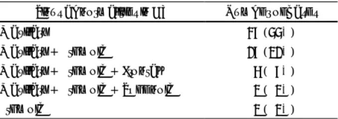
A paranasal sinus computerized tomography was car- ried out in all cases and among them calcification of the paranasal sinus was observed in 72 cases and among all 85 patients soft tissue density was observed in 3 cases with 1 case of chronic paranasal sinusitis caused by a nasal polyp of the opposite side, 1 case of retention cyst of the opposite side and 1 case of fungal sinusitis of both sides. The distribution of paranasal sinuses that showed soft tissue density is as follows;maxillary sinus in 47 cases (55%), maxillary and ethmoid sinus is 27 (32%), maxillary,ethmoid and frontal sinus in 6 (7%), maxillary,ethmoid and sphenoid sinus in 3 (4%) and sphenoid sinus in 3 cases (4%). There were no cases where all sinuses showed soft tissue density (Table 4).
According to histopathologic examinations, there
Table 2. Presenting symptoms in fungal sinusitis
Symptoms Number of the patients
Nasal obstruction 30 (35%)
Rhinorrhea 14 (16%)
Headache 11 (13%)
Post nasal dripping 09 (11%)
Foul odor 09 (11%)
Pain on cheek 05 (06%)
No symptom 07 (08%)
Table 1. Age and sex distribution of 85 patients with fungal sinusitis
Number of patients Age (yr)
Male Female Total
20-29 00 01 01
30-39 02 10 12
40-49 12 13 25
50-59 09 18 27
60-69 06 11 17
70-79 01 02 03
Total 30 55 85
Table 3. Duration of symptoms prior to surgery
Duration Number of cases
-3 month 11 (13%)
3-6 month 36 (43%)
6-12 month 24 (28%)
1 year- 14 (16%)
64 / J Rhinol 11(1,2), 2004
were 62 cases (72%) of Aspergillosis and 26 cases (28%) of unverified fungal diseases. It was fungal ball in all 86 cases with 1 case of aspergillosis that was accompanied by an inverted papilloma.
Among the 86 cases that were treated with transna- sal endoscopic sinus surgery, sole invasion of the ma- xillary sinus occurred in 67 cases (78%), simultaneous invasion of the maxillary and ethmoid sinus in 16 cases (19%) and sole invasion of the sphenoid sinus occurred in 3 cases (4%). Complication or recurrence findings were not noticed durng follow up in all cases that underwent transnasal endoscopic sinus surgery There were a total of 18 cases (21%) that showed a fungal ball from the point of view of transnasal endos- cope or nasal cavity and paranasal sinus irrigation during primary follow up after discharge. 15 cases were in the maxillary sinus and 3 in the maxillary and eth- moid sinuses. There were no cases of fungal debris in the sphenoid sinus. All cases improved through nasal cavity and paranasal sinus irrigation. After an average of 1.7 weeks of irrigation treatment, the fungal ball no longer existed. There was also no lesion observed on computerized tomography that was carried out during follow up.
DISCUSSION
Fungal infection of the nasal cavity and paranasal sinus was a rare disease since Mackenzie1) first repor- ted it 1893, however it is gradually increasing. Fun- guses that cause fungal sinusitis are Aspergilus, Mu- cormycose, Candida, Histoplasma and so forth2) where Aspergilus is the most common. In this study, 62 among 86 cases were aspergillosis. It has been reported that the occurrence rate is higher in women and in this study the male:female ratio was 1.8:1 with a higher occurrence rate in women as well which showed results that were in concurrence to a study reported by Lee,3)
Park4) et al.
Clinical symptoms are mostly asymptomatic or uni- lateral nasal obstruction, postnatal dripping, cheek pain, purulent rhinorrhea, headache and so forth. In this study, nasal obstruction was the most common symptom that was complained by 30 (35%) out of 85 patients. Nasal obstruction was also the most common symptom with 64% in a study carried out by Kim5) et al. In acute or chronic paranasal sinusitis patients whose symptoms continue without improvement and do not respond to an appropriate antibiotic treatment, fungal infection must be suspected. Fungal sinusitis is classified into allergic fungal sinusitis, non-invasive fungal ball, in- vasive type, and acute fulminant type.6)7) All 86 cases that underwent a transnasal endoscopic sinus surgery were the non invasive type. The invasive type appears as a chronic granulomatous infection that crosses the boundary of the paranasal sinus and slowly progresses to gradually destroy the anatomical boundary of the eyeball, cheekbone, brain, palate and so forth and tissue invasion must be histologically proven. The acute ful- minant type mostly occurs in metabolism disorders such as diabetes mellitus or leukemia, acquired immune de- ficiency syndrome or immune deficiency patients that have undergone organ transplantation.
Computerized tomography is important role in the diagnosis of fungal sinusitis. A partial irregularly cal- cified multiple high density of the paranasal sinus or partial bone erosion can be observed.6)8) In this study, calcification on computerized tomography was obse- rved in 72 cases (86%) as well which was similar to the results of 87.5% reported by Patel9)et al., Domes- tically, Lee3) et al., reported that 64.3% and Park4) et al., 96.4% showed calcification on CT. Such calcifi- cation has been evaluated as specific findings in the diagnosis of fungal sinusitis. Removal of fungal through a Caldwell-Luc operation and inferior meatal antros- tomy for ventilation of the paranasal sinus has been used as operative methods for fungal sinusits in the past.10) However, since Stemmberger11) first introduced surgery using transnasal endoscope in 1985, this me- thod has been primarily used for fungal sinusitis treat- ment.
Also in this study, all cases were treated through a transnasal endoscopic sinus surgery using the Stamm- berger technique.12) The authors carried out nasal cavity
Table 4. Involved paranasal sinuses in CT
Sinus abnormalities in CT Number of cases
Maxillary 47 (55%)
Maxillary + Ethmoid 27 (32%)
Maxillary + Ethmoid + Frontal 06 (07%) Maxillary + Ethmoid + Sphenoid 03 (03%)
Ethmoid 03 (03%)
Lee et al:Non-Invasive Fungal Sinusitis / 65
and paranasal sinus irrigation in order to remove the lesion and prevent recurrence, and among the 18 cases where a fungal ball was left over, none of the cases reoccurred and ther was no lesion observed on CT during follow up. Based on these findings, we believe that nasal cavity and paranasal sinus irrigation plays an important role in the treatment and prevention of reoccurrence of fungal sinusitis.
CONCLUSION
The authors successfully treated 86 cases of fungal sinusitis patients without reoccurrence and development of complications using transnasal endoscopic sinus surgery from October 1993 to June 2004. Nasal cavity and paranasal sinus irrigation carried out after transna- sal endoscopic sinus surgery played an important role in the removal of fungal debris and prevention of reoccurrence. In conclusion, we believe that transnasal endoscopic sinus surgery carried out by a skillful ope- rator that is followed by postoperative nasal cavity and paranasal sinus irrigation is sufficiently effective as a primary treatment method for non-invasive fungal sinusitis.
REFERENCES
1) Mackenzie JJ. Preliminary report on Aspergillus mycosis of the antrum maxillare. Johns Hopkins Hospital Bulletin 1893;4:9-10.
2) Schell WA. Histopathology of fungal rhinosinusitis. Otolaryngol Clin North Am 2000;33:251-76.
3) Lee BJ, Kim H, Kim JH, Kim YJ. Fungal sinusitis: Clinical fea- tures and treatment outcomes with emphasis on endoscopic sinus surgery. Korean J Otolaryngol 1998;41:318-22.
4) Park JH, Lee KC, Lee JH, Lee SD, Lee YB. Clinical evaluation of fungal sinusitis. Korean J Otolaryngol 1994;37:511-6.
5) Kim YD, Bai CH, Kwon OC, Choi CG, Suh JS, Song KW. Endos- copic sinus surgery of aspergillus sinusitis. Korean J Otolaryngol 1997;40:1531-6.
6) Renato R, Lalitha S, Micheael H, Jerry C, Edward K, Arnod N.
Diagnostic imaging of fungal sinusitis: Eleven new cases and li- terature review. Rhinology 1995;33:104-10.
7) Morpeth JF, Rupp NT, Dolen WK, Bent JP IV, Kuhn FA. Fungal sinusitis an update. Ann Allergy Asthma Immunol 1996;76:128-40.
8) Zinreich SJ, Kennedy DW, Malat J, Curtin HD, Epstein JI, Huff LC, et al. Fungal sinusitis-Diagnosis with CT and MR imaging.
Radiology 1988;169:439-44.
9) Patel PJ, Kolawole TM, Malabarey TM, Hulailah A, Hamid F, Chakaki M. CT finding in paranasal aspergillosis. Clinical Radilogy 1992;45:319-21.
10) McGuirt WF, Horrill JA. Paranasal sinus aspergillosis. Laryngoscope 1979;89:1563-9.
11) Stammberger H. Endoscopic surgery for mycotic and chronic re- curring sinusitis. Ann Otol Rhinol Laryngol 1985;94:1-11.
12) Stammberger H. In: Funtional endoscopic sinus surgery. Phila- delphia: Decker BC;1991. p.398-424.