급성 췌장염으로 발견된 췌장의 관내 관유두상 종양
김은영, 신재욱
1, 주연호
1, 이주용
1, 김지훈
2, 박윤정, 배명남, 배상묵
가톨릭대학교 의과대학 내과학교실, 1창원파티마병원 소화기내과, 2울산대학교 의과대학 서울아산병원 병리과
Acute Pancreatitis Associated with Intraductal Tubulopapillary Neoplasm of the Pancreas
Eun Young Kim, Jae Uk Shin
1, Yeon Ho Joo
1, Jue Yong Lee
1, Ji Hun Kim
2, Yun Jung Park, Myeng Nam Bae, Sang Mook Bae
Department of Internal Medicine, The Catholic University of Korea College of Medicine, Seoul, 1Division of Gastroenterology, Department of Internal Medicine, Changwon Fatima Hospital, Changwon, 2Department of Pathology, Asan Medical Center, University of Ulsan College of Medicine, Seoul, Korea
서 론
췌장의 관내 관유두상 종양(intraductal tubulopapillary neo- plasm of pancreas)은 최근에 새롭게 제시되고 있는 췌관 내 종양 으로 기존의 관내 유두상 점액성 종양(intraductal papillary mu- cinous neoplasm of pancreas)과는 달리 점액 분비가 없으며 관 내 폐쇄를 유발하는 종괴가 육안적으로 관찰된다. 현미경 관찰 시 관유두상 성장 형태를 취하며 고도의 이형성을 나타낸다. 저자 등 은 급성 췌장염으로 진단된 59세 여자 환자에서 복부 전산화 단 층 촬영 검사와 내시경적 역행성 췌담관 조영술을 통하여 췌관 내
종양 의심하에 췌십이지장 절제술(Whipple’s operation)을 시행 하였으며, 수술 후 4년 뒤 남은 췌장 부위에 종괴가 재발하여 전 췌장절제술 및 비장절제술을 시행 받은 췌장의 관내 관유두상 종 양 1예를 경험하였기에 보고하는 바이다.
증 례
59세 여자 환자가 내원 1일 전부터 발생한 상복부 통증을 주소 로 내원하였다. 통증은 매우 심하였으며 지속적인 양상이었다. 환 자는 내원 6개월 전부터 간헐적인 상복부 통증이 있어 개인 병원 Intraductal tubulopapillary neoplasm (ITPN) of the pancreas has been recently
reported. It is very rare, therefore clinical behavior and prognosis has not yet been characterized. We experienced a case of ITPN of the pancreas which presented with acute pancreatitis and treated with Whipple’s operation. Histopathologic finding showed papillary hyperplasia with carcinomatous change. The tumor recurred after 47 month of operation, and she underwent total pancreatectomy. Pathologic find- ing revealed tubulopapillary growth with high grade dysplasia. Immunohistochemial staining was not performed, however gross and microscopic findings were compatible with ITPN of the pancreas. We report a case of ITPN of the pancreas. (Ewha Med J 2013;36(Suppl):S9-S13)
Received June 15, 2013 Accepted July 25, 2013 Corresponding author Yeon Ho Joo
Division of Gastroenterology, Department of Internal Medicine, Changwon Fatima Hospital, 45, Changi-daero, Uichang-gu, Changwon 641-560, Korea
Tel: 82-55-270-1000, Fax: 82-55-265-7766 E-mail: jyhyhj@chol.com
Key Words
Intraductal tubulopapillary neoplasm;
Pancreas
에서 위염에 대한 약물치료를 시행 받았었다. 과거력에서 10년 전 부터 고혈압을 진단 받고 투약 중이었으며, 4년 전부터 당뇨에 대 한 경구 혈당강하제를 복용 중이었다. 알코올이나 흡연은 하지 않 으며, 가족력에서 특이사항은 없었다. 내원 당시 의식은 명료하였 으며, 급성 병색을 띄었다. 활력 징후는 정상이었으며, 진찰소견에 서 복부는 편평하고 부드러웠으며, 장음은 다소 감소되어 있었다.
복부에 종괴는 촉지되지 않았으며, 우상복부에 압통이 관찰되었 다. 결막은 창백하지 않았으며, 공막 황달은 없었다. 말초 혈액 검사에서 백혈구 수 18,260/mm3 (다핵구 83.5%), 혈색소 14.4 g/dL, 혈소판 수 260,000/mm3이었으며, 혈청 전해질 검사에서 Na 141 mmol/L, K 4.0 mmol/L, Cl 99 mmol/L였다. 혈청 생 화학검사에서 glucose 261 mg/dL, BUN 18.3 mg/dL, Cr 0.6 mg/dL, AST 19 IU/L, ALT 24 IU/L, total bilirubin, 0.5 mg/
dL, direct bilirubin 0.1 mg/dL, amylase 1,669 U/dL, lipase 1,165 U/dL, triglyceride 261 mg/dL, ALP 151 IU/L, LDH 394 IU/L이었다. 혈청 종양표지자 검사에서 CA 19-9 37.19 U/dL, CEA 1.24 U/dL, CA-125 29.6 U/dL이었다.
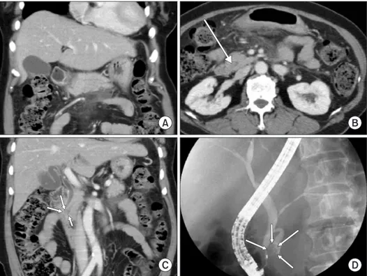
복부 컴퓨터 단층촬영에서 췌장 실질의 미만성 부종과 췌장 주 위의 지방 침윤, 액체 고임 등 급성 췌장염의 소견이 관찰되었으며 (Fig. 1A), 췌장 두부의 췌관 내부에 결절 음영이 의심되면서 췌 장 두부 췌관의 미세한 확장이 관찰되었다(Fig. 1B, C). 췌관 내 부의 이상 유무를 확인하기 위해 내시경적 역행성 췌담관 조영술 을 시행하였으며, 육안적으로 십이지장 유두부에 점액은 관찰되
지 않았다. 췌담관 조영술에서 췌장 두부의 췌관과 총담관의 확 장 소견과, 췌관 기시부의 음영 결손이 관찰되었다(Fig. 1D). 내 시경적 유두 괄약근 절제술 및 쇄모세포진 검사를 시행하였고, 쇄 모세포진 검사 결과 다염색성 핵과 핵눌림 소견이 관찰되어 종양 성 병변이 의심되었다(Fig. 2A). 내시경적 역행성 췌담관 조영술 및 세포진 검사 결과 췌두부암이 의심되어 췌십이지장 절제술을 시행하였다.
수술 소견에서 복강 내 복수는 없었으며 전이를 의심할 만한 소 견은 관찰되지 않았다. 췌두부에서 1.6×1.1 cm 크기의 종괴가 촉 지 되었으며(Fig. 2B), 현미경 소견에서 유두상 과증식 및 비정형 세포를 포함한 암성 변화 소견을 보였으며 주변 림프절 전이는 관 찰되지 않았다(Fig. 2C, D).
수술 시행 이후 복부 컴퓨터 단층촬영과 CA 19-9 검사로 추적 관찰을 시행하였다. 수술 후 47개월째 시행한 복부 컴퓨터 단층촬 영에서 잔존 췌장의 췌관 확장 소견 및 췌관 내부에 조영 증강이 되는 종괴 음영이 관찰되었다(Fig. 3). CA 19-9 수치는 수술 1년 후 10 IU/dL에서 53.38 IU/dL으로 증가되어 있었다. 당시 환자는 복통, 체중 감소, 황달 등의 증상은 관찰되지 않았으나 췌장암의 재발로 의심되어 전췌장적출술 및 비장적출술을 시행하였다. 수술 소견에서 남아있는 췌관의 전반적인 확장과 늘어난 췌관 내에 1.7
×1.0 cm 크기의 갈색 유두상 종괴가 관찰되었다 (Fig. 4A). 현 미경 소견에서 관유두상 증식과 고도의 이형성이 관찰되어 췌장의 관내 관유두상 종양으로 진단되었고(Fig. 4B, C), 4년 전 검체를
Fig. 1. Abdomen computed tomography (CT) findings. (A) Diffuse parenchymal swellling of pancreas, peripancreatic fatty infiltration and fluid collection are seen.
(B, C) A small nodule and scanty dilata- tion of pancreatic duct in the pancreas head are seen on the CT scan (arrows). (D) Endoscopic retrograde cholangiopancrea- tography findings. A nodular filling defect and duct dilatation of pancreatic head portion and common bile duct dilatation are seen on the cholangiopancreatogram (arrows).
다시 확인하여 같은 종양이었던 것으로 확인하였다. 수술 이후 1년 3개월째 시행한 추적 검사에서 재발 소견은 관찰되지 않았다.
고 찰
췌장의 관내 관유두상 종양은 최근에 새롭게 제시되고 있는 질 환으로 췌장 종양의 1% 미만, 췌관내 종양의 3% 정도로 매우 드 물게 발생한다고 알려져 있다[1]. 2000년 세계보건기구 분류에서 는 원발성 췌관내 종양을 췌장 상피내 종양(pancreatic intraepi- thelial neoplasia), 관내 유두상 점액 종양(intraductal papillary mucinous neoplasms), 췌관내 관상 종양(intraductal tubular neo- plasm)의 세가지로 분류하였다[2]. 하지만 최근 점액이 관찰되지
않고, 선방 세포 분화를 보이지 않으면서 관유두상 증식을 보이는 췌관내 종양의 증례들이 관찰되면서 새롭게 관유두상 종양으로 분류되고 있다[1,3].
현재까지 전 세계에 13개의 관유두상 종양 증례가 보고되었 다[1,4-6]. 그 중 10명의 환자들을 대상으로 Yamaguchi 등[1]이 관유두상 종양의 특징을 연구하여 보고하였으며 그 특징은 다음 과 같다. 1) 육안적 소견상 고형의 종양으로 췌관의 폐색 및 확장 을 일으킨다. 2) 육안적인 점액 분비가 없다. 3) 현미경적으로 관 유두상 증식을 보인다. 4) 종양 대부분에서 고도이형성이 관찰된 다. 5) 괴사 반점을 쉽게 찾을 수 있다. 6) 관내 분화를 의미하는 CK7 또는 CK 19를 발현한다. 7) 선방 세포 분화의 결여로 인한 trypsin 면역 조직 화학 염색 음성. 8) MUC2, MUC5AC, fascin Fig. 2. Histopathologic findings. (A) Brush cytology picture shows hyperchromatic nucleus and nuclear molding, therefore those are suspected to be malignant cells. (B) Gross finding after resection shows a 1.6×1.1 cm sized polypoid mass (red circle) is seen on the proximal portion of di- lated pancreatic duct. (C) Tubulopapillary proliferations are seen (H&E, ×40). (D) Cells with nuclear polymorphism, hyperchromatism, and mitosis are seen, therefore high grade dysplasia or carcinomatous change is suspected (H&E, ×250).
음성. 9) KRAS와 BRAF 돌연변이 결여이다.
Yamaguchi 등[1]의 보고에서는 10명의 환자 중 3명에서 췌장 실질 침윤이 관찰되었으며, 그 중 한 명은 수술적 절제 7개월 후에 간전이가 발생하여 사망하였다. 그리고, 본 증례와 유사하게 한
명의 환자는 수술적 절제 12개월 후 남은 췌장에서 재발하여 재수 술을 받고, 이후 18개월간 재발 없이 생존하고 있었다. 이외의 환 자들은 국소 절제 시행 후 재발 없이 생존해 있어 췌장의 관내 관 유두상 종양이 진단되었을 경우, 국소 절제만 시행하고 추적 관찰 Fig. 3. (A, B) Computed tomography (CT) findings after 47 months of resection.
Axial and coronal CT scan shows diffuse pancreatic duct dilatation with enhanc- ing mass (arrow) inside of the duct. (C, D) Magnetic resonance imaging findings after 47 month of resection. T2 weighted images also shows mass like lesion (arrow) inside of dilated pancreatic duct after 47 months of resection.
Fig. 4. (A) Gross sample of 2nd operation (total pancreatectomy and splenectomy).
A brownish mass like lesion mixed with brownish sludge material (red circle) is seen inside of dilated pancreatic duct. (B) Tubulopapillary proliferation is seen (H&E,
×40). (C) Nuclear polymorphism and hy- perchomatic nucleus suggests high grade dysplasia (H&E, ×250).
할 것인지, 처음부터 근치적인 수술을 시행할 것인지에 대한 더 많 은 연구와 논의가 필요하겠다.
상당수의 췌장의 관내 관유두상 종양이 다른 췌관 내 종양으로 오인되고 있는 것으로 추정된다[7]. 췌장 상피 내 종양은 입방세 포 또는 원주세포로 이루어진 췌관 내 종양으로 비정형의 정도는 다양하게 나타나며, 면역조직화학 검사에서 MUC1, MUC5AC, fascin 등이 발현되고, KRAS 돌연변이도 흔하게 관찰된다[1,8].
췌장의 관내 유두상 점액 종양은 점액으로 인해 췌관이 확장되 고, 종양 세포가 유두상을 형성하며, 관을 따라 증식한다. 췌장의 관내 관유두상 종양이 균등하게 고도이형성을 보이는 반면, 췌관 내 유두상 점액 종양은 다양한 정도의 이형성을 나타내며 MUC2, MUC3, MUC5AC가 종종 발현되기도 한다[9]. 췌관 내 관상 종 양은 췌장의 관내 관유두상 종양과 유사하게 MUC1양성, MUC2 와 MUC5에서 음성을 나타내지만 관유두상 증식 소견은 췌장의 관내 관유두상 종양에서만 관찰된다[1,10].
본 증례에서는 면역조직화학 검사는 시행하지 않았으나 육안적 으로 점액이 관찰되지 않았고, 현미경 소견상 관유두상 증식과 고 도 이형성 소견이 관찰되어 췌장의 관내 관유두상 종양으로 진단 하였다.
국내에서는 담관에서 발생한 관유두상 종양의 증례 보고만 있 을 뿐, 췌장에서 발생한 증례는 현재까지 보고되지 않고 있다[11].
췌장의 관내 관유두상 종양을 췌관 내 유두상 점액 종양의 아형 으로 보는 견해도 있으나, 앞서 언급한 것처럼 이 두 가지는 분명 히 구분되는 다른 특징을 가지고 있기 때문에 새로운 범주로 분류 되고 있다. 췌장의 관내 관유두상 종양으로 진단된 증례보고가 많 지 않고, 임상 추적기간이 짧아 추후 예후와 임상양상에 대한 더 많은 연구가 필요할 것이다.
참고문헌
1. Yamaguchi H, Shimizu M, Ban S, Koyama I, Hatori T, Fujita I, et al. Intraductal tubulopapillary neoplasms of the pancreas dis- tinct from pancreatic intraepithelial neoplasia and intraductal
papillary mucinous neoplasms. Am J Surg Pathol 2009;33:1164- 1172.
2. Longnecker DS, Adler G, Hruban RH, Kloppel G. Intraductal papillary-mucinous neoplasms of the pancreas. In: Hamilton SR, Aaltonen LA, editors. Pathology and genetics of tumours of the digestive system WHO classification of tumours. Lyon, France:
IARC Press; 2000. p.237-240.
3. Bosman FT, Carneiro F, Hruban RH, Theise ND. WHO classifica- tion of tumours of the digestive system. 2nd ed. Lyon, France:
IARC Press; 2010.
4. Bhuva N, Wasan H, Spalding D, Stamp G, Harrison M. In- traductal tubulopapillary neoplasm of the pancreas as a ra- diation induced malignancy. BMJ Case Rep 2011;2011. pii:
bcr0920114777. 10.1136/bcr.09.2011.4777.
5. Guan H, Gurda G, Marie Lennon A, Hruban RH, Erozan YS.
Intraductal tubulopapillary neoplasm of the pancreas on fine needle aspiration: case report with differential diagnosis. Di- agn Cytopathol 2012 Jul 16 [Epub]. http://dx.doi.org/10.1002/
dc.22890.
6. Jokoji R, Tsuji H, Tsujimoto M, Shinno N, Tori M. Intraductal tu- bulopapillary neoplasm of pancreas with stromal osseous and cartilaginous metaplasia; a case report. Pathol Int 2012;62:339- 343.
7. Tajiri T, Tate G, Matsumoto K, Hoshino H, Iwamura T, Kodaira Y, et al. Diagnostic challenge: intraductalneoplasms of the pan- creatobiliary system. Pathol Res Pract 2012;208:691-696.
8. Hruban RH, Adsay NV, Albores-Saavedra J, Compton C, Garrett ES, Goodman SN, et al. Pancreatic intraepithelial neoplasia: a new nomenclature and classification system for pancreatic duct lesions. Am J Surg Pathol 2001;25:579-586.
9. Furukawa T, Kloppel G, Volkan Adsay N, Albores-Saavedra J, Fukushima N, Horii A, et al. Classification of types of intraductal papillary-mucinous neoplasm of the pancreas: a consensus study. Virchows Arch 2005;447:794-799.
10. Tajiri T, Tate G, Inagaki T, Kunimura T, Inoue K, Mitsuya T, et al.
Intraductal tubular neoplasms of the pancreas: histogenesis and differentiation. Pancreas 2005;30:115-121.
11. Park HJ, Jang KT, Heo JS, Choi YL, Han J, Kim SH. A potential case of intraductal tubulopapillary neoplasms of the bile duct.
Pathol Int 2010;60:630-635.