REPO RT
Int J Thyroidol 2017 November 10(2): 102-106 https://doi.org/10.11106/ijt.2017.10.2.102Received August 31, 2017 / Revised October 27, 2017 / Accepted October 30, 2017
Correspondence: Ji Sun Park, MD, Department of Nuclear Medicine, Busan Paik Hospital, Inje University College of Medicine, 75 Bokji-ro, Busanjin-gu, Busan 47392, Korea
Tel: 82-51-890-8753, Fax: 82-51-896-6458, E-mail: [email protected]
Copyright ⓒ 2017, the Korean Thyroid Association. All rights reserved.
This is an open-access article distributed under the terms of the Creative Commons Attribution Non-Commercial License (http://creative- commons.org/licenses/by-nc/4.0/), which permits unrestricted non-commercial use, distribution, and reproduction in any medium, provided the original work is properly cited.
Development of Graves’ Ophthalmopathy after Radioactive Iodine Ablation Using Recombinant Human Thyrotropin for Incidentally Discovered Papillary Thyroid Carcinoma
Ji Sun Park
Department of Nuclear Medicine, Busan Paik Hospital, Inje University College of Medicine, Busan, Korea
Several studies have revealed an association between radioactive iodine (RAI) therapy for Graves’ disease and new or worsening Graves’ ophthalmopathy. In the present case, thyroid papillary cancer was incidentally detected in a 43-year-old woman who was receiving medication for Graves’ disease. This patient had undergone RAI ablation using recombinant human thyrotropin (rhTSH) after total thyroidectomy. The patient subsequently complained of a unilateral eyelid abnormality at approximately 6 months after the RAI ablation, and was diagnosed with bilateral Graves’ ophthalmopathy after a thorough ophthalmological examination.
I report this case for its interesting clinical features, rarity of occurrence and to highlight the importance of careful observation for appropriate management of Graves’ ophthalmopathy developing or worsening after RAI ablation in differentiated thyroid cancer patients.
Key Words: Graves’ ophthalmopathy, Thyroid cancer, Recombinant human thyrotropin, Radioactive iodine
Introduction
Graves’ ophthalmopathy is also known as thy- roid-associated ophthalmopathy, and is an in- flammatory autoimmune disorder of the orbit that is clinically relevant in 25-50% of patients with Graves’
disease.1,2) Although the pathogenesis of Graves’
ophthalmopathy remains unclear, thyroid-stimulating hormone receptors (TSHRs) in orbit cells and anti- bodies to TSHRs (TRAb) are considered the primary targets of the autoimmune reactions in this disease.3-5) Anti-thyroid drug therapy and thyroidectomy do not substantially affect the natural course of Graves’ oph- thalmopathy, and radioactive iodine (RAI) ablation is
thought to induce or aggravate Graves’ ophthal- mopathy.6-10) However, RAI ablation after thyroi- dectomy can provide a substantial early improvement in Graves’ ophthalmopathy, or at least outcomes that are similar to thyroidectomy alone.8,11-15)
Differentiated thyroid cancer (DTC) is rare among patients with Graves’ disease, and patients with a his- tory of Graves’ disease or Graves’ ophthalmopathy are not common among patients who receive RAI ablation for DTC.15-17) Herein I report a case of newly developed Graves’ ophthalmopathy in a patient with a history of Graves’ disease, who underwent total thy- roidectomy plus RAI ablation with recombinant human thyrotropin (rhTSH) for incidentally discovered papillary thyroid cancer.
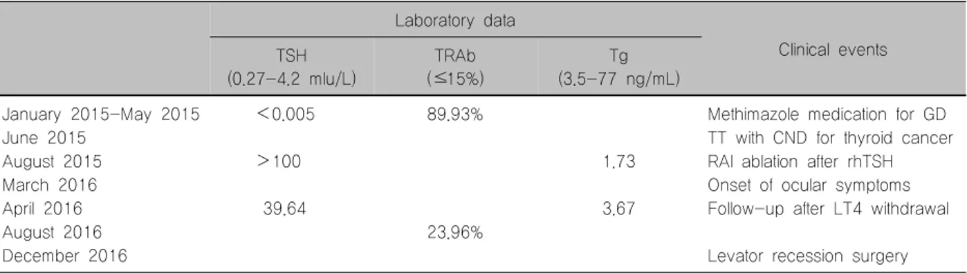
Table 1. Case summary
Laboratory data
Clinical events TSH
(0.27-4.2 mIu/L)
TRAb (≤15%)
Tg (3.5-77 ng/mL)
January 2015-May 2015 <0.005 89.93% Methimazole medication for GD
June 2015 TT with CND for thyroid cancer
August 2015 >100 1.73 RAI ablation after rhTSH
March 2016 Onset of ocular symptoms
April 2016 39.64 3.67 Follow-up after LT4 withdrawal
August 2016 23.96%
December 2016 Levator recession surgery
The values in parentheses indicate the normal reference ranges.
CND: central node dissection, GD: Graves’ disease, rhTSH: recombinant human thyrotropin, RAI: radioactive iodine, Tg:
thyroglobulin, TSH: thyroid-stimulating hormone, TRAb: TSH receptor antibody, TT: Total thyroidectomy
Case Report
In July 2015, a non-smoking 43-year-old woman with papillary thyroid cancer was referred for RAI ablation after total thyroidectomy. The cancer had been detected incidentally during ultrasonography, and she had previously been diagnosed with Graves’
disease in January 2015. At that time, she had a se- rum thyroid-stimulating hormone (TSH) level of
<0.005 mIu/L (normal: 0.27-4.2 mIu/L), a free thy- roxine (fT4) level of 6.84 ng/dL (normal: 0.93-1.71 ng/dL), a triiodothyronine (T3) level of 481.4 ng/dL (normal: 80-200 ng/dL), and was positive for TRAb (89.93%, normal: ≤15%). The patient had received methimazole for 5 months before the total thyroi- dectomy, which was performed with central lymph node dissection in June 2015. Table 1 shows a sum- mary of the clinical events in this case. The post- operative pathology findings revealed multifocal papil- lary thyroid carcinomas involving both lobes, with ex- trathyroidal extension and central lymph node metastasis. The patient started levothyroxine (LT4) re- placement on the day after surgery, and 1 month later had a serum TSH level of 0.628 mIu/L (normal:
0.27-4.2 mIu/L) and a fT4 level of 1.88 ng/dL (normal: 0.93-1.71 ng/dL).
In August 2015, the patient underwent RAI ablation with 3700 MBq I-131 after the rhTSH preparation, and the LT4 treatment was discontinued for 4 days before
the RAI ablation. On the day of that admission, blood tests revealed a TSH level of >100 mIu/L, a thyro- globulin (Tg) level of 1.73 ng/mL (normal: 3.5-77 ng/mL), an anti-thyroglobulin antibody level of 2.68 IU/mL (normal: <60 IU/mL), a fT4 level of 1.17 ng/dL, and a T3 level of 78.69 ng/dL (normal: 80-200 ng/dL). The whole body I-131 scan on day 7 after the RAI ablation revealed focal uptake in a thyroid remnant, and thyroid hormone replacement was re- started on the day after RAI ablation. A follow-up evaluation in October 2015 revealed a TSH level of
<0.011 mIu/L, an fT4 level of 1.84 ng/dL, and a Tg level of 1.06 ng/mL. Neck ultrasonography revealed no evidence of residual disease.
In April 2016 (9 months after the RAI ablation), a whole body I-123 scan was performed with single- photon emission computed tomography/computed to- mography of the neck and stimulated Tg testing.
Remnant thyroid tissue was detected in the thyroid bed (Fig. 1), and the stimulated Tg level was 3.67 ng/mL when the TSH level was 39.64 mlu/L after LT4 withdrawal for 3 weeks. The patient underwent fluo- rine-18-fluorodeoxyglucose positron emission to- mography/computed tomography (F-18 FDG PET/CT), which did not reveal evidence of recurrent or meta- static disease. Unexpectedly, the patient complained of a recent abnormality in her left eye, as well as redness in both eyes during the last 2 weeks. An ophthalmo- logical examination revealed left upper lid retraction and color vision abnormality, with exophthalmometer
Fig. 1. Whole body planar I-123 scanning (A) and subsequent neck single photon emission computed tomography/computed tomography (B, C) reveal remnant thyroid tissue in the left thyroid bed.
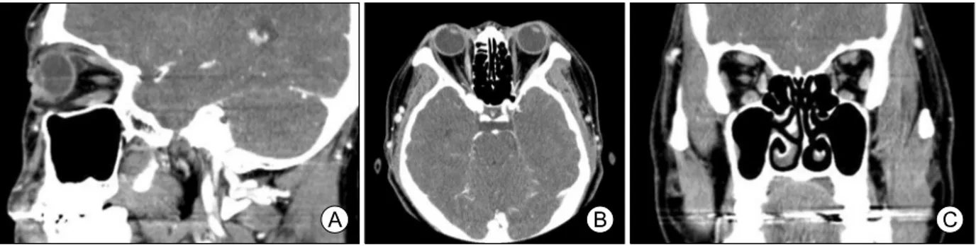
Fig. 2. Sagittal (A), axial (B) and coronal computed tomography of the orbits reveal enlargement of the bilateral extraocular muscles.
readings of 16 mm for the right eye and 16.5 mm for the left eye. There were no oculomotor abnormalities or diplopia. Orbital computed tomography revealed enlargement of the bilateral extraocular muscle bellies and bilateral exophthalmos (Fig. 2). Unfortunately, the patient was not tested for TRAbs. The LT4 therapy was continued with regular follow-ups, and the serum TRAb titer in August 2016 was 23.96%. The patient requested surgery for the persistent left upper eyelid retraction, and underwent levator recession surgery in December 2016.
Discussion
The incidence of thyroid cancer has been increas- ing in South Korea and throughout the world, which may be related to the increased use of diagnostic ultrasonography.18) However, clinicians infrequently perform RAI ablation for DTC patients with a history of Graves’ disease, because of the low frequency of thyroid cancer among patients with Graves’ disease patients (1.3-9.8%).17)
Several studies have revealed an association be-
tween RAI ablation for Graves’ disease and new or worsening Graves’ ophthalmopathy.7-10) A systematic review concluded that RAI ablation for Graves’ dis- ease was associated with a small but increased risk of new or worsening Graves’ ophthalmopathy, com- pared to anti-thyroid drug therapy.6) However, the ef- fect of RAI ablation on Graves’ ophthalmopathy in pa- tients after thyroidectomy differs from that in patients with an intact thyroid gland, and RAI ablation after total or near total thyroidectomy provides a substantial early improvement in Graves’ ophthalmopathy, or at least outcomes that were similar to thyroidectomy alone.8,11-15)
The present case is one of only a few reported cases with new or worsened Graves’ ophthalmopathy after RAI ablation plus rhTSH for DTC. One in vitro study reported that the TSHRs expressed in some or- bital cells may be functional and respond to high TSH levels, activating intracellular signaling pathways.19) Daumerie et al.20) reported 3 cases of Graves’ oph- thalmopathy reactivation after RAI ablation plus rhTSH for incidentally detected DTC after thyroidectomy for Graves’ disease. In those cases, the reactivation of Graves’ ophthalmopathy appeared within 3-6 weeks
after rhTSH administration during the euthyroid state.
Based on their observations, the researchers sug- gested the potential pathogenic role of exogenous high TSH reported in vitro is mirrored in vivo.20) However, unlike the previous cases, our patient de- veloped clinically overt eye symptoms after 6 months of rhTSH administration. In many cases, patients with thyroid cancer receive lifelong LT4 replacement ther- apy after resection of the thyroid gland, even with partial resection. Thyroid hormone levels can neg- atively affect the outcomes of Graves’ ophthalmop- athy, so care is needed when interpreting the relation- ship between rhTSH administration and worsening of Graves’ ophthalmopathy.15) Further well-designed large-scale studies are necessary to investigate the relationship between them.
High TRAb titers are an independent risk factor for new or worsening Graves’ ophthalmopathy, in addition to smoking, thyroid dysfunction, and RAI ablation.15) In the present case, the patient had high TRAb titers at the diagnosis of Graves’ disease, and the high titers persisted until the development of ophthalmopathy, despite being slightly lower than the titers at the diagnosis. Similarly high TRAb titers have been re- ported in previous cases.20) However, Louvet et al.8) suggested that non-autoimmunity factors might be in- volved in the worsening of Graves’ ophthalmopathy, as RAI ablation for microscopic thyroid tissue after thy- roidectomy would reduce TRAb titers levels by re- moving autoantigens.
Clinically apparent thyroid ophthalmopathy is pres- ent in approximately 25-50% of patients with Graves’
disease, although >70% of patients exhibit thyroid ophthalmopathy when examined using sensitive imag- ing techniques.2,4) Approximately 5% of patients with Graves’ ophthalmopathy have severe ocular symp- toms, although most patients experience mild or sub- clinical ocular involvement.3,4) Our patient had clear unilateral upper eyelid retraction at 6 months after RAI ablation, and bilateral extraocular muscle involvement was detected during orbital CT. Even though there were no clinical signs of Graves’ ophthalmopathy at or immediately after the RAI ablation, it is possible that she may have already had mild or subclinical eye
involvement. Several studies have indicated that Graves’ ophthalmopathy peaks at 6 months after RAI ablation, it can develop any time within the first 2 years after RAI ablation among patients with Graves’
disease.6) Therefore, a specialist follow-up is recom- mended for at least 12 months after RAI ablation, as the onset or aggravation of Graves’ ophthalmopathy varies among patients who receive RAI ablation after thyroidectomy for DTC.6,15)
Several studies assessed the role of steroid pro- phylaxis in the prevention of occurrence or pro- gression of Graves’ ophthalmopathy when used along with RAI. According to a systematic review reported by Acharya et al.,6) prednisolone prophylaxis was highly effective in preventing worsening of eye dis- ease in patients with pre-existing ophthalmopathy compared with RAI ablation without prednisolone.
Louvet et al.8) confirmed that patients with moderate to severe Graves’ ophthalmopathy were more treated by prophylactic glucocorticoids after RAI ablation and pa- tients with active Graves’ ophthalmopathy were more treated by prophylactic glucocorticoids after RAI abla- tion than patients with inactive ophthalmopathy.
In conclusion, despite remaining relatively rare, the incidental diagnosis of thyroid cancer has been in- creasing, and it is increasingly possible that clinicians will encounter thyroid cancer in patients with Graves’ disease. Thus, clinicians should be aware that patients with thyroid cancer and a history of Graves’ disease or risk factors for Graves’ ophthalmopathy require ap- propriate prophylaxis, even if they have no overt ocu- lar symptoms at the RAI ablation and receive rhTSH.
Careful observation and follow-up are needed to fa- cilitate the early detection and treatment of Graves’
ophthalmopathy.
References
1) Iyer S, Bahn R. Immunopathogenesis of Graves' ophthalmo- pathy: the role of the TSH receptor. Best Pract Res Clin Endocrinol Metab 2012;26(3):281-9.
2) Hiromatsu Y, Eguchi H, Tani J, Kasaoka M, Teshima Y.
Graves' ophthalmopathy: epidemiology and natural history. Intern Med 2014;53(5):353-60.
3) Bahn RS. Graves' ophthalmopathy. N Engl J Med 2010;362(8):
726-38.
4) Wiersinga WM, Bartalena L. Epidemiology and prevention of Graves' ophthalmopathy. Thyroid 2002;12(10):855-60.
5) Chong K. Thyroid eye disease: a comprehensive review. Hong Kong Fed Med Soc 2010;15:4-8.
6) Acharya SH, Avenell A, Philip S, Burr J, Bevan JS, Abraham P. Radioiodine therapy (RAI) for Graves' disease (GD) and the effect on ophthalmopathy: a systematic review. Clin Endocrinol (Oxf) 2008;69(6):943-50.
7) Bartalena L, Marcocci C, Bogazzi F, Manetti L, Tanda ML, Dell'Unto E, et al. Relation between therapy for hyperthyroidism and the course of Graves' ophthalmopathy. N Engl J Med 1998;
338(2):73-8.
8) Louvet C, De Bellis A, Pereira B, Bournaud C, Kelly A, Maqdasy S, et al. Time course of Graves' orbitopathy after total thyroidectomy and radioiodine therapy for thyroid cancer.
Medicine (Baltimore) 2016;95(48):e5474.
9) Tallstedt L, Lundell G, Torring O, Wallin G, Ljunggren JG, Blomgren H, et al. Occurrence of ophthalmopathy after treat- ment for Graves' hyperthyroidism. The Thyroid Study Group. N Engl J Med 1992;326(26):1733-8.
10) Traisk F, Tallstedt L, Abraham-Nordling M, Andersson T, Berg G, Calissendorff J, et al. Thyroid-associated ophthal- mopathy after treatment for Graves' hyperthyroidism with anti- thyroid drugs or iodine-131. J Clin Endocrinol Metab 2009;
94(10):3700-7.
11) Menconi F, Marino M, Pinchera A, Rocchi R, Mazzi B, Nardi M, et al. Effects of total thyroid ablation versus near-total thyroidectomy alone on mild to moderate Graves' orbitopathy treated with intravenous glucocorticoids. J Clin Endocrinol Metab 2007;92(5):1653-8.
12) Moleti M, Violi MA, Montanini D, Trombetta C, Di Bella B, Sturniolo G, et al. Radioiodine ablation of postsurgical thyroid remnants after treatment with recombinant human TSH
(rhTSH) in patients with moderate-to-severe graves' orbitopathy (GO): a prospective, randomized, single-blind clinical trial. J Clin Endocrinol Metab 2014;99(5):1783-9.
13) Moleti M, Mattina F, Salamone I, Violi MA, Nucera C, Baldari S, et al. Effects of thyroidectomy alone or followed by radioiodine ablation of thyroid remnants on the outcome of graves' ophthalmopathy. Thyroid 2003;13(7):653-8.
14) Leo M, Marcocci C, Pinchera A, Nardi M, Megna L, Rocchi R, et al. Outcome of Graves' orbitopathy after total thyroid ablation and glucocorticoid treatment: follow-up of a randomized clinical trial. J Clin Endocrinol Metab 2012;97(1):E44-8.
15) Stan MN, Bahn RS. Risk factors for development or deteriora- tion of Graves' ophthalmopathy. Thyroid 2010;20(7):777-83.
16) Ergin AB, Saralaya S, Olansky L. Incidental papillary thyroid carcinoma: clinical characteristics and prognostic factors among patients with Graves' disease and euthyroid goiter, Cleveland Clinic experience. Am J Otolaryngol 2014;35(6):784-90.
17) Belfiore A, Russo D, Vigneri R, Filetti S. Graves' disease, thyroid nodules and thyroid cancer. Clin Endocrinol (Oxf) 2001;
55(6):711-8.
18) Kim SH, Jung SL, Moon WJ, Park MS, Kim YS, Lee HJ, et al. The prevalence of thyroid nodules and thyroid cancers in the Koreans: the nationwide data analysis of thyroid ultra- sonography in 2004. J Korean Thyroid Assoc 2009;2(1):33-7.
19) van Zeijl CJ, Fliers E, van Koppen CJ, Surovtseva OV, de Gooyer ME, Mourits MP, et al. Thyrotropin receptor- stimulating Graves' disease immunoglobulins induce hyaluronan synthesis by differentiated orbital fibroblasts from patients with Graves' ophthalmopathy not only via cyclic adenosine monophos- phate signaling pathways. Thyroid 2011;21(2):169-76.
20) Daumerie C, Boschi A, Perros P. Is recombinant human TSH a trigger for Graves' orbitopathy? Eur Thyroid J 2012;1(2):
105-9.