IInnttrroodduuccttiioonn
Femoral varus osteotomy (FVO) has been widely used in the surgical treatment of Legg-Calve-Perthes disease (LCPD). Axer1) in 1965 reviewed FVO as a treatment of LCPD and reported a very favorable results comparing with those observed after conservative treatment. Since then, many authors5,6,12,13,17,18) have demonstrated that it can prevent femoral head deformity and restore spherical congruity, provided that the femoral head can be contained in the acetabulum.
The advantages of FVO are that the operation is done on the affected bone, it provides better lateral coverage than does innominate osteotomy, and it decreases the force across the joint. However, the disadvantages of FVO are also reported that it may shorten the limb and may create excessive varus angulation, leading to weakness of the abductors of the hip19,21,24).
However, only few papers have been reported the long term results of FVO which followed up to the full skeletal maturity. We selected 35 patients who had clinical and radiological examinations at the full skeletal maturity and analyzed the results with special emphasis on the sphericity of femoral head and the leg length discrepancy (LLD).
Submitted: July 14, 2009 1st revision: August 17, 2009 2nd revision: August 26, 2009 Final acceptance: August 26, 2009
�Address reprint request to Sung Man Rowe, MD
Department of Orthopedic Surgery, St.Carollo Hospital, 1742, Jorye-dong, Suncheon-si, 540-718, Jeollanam-do
TEL: +82-61-720-6401 FAX: +82-61-720-6363 E-mail: [email protected]
The Long Term Results of Femoral Varus Osteotomy in Patients with Legg-Calve-Perthes Disease
Jin Sang Wie, MD, Sung Man Rowe, MD, El O Jung, MD, Young Jin Lim, MD, Ji Hun Song, MD, Myung Guk Jung, MD Department of Orthopedic Surgery, St. Carollo Hospital, Suncheon, Korea
Purpose: The purpose of this study was to evaluate the long term results of performing femoral varus osteotomy (FVO) for the treatment of Legg-Calve-Perthes disease (LCPD).
Materials and Methods: We selected 35 LCPD patients who received FVO and they were followed up to the time their skeletons’ matured. The inclusion criteria were patients in a fragmentation stage, the patients were in Catterall group Ⅲ or Ⅳ, and the patients underwent a teleoroentgenographic examination at the time of full skeletal maturity.
Results: The radiological outcome at the time of skeletal maturity was assessed using Stulberg’s classification. The final results were 4 hips in class I, 17 hips in class II, 13 hips in class III, one hip in class IV and none in class V. The satisfactory results (good+fair hips) were 34 hips (97%). Significant shortening (>10 mm) was observed in 12 hips (34%). In 35 patients, 5 (14%) had same leg length (less than 2 mm difference), 27 (77%) had shortening of 2 mm or more, and 3 had lengthening of 2 mm or more in the operated limb. Of these 12 patients with significant shortening, only 3 patients (9%) showed shortening of 21 mm or more.
Conclusion: FVO is a reliable method for managing LCPD in patients who are in Catterall group III or IV and who are in the fragmentation stage of disease.
Key Words: Legg-Calve-Perthes disease, Femoral varus osteotomy
M
Maatteerriiaallss aanndd MMeetthhooddss
We selected 35 LCPD patients who received FVO for the treatment of LCPD and followed up to the full
skeletal maturity (Table 1). The inclusion criteria were unilateral involvement, patient in fragmentation stage, Catterall group Ⅲ or Ⅳ, and patients who had the teleoroentgenographic examination at the full
Table 1. Patients Data.
Patient Number 35
Gender (Male/Female) 32/3
Age at Disease Onset 07.3 Years (05~11 Years).
Age at Surgery (Femoral Varus Osteotomy) 08.6 Years (6.5~12 Years)
Age at Final Follow-up 24.9 Years (18~34 Years).
Period of Follow-up 16.2 Years (10~23 Years).
Severity of Disease Catterall Classification3)
I+II 0
III 23 Hips
IV 12 Hips
Herring Classification7)
A 0
B 24 Hips
C 11 Hips
Mode of Osteotomy
Subtrochanteric Open Wedge Osteotomy 11 Hips
Lloyd-Roberts Intertrochanteric Osteotomy 24 Hips
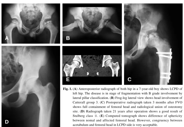
Fig. 1. (A) Anteroposterior radiograph of both hip in a 7-year-old boy shows LCPD of left hip. The disease is in stage of fragmentation with B grade involvement by lateral pillar classification. (B) Frog-leg lateral view shows head involvement of Catterall group 3. (C) Postoperative radiograph taken 3 months after FVO shows full containment of femoral head and radiological union of osteotomy site. (D) Radiograph taken 21 years after operation shows a good result of Stulberg class Ⅱ. (E) Computed tomograph shows difference of sphericity between nomal and affected femoral head. However, congruency between acetabulum and femoral head in LCPD side is very acceptable.
A B
C E
D
skeletal maturity.
FVO was performed with subtrochanteric open- wedge osteotomy using straight plate fixation in 11 hips (Fig. 1) and with Lloyd-Roberts technique15) of intertrochanteric oblique osteotomy (semi-open technique) using the angled blade plate fixation in 24 hips (Fig. 2).
The final outcomes were assessed radiologically with classification system of Stulberg et al25). based on the sphericity and congruency of the hip.
Stulberg et al25). defined five patterns of hip structure based on plain radiographic appearances at skeletal maturity following childhood LCPD. The classification indicates the extent of hip malformation and remodeling reached at the termination of the repair phase at skeletal maturity. The types of residual appearances are graded as follows: class I, completely normal hip joint; class Ⅱ, spherical femoral head associated with one or more of coxa magna (larger than normal although spherical femoral head), short neck, or dysplastic, abnormally steep acetabulum; class Ⅲ, nonspherical, mushroom- shaped, ovoid head plus coxa magna, short neck,
and steepened acetabulum; class Ⅳ, flat femoral head with coxa magna, short neck, and steepened acetabulum; and class Ⅴ, flat femoral head of normal size with a normally shaped neck and acetabulum.
We measured the leg length on radiograph of both entire legs (teleoroentgenograms) to compare.
Teleoroentgenograms of a single exposure of both legs on a long, 35×90 cm (ie, 14×36 in) film were taken from a distance of 2 m with a radiopaque ruler placed on the film cassette. LLD was measured from the most proximal point of the femoral head to the mid point of the lower tibial surface on teleoroentgenograms.
We also measured the neck shaft angle of the affected femur at skeletal maturity to evaluate the residual coxa vara.
Statistical analysis was performed using SPSS for Windows Release 10.0 (SPSS Inco, Chicago, U.S.A).
All analyses were set at the 95% confidence interval for significance.
R Reessuullttss
SSpphheerriicciittyy aanndd ccoonnggrruueennccyy ooff tthhee FFeemmoorraall ooff hheeaadd
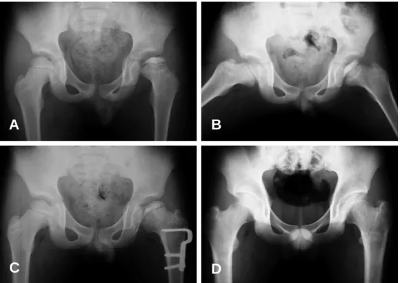
Fig. 2. (A) Anteroposterior radiograph in a 8-year-old boy shows LCPD finding of left hip with fragmentation stage and lateral pillar B involvement. (B) Frog-leg lateral radiograph shows head involvement of Catterall group 4. (C) Postoperative radiograph taken 4 months after FVO shows acceptable varus and bony heading of osteotomy site. (D) Final follow-up radiograph taken 14 years after operation shows a good result of Stulberg class II.
A B
C D
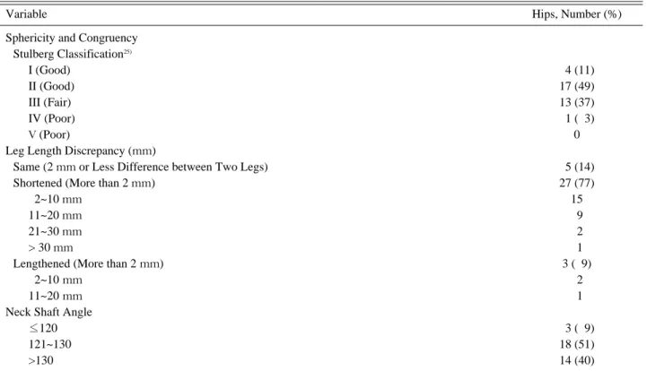
Radiographic outcome was assessed with classification system of Stulberg25) to grade residual deformity at skeletal maturity. The final results were 4 hips in class I (11%), 17 hips in class Ⅱ (49%), 13 hips in class Ⅲ (37%), one hip in class Ⅳ (3%) and none in class Ⅴ (0%). According to these results, good hips (class I+Ⅱ) were in 21 hips (60%), fair hips (class Ⅲ) in 13 hips (37%), and poor hip in only one hip (3%).
Therefore, of the total 35 hips with FVO, the hips showing satisfactory results (good + fair hips) were 34 hips (97%).
LLeegg lleennggtthh ddiissccrreeppaannccyy ((LLLLDD))
We measured the leg lengths of both legs on teleoroentgenogram. Mean value of LLD was 9.2 mm, ranging from a shortening of 31 mm to a lengthening of 13 mm. In 35 patients who received FVO, 5 (14%) had same length (less than 2 mm difference), 27 (77%) had shortening of 2 mm or more, and 3 had lengthening of 2 mm or more in the operated limb.
Significant shortening (>10 mm) was observed in 12 patients (34%). Of these 12 patients, only 3 (9%) showed shortening of 21 mm or more.
In addition, correlation study between the
observed amount of leg shortening and the mode of femoral osteotomy showed a significant correlation (P=0.012). Of 35 patients, shortening of the affected leg appeared in 27 patients (77%). However, the amount of shortening was different according to the mode of osteotomy. Mean value of shortening were 6.2 mm in subtrochanteric open-wedge osteotomy group and 12.4 mm in intertrochanteric oblique osteotomy group (P=0.012).
N
Neecckk sshhaafftt aannggllee
The mean value of neck shaft angle at final follow- up was 126�at skeletal maturity and excessive coxa vara (≤120�) was found only in 3 hips (9%) of the 35 hips (Table 2).
D
Diissccuussssiioonn
Surgical containment is the most recent step in containment management of LCPD. The common methods of surgical containment are the femoral varus osteotomy and the innominate osteotomy of Salter23). Surgical containment is surgical repositioning of the femoral head or the acetabulum to redirect the
Table 2. Final Results at the Full Skeletal Maturity
Variable Hips, Number (%)
Sphericity and Congruency Stulberg Classification25)
I (Good) 04 (11)
II (Good) 17 (49)
III (Fair) 13 (37)
IV (Poor) 01 (03)
V (Poor) 0
Leg Length Discrepancy (mm)
Same (2 mm or Less Difference between Two Legs) 05 (14)
Shortened (More than 2 mm) 27 (77)
02~10 mm 15
11~20 mm 09
21~30 mm 02
> 30 mm 01
Lengthened (More than 2mm) 3 (09)
02~10mm 02
11~20mm 01
Neck Shaft Angle
≤120 03 (09)
121~130 18 (51)
>130 14 (40)
hip geometry. The concept of FVO is that the osteotomy place the femoral head deeper within the confines of the acetabulum, thereby preventing flattening.
However, the postoperative results of FVO varied widely according to the reported authors3,15,23). Lloyd- Roberts et al.15) concluded that after reviewing the controlled study of the FVO in 48 LCPD patients containment by FVO was the treatment of choice in patients with “at risk”signs provided that severe deformity has not already occurred. Of 48 LCPD patients, the results were 58% good, 23% fair and 19% poor. Friedlander and Weiner4)reviewed a large series of 116 LCPD patients and concluded that FVO was a reliable method of treatment regardless of age.
They had 51% good hips, 40% fair hips and 9% poor hips. All patients showing poor results (Stulberg V) were patients with Herring class C involvement and older than 9 years of age. Hoikka et al.8)reported the results of 112 FVO performed on 102 children and had 52% good hips, 24% fair hips and 24% poor hips.
Their results worsened with the increasing age of the patients at operation. We could find several papers reporting the mid-term or long term results of FVO in Korean LCPD children11,19). Kim et al.11) reported his experiences of 13 FVO and their result with mid-term follow up were 31% good, 61% fair and 8% poor.
Park and Choi19) reported mid-term follow-up results in 23 FVO. Their results were 44% good hips, 52%
fair hips and 4% poor hips.
Our results showing 60% good hips, 37% fair hips and 3% poor hips were better than previous reports.
We thought that our better outcomes were resulted from careful selection of patients who were in fragmention stage at operation. In addition, the difference of final results between papers could be caused by the conditions of the operated patients including age at disease onset, severity of disease and stage of disease.
Herring et al.7) stated that the lateral pillar classification had strong correlation with final outcome. In this study, however, only Herring group B and C are subject of inclusion and study volume is too small to compare (24 hips in Group B and 11 in Group C). Therefore, we can not find statistically significant correlation.
Many excellent results have been reported with the
use of FVO. One concern, however, was that it further shortened the affected leg and thus increased the need for the epiphyseal arrest of the normal opposite leg9,20-22). It is generally accepted that the amount of the shortening of the affected legs in LCPD depends almost exclusively on the severity of inhibition of endochondral ossification in the proximal femoral growth plate. In addition, the loss of epiphyseal height, disuse atrophy of the diseased limb, and performing FVO in older children are considered to contribute to the shortening of the affected leg21,22).
However, the incidence and the amount of residual shortening following LCPD vary widely in the literature.
The effect of FVO on residual shortening in LCPD patients remains to be resolved. Some investigators have suggested that FVO causes or aggravates shortening in the operated limb. Canale2) stated that FVO could result in further shortening of an already shortened extremity, and Leitch et al.14) reported that FVO should be avoided in patients over 8 years of age because of its adverse effects on LLD. In contrast, Mirovsky et al.16) investigated the residual shortening of affected limbs in 55 patients treated by FVO and compared these findings with those of 71 patients treated conservatively; when last examined, the mean shortening (0.9 cm) was identical in both groups. Karpinski et al.10) reported that LLD in patients treated by bracing was greater than that in a matched osteotomy group. They also stated that they considered limb disuse to be a major etiologic factor of orthotic-induced shortening, and that overgrowth of the affected limb may have resulted from accelerated growth after surgery, resembling the lengthening that occurs after a fracture in a child.
In my previous study20), our measurements showed that the mean total femoral and tibial shortening was 15.0 mm in the abduction orthosis (AO) group and 9.2 mm in the FVO group (P<0.001). A shortening of 10 mm or more was observed in 66% of the AO group and 40% of the FVO group. Our results showed that FVO produced a better result than AO in terms of the incidence and the level of residual shortening following LCPD. Interestingly, when we measured the tibial lengths to determine the response of the ipsilateral tibia, we observed a
significant difference between the tibial lengths in two treatment groups?a 2.5-mm shortening in patients treated by AO and a 0.9-mm lengthening in patients treated by FVO (P<0.001). In the case of FVO, all 30 patients showed either the same or greater tibial length, except three patients with mild shortening. However, in the AO group, only 20 (53%) patients had the same or greater tibial length.
Our conclusion was that the residual shortening in patients treated by FVO (mean 9.2 mm) was less than patients treated by AO (mean 15.0 mm). We inferred that the difference was due to the overgrowth of the ipsilateral tibia in patients treated by FVO.
In conclusion, in terms of sphericity and congruency we had very satisfactory results in 34 children of the total 35 LCPD patients who had head involvement of 50% or more. Significant shortening (>10 mm) was appeared in 12 patients (12%).
However, of these 12 patients only 3 patients had shortening more than 20 mm. Based on literature review of 35 LCPD children treated by FVO, FVO is a reliable method of management in patients with Catterall group Ⅲ and Ⅳ.
REFERENCES
01. Axer A. Subtrochanteric osteotomy in the treatment of perthes’ disease. J Bone Joint Surg, 47-B: 489-499, 1965.
02. Canale ST, Beaty JH. Campbell’s operative orthopedics.
Vol. 2. 11th ed. Philadelphia, Mosby: 1262-1281, 2008.
03. Catterall A. Legg-Calve´-Perthes disease. London, Churchill Livingstone 1982.
04. Friedlander JK, Weiner DS. Radiographic results of proximal femoral varus osteotomy in legg-calve´-Perthes disease. J Pediatr Orthop, 20: 566-571, 2000.
05. Geretschla¨ger A, Lauen J, Zichner L. Measurement of varus angulation after femoral varus osteotomy in Legg- Calve´-Perthes disease. J pediatr orthop, 14-B: 262~265, 2005.
06. Herceg MB, Cutright MT, Weiner DS. Remodeling of the proximal femur after upper femoral varus osteotomy for the treatment of Legg-Calve´-Perthes disease. J Pediatr Orthop, 24: 654~657, 2004.
07. Herring JA, Neustadt JB, Williams JJ, Early JS, Browne RH. The lateral pillar classification of Legg-Calve´-Perthes disease. J Pediatr Orthop, 12: 143-150, 1992.
08. Hoikka V, Lindholm TS, Poussa M. Intertrochanteric varus osteotomy in legg-calve´-Perthes disease: a report on 112 hips. J Pediatr Orthop, 6: 600-604, 1986.
09. Joseph B, Rao N, Mulpuri K, Varghese G, Nair S. How does a femoral varus osteotomy alter the natural evolution
of Perthes’ disease?. J Pediatr Orthop, 14-B: 10-15, 2005.
10. Karpinski MR, Newton G, Henry AP. The results and morbidity of varus osteotomy for Perthes’ disease. Clin Orthop Relat Res, 209: 30-40, 1986
11. Kim HT, Yun PJ, Yoo CI. Prognostic indices in surgical treatments of Legg-Calve´-Perthes disease. J Korean Orthop Assoc, 32: 1189-1198, 1997.
12. Kitakoji T, Hattori T, Iwata H. Femoral varus osteotomy in Legg-Calve´-Perthes disease: points at operation to prevent residual problems. J Pediatr Orthop, 19: 76-81, 1999.
13. Kitakoji T, Hattori T, Kitoh H, Katoh M, Ishiguro N.
Which is a better method for perthes’ disease. Clin Orthop, 430: 163-170, 2005.
14. Leitch JM, Paterson DC, Foster BK. Growth disturbance in Legg-Calve´-Perthes disease and the consequences of surgical treatment. Clin Orthop, 262: 178-184, 1991.
15. Lloyd-Roberts GC, Catterall A, Salamon PB. A controlled study of the indications for and the results of femoral osteotomy in Perthes’ disease. J Bone Joint Surg, 58-B:
31-36, 1976.
16. Mirovsky Y, Axer A, Hendel D. Residual shortening after osteotomy for Perthes’ disease. A comparative study. J Bone Joint Surg, 66-B: 184-188, 1984.
17. Moberg A, Hansson G, Kaniklides C. Results after femoral and innominate osteotomy in Legg-Calve´-Perthes disease. Clin Orthop, 334: 257-264, 1997.
18. Noonam KJ, Price CT, Kupiszewski SJ, Pyevich M.
Results of femoral varus osteotomy in children older than 9 years of age with Perthes disease. J Pediatr Orthop, 21:
198-204, 2001.
19. Park SS, Choi IH. Femoral varus osteotomy versus salter innominate osteotomy in the treatment of Legg-Calve´- Perthes desease. J of Korean Orthop Assoc, 33: 557-567, 1998.
20. Rowe SM, Moon ES, Song EK, Yoon TR, Kim MS, Cho SB. Residual shortening after legg-calve-perthes disease, focusing on the response of the lpsilateral tibia. J Pediatr Orthop, 25: 296-299, 2005.
21. Rowe SM, Kim MS, Moon ES, Song EK, Yoon TR, Cho SB. Developmental pattern of femoral shortening following devascularization of the capital femoral epiphysis in piglets. J Pediatr Orthop, 25: 300-304, 2005.
22. Rowe SM, Moon ES, Kim MS, Lee JY, Hur CI, Ha TY.
Pathoanatomical factors responsible for femoral shortening in Legg-Calve´-Perthes disease. J Korean Orthop Assoc, 40: 539-543, 2005.
23. Salter RB. The present status of surgical treatment for Le´gg- Perthes disease. J Bone Joint Surg, 66-A: 961-966, 1984.
24. Sponseller PD, Desai SS, Millis MB. Comparison of femoral and innominate osteotomies for the treatment of Legg-Calve´ -Perthes disease. J Bone Joint Surg, 70-A:
1131-1139, 1988.
25. Stulberg SD, Cooperman DR, Wallensten R. The natural history of Legg-Calve´ -Perthes disease. J Bone Joint Surg, 63-A: 1095-1108, 1981.
Legg-Calve-Perthes 병에 있어서 대퇴골 내반 절골술의 장기 성적
위진상∙노성만∙정을오∙임영진∙송지훈∙정명국 성가롤로병원 정형외과
목적: Legg-Calve-Perthes 병의 치료를 위한 대퇴골 내반 절골술의 장기 성적을 보고하고자 하였다.
대상 및 방법: 대퇴골 내반 절골술을 시행하고 골성숙 시기까지 추시가 가능하였던 35명의 환자를 대상으로 하였다. 수술 당시 연령은 평균 8.6년(6.5~12.0년)이었다. 최종 결과의 분석은 Stulberg 분류를 사용하였으며 양하지 길이의 측정을 위하여 Teleoroentgenogram을 촬영하였다.
결과: Stulberg 분류에 의한 결과는 제 I군 4예 (11%), 제 II군 17예(49%), 제 III군 13예(37%), 제 IV군 1예(3%), 그리고 제 V군은 1예도 없었다. Stulberg 분류상 우수 21예(60%), 양호 13예 (37%), 그리고 불량 1예(3%)였다. 양하지 길이의 차이는 환지가 더 길어진 경우 3예(9%), 양하 지의 길이가 같은 경우 5예(14%)를 제외한 27예(77%)에서 지단축을 보였다. 그러나 21 mm 이 상의 지단축은 3예(9%)에서만 관찰되었다.
결론: Legg-Calve-Perthes 병에 있어서 수술적 유치를 목적으로 사용하는 대퇴골 절골술은 장 기 추시결과 우수(60%)와 양호(37%)율이 매우 높았으며 지단축의 후유증도 경미하였다.
색인 단어: Legg-Calve-Perthes 병, 대퇴골 내반 절골술
국 국문문초초록록