ABSTRACT
This case report showed a young soldier complained of low back pain during military training. Intramuscular hematoma accompanied by the lumbar compression fracture was observed in computed tomography. However, the possibility of intramuscular tumors could not be ruled out through additional examinations, and thus surgically removed, and was diagnosed as cavernous hemangioma. This report is a rare and instructive case in which a hemangioma mimicked bleeding with the lumbar fracture.
Keywords: Military training; Compression fractures; Intramuscular, Hematoma;
Cavernous hemangioma
INTRODUCTION
Intramuscular hemangioma is a rare benign tumor that accounts for 0.8% of all cases of hemangioma. In the previous report, the mean age at presentation was 17.6 years and 85%
of the cases presented before age 30; and the male: female ratio was 1.4:1.3) The anatomical distribution of hemangiomas included the lower limb in 32%, the head and neck in 27%, the upper limb in 24%, and the trunk in 17%.3,19) In many cases, intramuscular hemangiomas are asymptomatic, and are often identified when investigating for other diseases.3) This case shows a patient with a cavernous hemangioma mimicking an organized hematoma, that was identified after military training.
CASE REPORT
A 20-year-old soldier fell from a height of 1 m during military training. Arriving at the hospital, the patient had no neurological deficits. He had back pain and tenderness on upper lumbar level. Computed tomography (CT) scan of the lumbar spine revealed compression fractures of the L1, L2, and L3 vertebral bodies, as well as the spinous process fracture at L2.
In addition, an approximately 4 cm sized, round, and iso-density lesion compared with soft tissue was identified in the paraspinal musculature at the L2–3 level beside the L2 transverse process. It had a micro-calcification. The authors suspected that the probability of abnormal
Case Report
Received: Jul 19, 2020 Revised: Aug 23, 2020 Accepted: Aug 28, 2020 Address for correspondence:
Sang Hoon Yoon
Department of Neurosurgery, Armed Forces Capital Hospital, 81, Saemaeul-ro 177beon-gil, Bundang-gu, Seongnam 13574, Korea.
E-mail: [email protected]
Copyright © 2020 Korean Neurotraumatology Society
This is an Open Access article distributed under the terms of the Creative Commons Attribution Non-Commercial License (https://
creativecommons.org/licenses/by-nc/4.0/) which permits unrestricted non-commercial use, distribution, and reproduction in any medium, provided the original work is properly cited.
ORCID iDs Chan Hee Shin
https://orcid.org/0000-0001-9653-3941 Byung-Kyu Cho
https://orcid.org/0000-0002-0578-6597 Sang Hoon Yoon
https://orcid.org/0000-0003-0212-4819 Sung Hwan Hwang
https://orcid.org/0000-0003-4232-4719 Joon Ho Yoon
https://orcid.org/0000-0001-7406-3459 Conflict of Interest
The authors have no financial conflicts of interest.
Chan Hee Shin , Byung-Kyu Cho , Sang Hoon Yoon , Sung Hwan Hwang , and Joon Ho Yoon
Department of Neurosurgery, Armed Forces Capital Hospital, Seongnam, Korea
Incidentally Found Intramuscular Hemangioma, Mimicking Traumatic Hematoma after Military Training:
A Case Report
findings observed after acute bleeding associated with a fracture was most likely, and additional images were necessary. That is why micro-calcification was not usual findings of traumatic hematoma. Magnetic resonance image (MRI) and 18F-fluorodeoxyglucose positron emission tomography (18F-FDG-PET) scan were preformed to differentiate benign or malignant mass from this lesion. T1-weighted MRI revealed this mass to be of mixed-signal, with contrast enhancement. MR gradient echo sequence showed very dark signal intensity at L2–3 level. The round-shape and lobulated mass was located between the longissimus and multifidus muscle (FIGURE 1).
In this patient, there was a hypermetabolic signal in the intramuscular lesion at the L2–3 level and the L2 vertebral body due to compression fracture in the 18F-FDG-PET CT scan (FIGURE 2).
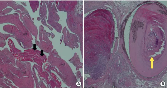
The differential diagnoses for this lesion included a hematoma secondary to trauma, and a malignant tumor with unusual calcification. The patient underwent complete resection macroscopically, and it looked grossly mulberry-like with abundant vascularity, a round- shaped, well-demarcated, and hard mass. Hematoxylin and eosin stains showed multiple size of vascular spaces lined by endothelial cell with striated muscle fibers with intraluminal
A B C D
L2 L2 L2
L M
FIGURE 1. MRI showed homogeneously dark signal intensity on T2 weighted image (A) and low signal intensity with spiculating enhancement on T1 weight gadolinium enhancement image (B). Computed tomography images showed a heterogeneous mass with calcification (C, yellow arrowhead). MRI gradient echo sequence image showed low signal intensity (white arrow) at the L2–3 level, round-shaped mass-like lesion between L and M muscle (D).
MRI: magnetic resonance image, L: longissimus, M: multifidus.
A B
FIGURE 2. A hyper-uptake at the L2 vertebral body on 18F-FDG-PET due to compression fracture of the L2 body (A). White arrow showed a hyper-metabolism at the intermuscular layer at L2–3 level in an 18F-FDG-PET scan (A) and (B).
organizing hematoma and phlebolith. The pathology report confirmed the lesion to be a cavernous hemangioma with phleboliths (FIGURE 3).
DISCUSSION
Cavernous hemangioma has been found in skeletal muscles of limb, neck, and trunk.
Approximately about >70%, It is found in limb and neck. It is a very rare case of cavernous hemangioma which found in lumbar muscle layers.19)
The etiology of cavernous hemangioma may be congenital, trauma, or hormone
imbalance, but the exact etiology is not clearly defined. Hemangiomas are benign vascular malformations that are thought to arise from the abnormal development of embryonic vascular structures.5) Intramuscular hemangiomas account for less than 1% of all
hemangiomas, and they are often diagnosed incidentally during an investigation for other diseases. Intramuscular hemangiomas are characterized by multicentric proliferation of endothelial cells.16)
Cavernous hemangiomas are composed of histologically the cavernous type and the mixed type and divide into the size of vessels of hemangiomas, clinical symptoms, and recurrence rates.6,12) The cavernous type is comprised of relatively larger vessels than the capillary type, and approximately 19% of cases of this type involve the head and neck area. This type is also associated with relatively longer disease duration and a larger size. Since pain is observed in most of these cases, it is easier to make an accurate diagnosis. The local recurrence rate after resection of it is 9%.19) The mixed type involves a histological mixture of capillary and cavernous types, with only 5% occurring in the head and neck area; the mixed type is associated with the highest recurrence rate among the 3 types with 28%.19) On histological analysis, the current case was the cavernous type of cavernous hemangioma, and its accompanying pain was helpful for diagnosis.
mf
mf
mf
A B
FIGURE 3. Histopathologic examination shows multiple vascular spaces lined by endothelial cells (black arrows) with striated mf (A, H&E stain, ×40). Organizing hematoma is seen in a vessel with phlebolith (yellow arrow) (B, H&E stain, ×40).
mf: muscle fibers, H&E: hematoxylin and eosin.
One-fifth of all hemangiomas can be linked to trauma.3,13,15) Cavernous hemangioma is a benign tumor and tumor-like lesion of blood vessels. The established course is growth, fibro-adipose replacement, intravascular clotting, atrophy, and involution, supported by 90%
occurring before the age of 30 years, as well as low incidence in older adults. They occur most commonly in subcutaneous adipose tissue but may also be found in muscle.2,3,13,17)
In patients with a history of trauma, the intramuscular hematoma is a common imaging finding. This patient also had the compression fracture with a spinous process fracture and the intramuscular hemangioma, which could have easily been misdiagnosed as a simple hematoma.
It is difficult to do a diagnosis of intramuscular hemangioma because cavernous
hemangioma has a low prevalence in the intramuscular area and usually shows non-specific clinical symptoms. The common symptoms of cavernous hemangioma include tenderness and swelling. In our case, the lesion did not cause any symptoms like pain, swelling, or tenderness. In our case, CT was taken to evaluate the trauma of this patient, there was a hematoma with very small microcalcification. It could not be a simple acute hematoma finding. It could be a finding of a tumor rather than a simple hematoma.
Calcification usually could be found histologically in a wide variety of tumors; soft-tissue osteosarcoma, liposarcoma, chondrosarcoma, and metastasis.10) In this case, the calcification of the lesion had characteristics of fine and stippled on CT images. The calcifications in liposarcoma tend to be large and coarse. The ossification of soft-tissue osteosarcoma may be dense in CT images.18) The microcalcifications on CT images may be a benign tumor sign, but it cannot be sure. Further studies to differentiate the hematoma with calcification and benign or malignant neoplasm were done.
MRI was performed to characterize this abnormal lesion, it is a contrast enhancement, diffusivity, tumor-normal tissue interface, or invasiveness to the surrounding muscles.
MRI provides useful information on the site and size of intramuscular tumors. MRI findings of IMH include high-signal intensity on both T1- and T2-weighted images. In T2-weighted images, the tumor can be clearly distinguished from the normal surrounding muscle structures. Characteristic findings within the tumor include the presence of heterogeneous signal intensity as a result of increased blood flow in the dilated tortuous vessels; this finding is quite important in the diagnosis process.8) T2 weighted image shows typical black signal of the lesion.
18F-FDG-PET CT scan was performed to differentiate a benign or malignant lesion.
Malignant tumor usually has hypermetabolism and no calcification. There are abundant vascular structures in malignancy. 18F-FDG-PET CT scan can show hypermetabolic activities in a malignant tumorous lesion or fractures. Malignant tumor usually shows hypermetabolic activity. There are abundant vascular structures in malignancy.14)
About 15% of hemangiomas are calcified.2) CT scan indicates the presence of an enhanced and well-circumscribed lesion with external structures, although identifying the
components within the tumor is known to be difficult.4) If calcification is accompanied by an intramuscular hematoma found during investigation following a traumatic injury, it is recommended MRI study to differentiate a simple hematoma from malignancy.8,11) Multiple
Treatment may involve radiotherapy, systemic steroid administration, intralesional steroid or sclerosant injection, cryotherapy, vascular ligation, embolization, and surgical excision.
It could be usually regular observation and follow up until patients with hemangiomas had functional impairments. If the patients with hemangiomas had impaired symptoms (i.e.
severe pain), total surgical excision is usually preferred, since the success rates of other methods are limited. The risk of recurrence can be minimized by total surgical excision.1,7,9,16) The authors find out that intramuscular cavernous hemangioma in current case is the first case report of hemangioma developed in lumbar muscle layers. In this case, the patients had not any symptoms, tenderness, swelling, and neurologic deficit. It could be possible to observe and regular MRI follow up. However, total surgical excision may be preferred when the possibility of a malignant tumor cannot be completely excluded. The authors explained to the patient about the risks and benefits of surgical excision. The patients sincerely hoped to remove the possible cavernous malformation.
CONCLUSION
This report describes a patient with an intramuscular hemangioma that developed in a rare location, the longissimus muscle at the lumbar level after fall down. MRI should be recommended in case of post-traumatic intramuscular hematoma with calcification to differentiate from neoplasms such as hemangioma and malignancy. Total surgical excision is recommended as the treatment of choice in the intramuscular hemangioma.
REFERENCES
1. Afsar FS, Oziz E, Hamdioglu Y, Karasoy I, Uguzi B. Intramuscular haemangioma of the masseter muscle in a 9-year-old girl. Acta Angiologica 13:42-46, 2007
2. Allen PW, Enzinger FM. Hemangioma of skeletal muscle. An analysis of 89 cases. Cancer 29:8-22, 1972 PUBMED | CROSSREF
3. Beham A, Fletcher CD. Intramuscular angioma: a clinicopathological analysis of 74 cases. Histopathology 18:53-59, 1991
PUBMED | CROSSREF
4. Buetow PC, Kransdorf MJ, Moser RP Jr, Jelinek JS, Berrey BH. Radiologic appearance of intramuscular hemangioma with emphasis on MR imaging. AJR Am J Roentgenol 154:563-567, 1990
PUBMED | CROSSREF
5. Calişaneller T, Ozdemir O, Yildirim E, Kiyici H, Altinörs N. Cavernous hemangioma of temporalis muscle:
report of a case and review of the literature. Turk Neurosurg 17:33-36, 2007 PUBMED
6. Clemis JD, Briggs DR, Changus GW. Intramuscular hemangioma in the head and neck. Can J Otolaryngol 4:339-346, 1975
PUBMED
7. Demir Z, Öktem F, Celebioğlu S. Rare case of intramasseteric cavernous hemangioma in a three-year-old boy: a diagnostic dilemma. Ann Otol Rhinol Laryngol 113:455-458, 2004
PUBMED | CROSSREF
8. Hawnaur JM, Whitehouse RW, Jenkins JP, Isherwood I. Musculoskeletal haemangiomas: comparison of MRI with CT. Skeletal Radiol 19:251-258, 1990
PUBMED | CROSSREF
9. Kale US, Ruckley RW, Edge CJ. Cavernous haemangioma of the parapharyngeal space. Indian J Otolaryngol Head Neck Surg 58:77-80, 2006
PUBMED | CROSSREF
10. Liu K, Tripp S, Layfield LJ. Heterotopic ossification: review of histologic findings and tissue distribution in a 10-year experience. Pathol Res Pract 203:633-640, 2007
PUBMED | CROSSREF
11. Makeieff M, Maurice N, Mondain M, Crampette L, Guerrier B. Intramuscular hemangioma of posterior neck muscles. Eur Arch Otorhinolaryngol 258:28-30, 2001
PUBMED | CROSSREF
12. Mulliken JB, Glowacki J. Hemangiomas and vascular malformations in infants and children: a classification based on endothelial characteristics. Plast Reconstr Surg 69:412-422, 1982 PUBMED | CROSSREF
13. Ramon F. Tumors and tumor-like lesions of blood vessels in de Schepper AM, Vanhoenacker FM, Parizel PM, Gielen JL (eds): Imaging of soft tissue tumors. Heidelberg: Springer-Verlag Berlin Heidelberg, pp263- 282, 2006
14. Shreve PD, Anzai Y, Wahl RL. Pitfalls in oncologic diagnosis with FDG PET imaging: physiologic and benign variants. Radiographics 19:61-77, 1999
PUBMED | CROSSREF
15. Tang P, Hornicek FJ, Gebhardt MC, Cates J, Mankin HJ. Surgical treatment of hemangiomas of soft tissue.
Clin Orthop Relat Res 399:205-210, 2002 PUBMED | CROSSREF
16. Top H, Barcın E. Posttraumatic intramuscular hemangioma of the left temporal muscle. Eur J Plast Surg 27:210-212, 2004
CROSSREF
17. Wild AT, Raab P, Krauspe R. Hemangioma of skeletal muscle. Arch Orthop Trauma Surg 120:139-143, 2000 PUBMED | CROSSREF
18. Wilkerson BW, Crim JR, Hung M, Layfield LJ. Characterization of synovial sarcoma calcification. AJR Am J Roentgenol 199:W730-W734, 2012
PUBMED | CROSSREF
19. Wolf GT, Daniel F, Krause CJ, Kaufman RS. Intramuscular hemangioma of the head and neck.
Laryngoscope 95:210-213, 1985 PUBMED | CROSSREF