Pediatr Gastroenterol Hepatol Nutr 2015 March 18(1):60-65
PGHN
Case Report
Disseminated Cytomegalovirus Infection and Protein Losing Enteropathy as Presenting Feature of Pediatric Patient
with Crohn’s Disease
Murat Cakir, Safak Ersoz* and Ulas Emre Akbulut
Department of Pediatric Gastroenterology Hepatology and Nutrition, *Department of Pathology, Faculty of Medicine, Karadeniz Technical University, Trabzon, Turkey
We report a pediatric patient admitted with abdominal pain, diffuse lower extremity edema and watery diarrhea for two months. Laboratory findings including complete blood count, serum albumin, lipid and immunoglobulin levels were compatible with protein losing enteropathy. Colonoscopic examination revealed diffuse ulcers with smooth raised edge (like "punched out holes") in the colon and terminal ileum. Histopathological examination showed active colitis, ulcerations and inclusion bodies. Immunostaining for cytomegalovirus was positive. Despite supportive man- agement, antiviral therapy, the clinical condition of the patient worsened and developed disseminated cytomegalovi- rus infection and the patient died. Protein losing enteropathy and disseminated cytomegalovirus infection a present- ing of feature in steroid-naive patient with inflammatory bowel disease is very rare. Hypogammaglobulinemia asso- ciated with protein losing enteropathy in Crohn's disease may predispose the cytomegalovirus infection in previously healthy children.
Key Words: Crohn disease, Cytomegalovirus infections, Protein-losing enteropathies
Received:November 11, 2014, Revised:December 25, 2014, Accepted:January 9, 2015
Corresponding author: Murat Cakir, Department of Pediatric Gastroenterology Hepatology and Nutrition, Faculty of Medicine, Karadeniz Technical University, Trabzon 35100, Turkey. Tel: +90-462-05326810318, Fax: +90-462-3775890, E-mail: [email protected]
Copyright ⓒ 2015 by The Korean Society of Pediatric Gastroenterology, Hepatology and Nutrition
This is an openaccess article distributed under the terms of the Creative Commons Attribution NonCommercial License (http://creativecommons.org/licenses/by-nc/3.0/) which permits unrestricted noncommercial use, distribution, and reproduction in any medium, provided the original work is properly cited.
INTRODUCTION
Gastrointestinal cytomegalovirus (CMV) infection in immunocompetent children is very rare. In im- munocompromised patients, it can involve the entire alimentary tract from esophagus to rectum [1,2]. In the colon, it can cause colitis-like syndrome includ- ing diarrhea, hemotochezia and abdominal pain as- sociated with fever and weight loss [2,3]. Although
the association of CMV infection with steroid-re- fractory or -dependent patients with inflammatory bowel disease (IBD) have been well-defined, it is very rare in steroid-naive patients. Protein losing en- teropathy (PLE) as an initial presentation of Crohn's disease (CD) is also very rare [4]. Herein, we reported a pediatric steroid-naive CD patients’ presented with disseminated CMV infection and PLE.
Fig. 1. (A) Colonoscopic finding of the patient on admission. Note the ulcer with smooth raised edge (like “punched out holes”).
(B) Aphthous ulcers in the cecum suggesting Crohn’s disease.
CASE REPORT
A 15-year-old girl was admitted to our unit with abdominal pain, abdominal distension, lower ex- tremity edema and watery diarrhea (6-10 times/day) for two months. Physical examination revealed mu- cocutaneous pallor, massive ascites and +2 edema in the lower extremities. She had a weight of 46.5 kg (3-10 percentile) and height of 157 cm (3-10 percen- tile). Laboratory analysis revealed hemoglobin, 8.1 g/dL; leukocytes, 3,500/mm3 (10% lymphocyte); plate- lets, 306,000/mm3; total protein, 3.4 g/dL (6.6-8.7 g/dL); albumin, 1.1 g/dL (3.4-5 g/dL); γ globulin, 2.2 g/dL; erythrocyte sedimentation rate, 20 mm/h;
C-reactive protein, 10.2 g/dL; and other laboratory analysis including serum electrolytes, liver enzymes and urea and creatinine were normal. Coagulation parameters were mildly increased. Serum im- munoglobulin (Ig) G, IgA, IgM and IgE levels were 489 mg/dL (608-1,472), 44.6 mg/dL (>50), 92.9 mg/dL (52-242) and 1.9 IU/mL, respectively. Serum lipid levels were as follows: low-density lip- oprotein-cholesterol, <4 mg/dL; high-density lip- oprotein-cholesterol, <3 mg/dL; and total cholester- ol, 42 mg/dL. Vitamin B12, folic acid and ferritine levels were normal. Urine analysis for the protei- nuria including spot and 24 hour collection was negative. The patient had blood, urine, stool culture, stool ova and parasite ×3, and stool Clostridium diffi-
cile toxin tested, all of which were negative.
Echocardiography was normal. Chest X-ray showed bilateral mild pleural effusion. Based on the clinical (ascites and edema in the lower extremities) and lab- oratory findings (hypoalbuminemia, leucopenia, low lipid, γ globulin and immunoglobulin levels, negative proteinuria); a diagnosis of PLE was made.
Due technical and laboratory inadequacies lympho- scintigraphy using 99mTc-human serum albumin and alpha-1 antitrypsin level in the stool did not studied. Abdominal computed tomography (CT) re- vealed massive ascites and diffuse thickening (1 cm) in intestinal wall including terminal ileum and whole colonic segments. Additionally, multiple lymph no- des with biggest one in 2.5×3 cm size in the celiac truncus, portal hilus and right parailiac region were seen. Thorax CT showed bronchopneumonia and lymph nodes on the para-tracheal and sub-aortic re- gion with the biggest one in 1 cm size. Upper gastro- intestinal endoscopy and colonoscopy was performed for the etiology of PLE. Endoscopy revealed edema and hypertrophy in the gastric mucosa and nodularity and mucosal fissures in the proximal duodenal mucosa. Colonoscopy revealed loss of mucosal vas- cular pattern, diffuse aphthous and mucosal ulcers with smooth raised edge (like “punched out holes”), mucosal fissures and fragility (Fig. 1). Ulcerated le- sions were also seen in the terminal ileum.
Anti-Saccharomyces cerevisiae antibodies IgA
Fig. 2. (A) Cyrptitis and active chronic inflammation in the lamina propria of the colonic mucosa suggesting active colitis (H&E,
×200). (B) Immunostaining for cytomegalovirus (CMV) antigen, brownish appearance in the vascular endothelial cells indicates CMV-positive cells (immunohistochemistry, ×400).
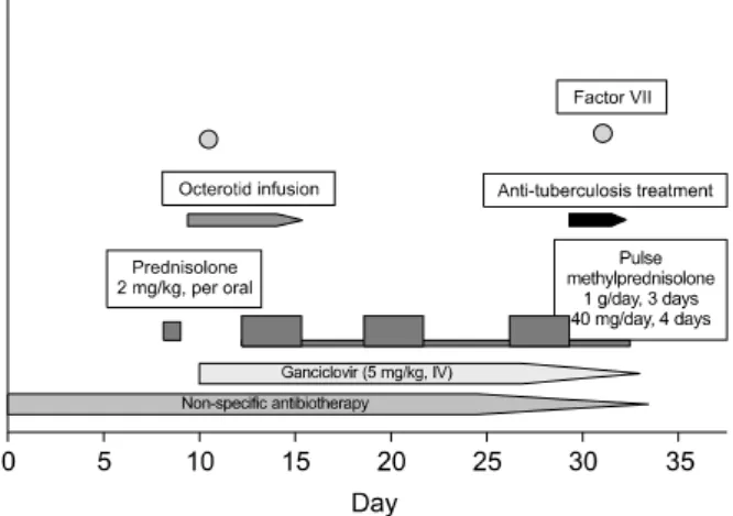
Fig. 3. Treatment regimen of the patient. IV, intra-venous.
was mildly positive. Purified protein derivative was negative. Celiac serology (tissue transglutaminase [tTG]-IgA and tTG-IgG) and HIV serology was negative. Endoscopic and laboratory diagnosis was compatible with IBD particularly CD. Supportive treatment including antibiotics, albumin, parenteral nutrition and IVIG was initiated for PLE. Mesalazine and oral prednisolone (2 mg/kg/day-2 doses) was prescribed for the colitis. Two days later (after the 4th doses of prednisolone); the patient had massive hematochezia. Hemoglobin levels dropped to 6.6 mg/dl. The patient was transferred to intensive care unit and given 2 units red blood cells Octreotide in- fusion and fresh frozen plasma (2 times/day) was in- itiated for the massive bleeding and mild coagulop- athy, respectively. Due to prolonged massive bleed- ing, the patient was also given Factor VII infusion. At the same day, the histopathological analyses of the colonic biopsies revealed severe active colitis and ul- ceration and CMV inclusions. Immunostaining of co- lonic biopsies for CMV antigens were positive (Fig. 2).
Anti-CMV IgM antibodies, anti-CMV IgG anti- bodies from the peripheral blood and CMV polymer- ase chain reaction from peripheral blood, bone mar- row, colonic tissue and tracheal sputum was positive. The patient’s prednisolone was dis-
continued, and she was placed on iv ganciclovir (5 mg/kg). Despite the supportive (red blood cell trans- fusion) and medical treatment (octreotide, fresh fro- zen plasma and Factor VII), massive bleeding was continued. Pulse methylprednisolone was begun on the 3rd day of ganciclovir and bleeding was mildly subsided. But, the patient needed mechanic ven- tilation support due to pneumonia. Ganciclovir treatment was continued to 21 days, pulse methyl- prednisolone was given three courses with the sup- portive treatment but massive bleeding was resumed
and the patient clinical condition was worsened.
Factor VII was given for the second time but no re- spond was achieved. Due to multisystem involve- ment (pulmonary, intestinal and huge abdominal lymphadenopathy), anti-tuberculous treatment was initiated but the patient died one day later due to massive pulmonary hemorrhage and shock (Fig. 3).
DISCUSSION
In this manuscript, we report an interesting case with CD. She was presenting with diffuse CMV in- fection and PLE. PLE as a presenting feature of CD is very rare. CMV infection particularly CMV colitis in the steroid-naive patient with IBD is also very rare [1-5].
CMV is a common infection that effects the 40-100% of all adults worldwide. Although it is asymptomatic in 90% of the cases, the classical man- ifestation of acute symptomatic CMV infection in- cludes mononucleosis-like syndrome. CMV remains latent within the host and can be reactivated later [1,2]. CMV infection may present as interstitial pneumonia, hepatitis, meningoencephalitis, my- ocarditis, retinitis, colitis and hemolytic anemia in immunocompromised patients [3-5]. Gastrointestinal CMV disease in immunocompromised patients can involve the entire alimentary tract from esophagus to rectum. In the colon, it can cause colitis-like syn- drome including diarrhea, hemotochezia and ab- dominal pain associated with fever and weight loss [3].
The association of CMV with IBD has been well-de- fined in steroid-refractory or -dependent adult patients.
Prevalence of CMV in endoscopic biopsies from the patients with steroid-refractory colitis varies from 3% to 66% depends on the methods used for diag- nosis [3]. The effect of CMV infection to the clinical course or prognosis is controversial. Kim et al. [2]
showed that ulcerative colitis (UC) patients with CMV infection have more severe disease and have longer hospitalization. But, none of the patients with CMV infection developed fulminant colitis, required surgery or died in their study. Similar results were re-
ported from Yoshino et al. [6] and Matsuoka et al.
[7]. Contrary to these reports, Berk et al. [8] reported high incidence of toxic megacolon, colectomy re- quirements and mortality rate in IBD patients with CMV infection. Pfau et al. [9] reviewed the 30 pa- tients with IBD and CMV infection and they found that 56.6% had pancolitis, 56.6% required surgery and 20% of the patients died. These findings are sug- gested that the CMV infection may cause serious complications and mortality in patients with IBD.
There have been little data about CMV infection in pediatric IBD patients. Ghidini et al. [10] reported 6 pediatric IBD patients with CMV infection; most of them had pancolitis and refractory to immuno- suppressive agents. They showed that early de- tection and antiviral treatment was associated with good outcome.
The association of CMV infection with ste- roid-naive IBD patients is very rare. Inoue et al. [11]
reported a steroid-naive IBD patient with CMV colitis and reviewed the 7 adult cases in the literature. Their patient had pancolitis and receiving 5-aminosalicylic acid for two months and admitted with diarrhea.
CMV infection was diagnosed by serology and histo- pathological examination. After antiviral treatment, symptoms did not improved and pulse methyl- prednisalone was administered. The patients’ CMV antigenemia was become negative on the following days but toxic megacolon developed and the patient underwent colectomy. Four of the other seven pa- tients with CMV colitis had pancolitis and all the pa- tients improved after the ganciclovir treatment. To date, we could not found any pediatric steroid-naive IBD patients with CMV infection in the literature.
Our patient had some clinical findings suggesting CMV infection such as huge lymph nodes in the ab- domen and thorax, and atypical ulcers (with smooth raised edge, like “punched out holes”) on colonoscopy.
Diagnosis of CMV was made based on the histological and serological examination. Despite the ganciclovir treatment, CMV infection progressed. Although sup- portive treatment was administered, massive colonic bleeding continued and pulse methylprednisalone was added to the treatment but the clinical condition
of the patient worsened and died.
PLE is a rare clinical condition characterized by loss of serum proteins via gastrointestinal tract. The main laboratory findings are reduced serum concen- trations of albumin, γ globulins and lipids. The di- agnosis is based on the exclusion of other causes of hypoproteinemia, and to demonstrate increase ex- cretion of alpha-1 antitrypsin [4]. Both UC and CD may lead the PLE. Protein loss in these conditions is related to enhanced leakage of protein-rich fluids across the eroded epithelium. The degree of mucosal involvement correlates with the severity of protein loss [12]. CMV associated Menetrier’s disease may also cause PLE [13]. Although enlarged and hyper- trophic gastric folds were seen in the upper endos- copy, the histopathological finding were incon- sonant with Menetrier’s disease in our patient.
The association of CMV infection with the new on- set IBD suggests the role of CMV in the pathogenesis of IBD. Orvar et al. [14] and van Dorp et al. [15] sug- gested that CMV infection may initiate an immune response and then autoimmune response in the sus- ceptible host can lead to IBD. But, there are many re- ports in the literature contrary to this hypothesis [2,3,16]. We thought that severe CD in our case lead the PLE, and hypogammaglobulinemia due to mas- sive and prolonged protein loss via gastrointestinal tract cause susceptible to CMV infection in our case.
It is still debate to test or evaluate the CMV in new- ly diagnosed IBD patients. CMV colitis is clinically very similar to IBD, and can occur concomitant to UC or CD regardless to the degree of colon involved. On a recent consensus report, evaluation of CMV is rec- ommended on the 3rd day when unresponsive to ini- tial steroid treatment in acute severe colitis in chil- dren [17].
In conclusion, we report a unique case of CD. She had PLE and disseminated CMV infection as initial presentation of the disease. Hypogammaglobulinemia associated with PLE in CD may predispose the CMV infection in previously healthy children.
REFERENCES
1. Crough T, Khanna R. Immunobiology of human cyto- megalovirus: from bench to bedside. Clin Microbiol Rev 2009;22:76-98.
2. Kim JJ, Simpson N, Klipfel N, Debose R, Barr N, Laine L. Cytomegalovirus infection in patients with active in- flammatory bowel disease. Dig Dis Sci 2010;55:1059-65.
3. Lawlor G, Moss AC. Cytomegalovirus in inflammatory bowel disease: pathogen or innocent bystander? Inflamm Bowel Dis 2010;16:1620-7.
4. Umar SB, DiBaise JK. Protein-losing enteropathy: case illustrations and clinical review. Am J Gastroenterol 2010;105:43-9.
5. Ho M. The history of cytomegalovirus and its diseases.
Med Microbiol Immunol 2008;197:65-73.
6. Yoshino T, Nakase H, Ueno S, Uza N, Inoue S, Mikami S, et al. Usefulness of quantitative real-time PCR assay for early detection of cytomegalovirus infection in patients with ulcerative colitis refractory to immunosuppressive therapies. Inflamm Bowel Dis 2007;13:1516-21.
7. Matsuoka K, Iwao Y, Mori T, Sakuraba A, Yajima T, Hisamatsu T, et al. Cytomegalovirus is frequently re- activated and disappears without antiviral agents in ul- cerative colitis patients. Am J Gastroenterol 2007;102:
331-7.
8. Berk T, Gordon SJ, Choi HY, Cooper HS. Cytomegalovirus infection of the colon: a possible role in exacerbations of inflammatory bowel disease. Am J Gastroenterol 1985;80:355-60.
9. Pfau P, Kochman ML, Furth EE, Lichtenstein GR.
Cytomegalovirus colitis complicating ulcerative colitis in the steroid-naive patient. Am J Gastroenterol 2001;
96:895-9.
10. Ghidini B, Bellaiche M, Berrebi D, Viala J, Hugot JP, Mougenot JF, et al. Cytomegalovirus colitis in children with inflammatory bowel disease. Gut 2006;55:582-3.
11. Inoue K, Wakabayashi N, Fukumoto K, Yamada S, Bito N, Yoshida N, et al. Toxic megacolon associated with cy- tomegalovirus infection in a patient with steroid-naïve ulcerative colitis. Intern Med 2012;51:2739-43.
12. Ferrante M, Penninckx F, De Hertogh G, Geboes K, D'Hoore A, Noman M, et al. Protein-losing enteropathy in Crohn's disease. Acta Gastroenterol Belg 2006;69:
384-9.
13. Sferra TJ, Pawel BR, Qualman SJ, Li BU. Ménétrier disease of childhood: role of cytomegalovirus and trans- forming growth factor alpha. J Pediatr 1996;128:213-9.
14. Orvar K, Murray J, Carmen G, Conklin J.
Cytomegalovirus infection associated with onset of in-
flammatory bowel disease. Dig Dis Sci 1993;38:2307-10.
15. van Dorp WT, Jonges E, Bruggeman CA, Daha MR, van Es LA, van Der Woude FJ. Direct induction of MHC class I, but not class II, expression on endothelial cells by cytomegalovirus infection. Transplantation 1989;
48:469-72.
16. Domènech E, Vega R, Ojanguren I, Hernández A, Garcia-Planella E, Bernal I, et al. Cytomegalovirus in- fection in ulcerative colitis: a prospective, comparative study on prevalence and diagnostic strategy. Inflamm
Bowel Dis 2008;14:1373-9.
17. Turner D, Travis SP, Griffiths AM, Ruemmele FM, Levine A, Benchimol EI, et al; European Crohn's and Colitis Organization; Porto IBD Working Group, European Society of Pediatric Gastroenterology, Hepatology, and Nutrition. Consensus for managing acute severe ulcerative colitis in children: a systematic review and joint statement from ECCO, ESPGHAN, and the Porto IBD Working Group of ESPGHAN. Am J Gastroenterol 2011;106:574-88.