Annals of Otology. Rhinohgy & Laryngology n9(9):646-650. © 2010 Annals Publishing Company. All rights reserved.
Sphenoid Sinus Pneumatization and Its Relation to Bulging of
Surrounding Neurovascular Structures
Jae Hoon Cho, MD, PhD; Jin Kook Kim, MD, PhD; Jeung-Gweon Lee, MD, PhD;
Joo-Heon Yoon, MD, PhD
Objectives: We investigated the bulging and dehiscence of neurovascular structures in the sphenoid sinus and their
rela-tionships to the pneumatization of the sphenoid sinus.
Methods: One hundred sagittally hemisected cadaveric heads were examined. The degree of pneumatization of the
sphe-noid sinus was determined. Bulging and dehiscence of the internal carotid artery (ICA), optic nerve, maxillary nerve, and vidian nerve were examined, and the distances between these structures and the anterior or superior wall of the sphenoid sinus were measured. Additionally, the degree of bony thickness over these structures was determined.
Results: The prevalences of bulging of the optic nerve, segments I and 3 of the ICA, and the maxillary and vidian nerves
were 56%, 34%, 65%, 4 1 % , and 52%, respectively. The greater the degree of pneumatization. the more frequently did the structures bulge into the sphenoid sinus. The optic nerve was found to be in close proximity to the anterior and su-perior walls of the sphenoid sinus. The bone over the surrounding structures was very thin, especially for the complete sellar type.
Conclusions: The prevalence of bulging of the optic nerve, the ICA, and the maxillary and vidian nerves increased in
proportion to the degree of sphenoid sinus pneumatization.
Key Words: internal carotid artery, maxillary nerve, optic nerve, sphenoid sinus, vidian nerve.
INTRODUCTION
The sphenoid sinus is located in the center of the head and is surrounded by several neurovascu-lar structures, including the optic nerve, the internal carotid artery (ICA), the maxillary nerve, and the vidian nerve. Since the development of endoscopie equipment and navigational systems, it has become much easier to access the sphenoid sinus and to treat resident diseases directly. However, even minimal damage to surrounding structures during an opera-tion can lead to an irrevocable outcome such as blindness or massive bleeding.' Therefore, careful preoperative evaluation of the sphenoid sinus and its relation to the surrounding structures is critical.
When evaluating the sphenoid sinus, particular attention should be paid to the bulging and dehis-cence of surrounding structures. The more the optic nerve and the ICA bulge into the sphenoid sinus or the greater their dehiscence, the greater is the chance of accidental datnage. Bulging and dehiscence of re-lated structures are likely to be closely rere-lated to the extent of sphenoid sinus pneumatization.
Logical-ly, the optic nerve and carotid artery should be able to bulge out easily when the sphenoid sinus is ex-tensively pneumatized, and dehiscence should only occur when these surrounding structures bulge sig-nificantly. Therefore, both pneumatization of the sphenoid sinus and bulging or dehiscence of the sur-rounding structures should be considered. However, most studies of the anatomy of the sphenoid sinus and its surrounding structures have dealt with these subjects separately.'"'"The purpose of this study was to investigate bulging and dehiscence of the neuro-vascular structures located in the sphenoid sinus and to evaluate their relationships to the pneumatization of the sphenoid sinus.
METHODS
One hundred eighteen sagittally hemisected fresh cadaveric heads from 59 Korean adults were ex-amined. For simplicity of analysis, we excluded 18 hemisected heads with Onodi cells. Once all of the mucosa in the sphenoid sinus had been carefully re-moved, the degree of pneumatization of the sphe-noid sinus was determined according to the
classi-From the Department of Otorhinolaryngology-Head and Neck Surgery, College of Medicine. Konkuk University (Cho. Kim), and the Department of Otorhinolaryngology (Lee. Yoon). The Airway Mucus Institute (Yoon), and the Brain Korea 21 Project for Medical Sci-ence (Yoon). Yonsei University College of Medicine. Seoul. Korea.
Correspondence: Joo-Heon Yoon. MD, PhD. Dept of Otorhinolaryngology, Yonsei University College of Medicine. 134
Shinchon-dong, Seodaemun-2U. Seoul. Korea 120-752.
Conchal type
Incomplete
sellar type
Complete
sellar type
Fig L Four types of sphenoid sinus pneumatization. In
conchal type, there is no air cavity in sphenoid sinus. In presellar type, air cavity does not expand beyond verti-cal plane passing through anterior clinoid process. Se!lar type is subdivided into complete and incomplete types. In complete sellar type, air cavity extends to clivus, and in incomplete type, it does not.
lication of Hammer and Radberg.^ Bulging and de-hiscence of the ICA, optic nerve, maxillary nerve, and vidian nerve were measured. Bulging was noted when the impression of a structure was clearly iden-tifiable, and dehiscence was documented when any bony defect was observed in the bulge. The distanc-es between thdistanc-ese structurdistanc-es and the anterior or supe-rior wall of the sphenoid sinus were determined with steel vernier calipers. To determine the bony thick-ness over these structures, we first identified the bone segment at the point of maximal bulging and then measured its thickness using calipers. Three experienced rhinologists performed the examina-tions, with the cases evenly allocated among them. When the degree of sphenoid sinus pneumatization
or the existence of a bulge or dehiscence was am-biguous, all 3 rhinologists discussed the case to ar-rive at a unanimous conclusion. The internal review board of Yonsei University Hospital exempted this study, because it did not come within the purview of their regulations.
Pneumatization of Sphenoid Sinus. We classified the sphenoid sinuses into .sellar, presellar, and con-chal types, as proposed by Hammer and Radberg.'' The sellar type was subdivided into complete and incomplete types. The complete type was identified when the pneumatization reached the clivus, and the incomplete type included cases in which the pneu-matization did not reach the clivus (Fig 1).
Optic Nerve. Bulging and dehiscence of the op-tic nerve were examined. The distances between the most anterior bulging point of the optic nerve and the superior and anterior walls of the sphenoid sinus were measured separately. Finally, the bony thick-ness of the point of maximum bulging of the optic nerve was measured (Fig 2).
Internal Carotid Artery. The ICA around the sphe-noid sinus was divided into 3 segments. Segment 1 was defined as the portion of the ICA from the pos-teroinferior part of the lateral wall to the posterior clinoid process. Segment 2 was the short horizontal portion inferior to the pituitary fossa. Segment 3 was the vertical portion between the pituitary fossa and the optic nerve. The bulging and dehiscence of each segment were examined separately. The distances between the anterior wall of the sphenoid sinus and the most anterior bulging points of segments 1 and 3 were measured separately. Finally, the bony thick-nesses of the points of maximum bulging on seg-ments 1 and 3 were measured (Fig 2).
Fig 2, Lateral wall of sphenoid sinus
(right side). A, C, and D indicate dis-tances between anterior wall of sphenoid sinus and most anterior bulging part of optic nerve (ON; A), segment I of in-ternal carotid artery (ICA I : C). and seg-ment 3 of internal carotid artery (1CA3; D). B and E indicate distances between superior wall of sphenoid sinus and most anterior bulging part of optic nerve (B) and between superior wall of sphenoid sinus and most superiorly bulging part of maxillary nerve (MN; E).
648 Cho et al. Sphenoid Sinus Pneumatization
Maxillary and Vidian Nerves. The bulging and dehiscence of the maxillary and vidian nerves were also examined. The distance between the most supe-riorly bulging point of the maxillary nerve and the superior wall was measured. The bony thicknesses of the points of maximum bulging on the maxillary and vidian nerves were measured separately (Fig 2).
RESULTS
Pneumatization of Sphenoid Sinus. The sellar type was the most predominant type (90%) and comprised almost equal numbers of incomplete (47%) and com-plete (43%) types. The presellar type was found in 9% of cases, and the conchal type was found in only 1% of cases. These results are summarized in Table 1.
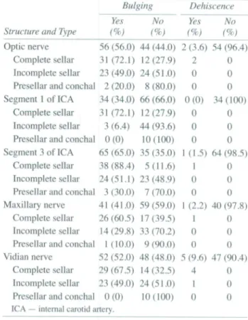
Bulging and Dehiscence of Surrounding Struc-tures. Overall, the incidence of bulging in the optic nerve, segments 1 and 3 of the ICA, the maxillary nerve, and the vidian nerve were 56%, 34%, 65%, 41%, and 52%, respectively. The presence of bulg-ing depended mainly on the degree of pneumatiza-tion of the sphenoid sinus. The more pneumatized the sphenoid sinus, the more frequently the struc-tures bulged out into the sphenoid sinus. For exam-ple, the optic nerve bulged in 72.1% of complete sellar-type cases, in 49.0% of incomplete sellar-type cases, and in only 20.0% of presellar- and conchal-type cases. Similar results were observed for the other structures.
Theoretically, dehiscence should occur only in cases in which bulging is present; the prevalence of dehiscence was therefore only calculated for cases in which bulging was present. The dehiscence rates were quite low for all structures: 3.6% for the optic nerve, 0% for segment 1 of the ICA, 1.5% for seg-ment 3 of the ICA, 2.2% for the maxillary nerve, and 9.6% for the vidian nerve. These results are summa-rized in Table 2.
Distances Between Surrounding Structures and Sphenoid Sinus Walls. Segment 3 of the ICA was fairly distant from the anterior wall of the sphenoid sinus, whereas the optic nerve was quite close to both the anterior and superior walls. For the
com-TABLE 1. PREVALENCE OF SPHENOID SINUS PNEUMATIZATION
TABLE 2. PREVALENCES OF BULGING AND DEHISCENCE OF SURROUNDING STRUCTURES
Type Conchal type Presellar type Sellar type Incomplete Complete Number (%) 1(1) 9(9) 90 (90) 47 (47) 43 (43)
Structure and Type
Optic nerve Complete sellar Incomplete sellar Presellar and conchal Segment 1 of ICA
Complete sellar Incomplete sellar Presellar and conchal Segment 3 of ICA
Complete sellar Incomplete sellar Presellar and conchal Maxillary nerve
Complete sellar Incomplete sellar Presellar and conchal Vidian nerve
Complete sellar Incomplete .sellar Presellar and conchal
Bulging Yes (%) 56 (56.0) 31 (72.1) 23 (49.0) 2 (20.0) 34 (34.0) 31 (72.1) 3 (6.4) 0(0) 65 (65.0) 38 (88.4) 24(51.1) 3 (30.0) 41 (41.0) 26 (60.5) 14(29.8) 1 (10.0) 52 (52.0) 29(67.5) 23 (49.0) 0(0) ICA — internal carotid artery.
No (%) 44 (44.0) 12(27.9) 24(51.0) 8 (80.0) 66 (66.0) 12(27.9) 44(93.6) 10(100) 35 (35.0) 5(11.6) 23 (48.9) 7 (70.0) 59 (59.0) 17(39.5) 33 (70.2) 9 (90.0) 48 (48.0) 14 (32.5) 24(51.0) 10(100) Dehiscence Yes (%) 2(3.6) 2 0 0 0(0) 0 0 0 1 (1.5) 1 0 0 1 (2.2) 1 0 0 5 (9.6) 4 1 0 No (%) 54 (96.4) 0 0 0 34(100) 0 0 0 64 (98.5) 0 0 0 40(97.8) 0 0 0 47 (90.4) 0 0 0
plete sellar type, the average distance from segment 3 of the ICA to the anterior wall was 9.5 ± 3.1 mm, whereas the distance from the optic nerve to the an-terior wall was only 1.9 ± 2.2 mm and that to the su-perior wall was 3.7 ± 3.4 mm. The optic nerve was in contact with the anterior wall in 45% of cases and with the superior wall in 34% of cases. These results are summarized in Table 3.
Bony Thicknesses Over Surrounding Structures. Overall, the bone over the surrounding structures was very thin. For the complete sellar type, the av-erage bone thickness was only 0.2 to 0.3 mm for all structures. For the incomplete sellar type, the bone
TABLE 3. DISTANCES BETWEEN SURROUNDING STRUCTURES AND SPHENOID SINUS WALLS
Complete Incomplete
Sellar Sellar Presellar
Optic nerve to anterior 1.9 ± 2.2 3.1 ±3.7 0 wall
Optic nerve to superior 3.7 + 3.4 3.1 ±3.4 2.62 wall Segment 1 of ICA to anterior wall Segment 3 of ICA to anterior wall Maxillary nerve to superior wall
Data are mean ± SD in millimeters.
19.3 ±3.3 18.6 ±6.2
9.5 ±3.1 9.2 ±3.3 3.5 ±2.8
TABLE 4. BONY THICKNESSES OVER SURROUNDING STRUCTURES Optic nerve Segment 1 of ICA Segment 3 of ICA Maxillary nerve Vidian nerve
Data are mean ± SD
Complete Sellar 0.2 ±0.1 0.3 ±0.2 0.3 ±0.1 0.2 ±0.2 0.2 ±0.2 in millimeters. Incomplete Sellar 0.6 ±0.5 0.2 ±0.2 0.3 ±0.1 0.4 ±0.3 0.3 ±0.1 Presellar 0.3 0.3 ± 0.4
over the optic nerve was marginally thicker than that found for the complete sellar type; 0.6 ± 0.5 mm versus 0.2 ± 0.1 mm, respectively.
For the rernaining structures, the thicknesses were nearly identical between the complete and incom-plete sellar types. These results are summarized in Table 4.
DISCUSSION
The sphenoid sinus is rarely pneumatized at birth. The sinus begins to pneumatize after 4 years of age and completes its pneumatization between the ages of 6 and 12 years.'" As mentioned previously, the degree of sphenoid sinus pneumatization varies be-tween the conchal and complete sellar types; this fact is very important when one is approaching the pituitary gland via the transsphenoidal route.** For the conchal and presellar types, access via the trans-sphenoidal route is very difficult, whereas access is much easier for the sellar type. There is also a dif-ference in ease of access between the complete and incomplete sellar types. It is possible to expose the entire floor of the pituitary gland only for the com-plete sellar type.
This classification of the sphenoid sinus is im-portant not only for pituitary surgery, but also for evaluation of the structures surrounding the sphe-noid sinus. As the sphesphe-noid sinus becomes better pneumatized, it is more likely that the surrounding structures will bulge into the sphenoid sinus; if this occurs, the probability that these structures will be damaged during surgery increases. Therefore, the overall prevalence of bulging and dehiscence in the surrounding structures is less meaningful than is the individualized prevalence based on sphenoid type. However, few studies have investigated this idea. This lack of literature was the major impetus for the present study.
The sphenoid sinuses were classified into 4 types in this study. However, the conchal and preseJlar types were so rare that we considered them together for analysis. As we predicted, the prevalence of bulg-ing of the surroundbulg-ing structures differed accordbulg-ing to sphenoid sinus type. Bulging was frequently
ob-served for the complete sellar type, whereas bulg-ing was seldom found for the presellar and conchal types. It is much easier to uncover the sphenoid si-nus once it has been well pneumatized, although the risk of damage also increases proportionally to the extent of pneumatization.
The optic nerve has been reported to bulge into the sphenoid sinus in 88% to 100% of cases--^-*; however, we observed bulging of the optic nerve in only 56% of cases in the present study. This discrep-ancy may be due to ethnic differences, as most of the previous studies were based on Caucasian popula-tions. Sethi et al** reported that the ICA bulged out in 93% of cases, whereas Tan and Ong'^ reported ICA bulging in 67.7% of cases. We subdivided the ICA into 3 segments and analyzed each segtnent sepa-rately. The overall bulging prevalence for segment I was 34%, and that for segment 3 was 65%. The re-sults for segment 3 are nearly identical to the rere-sults reported by Tan and Ong.'' The prevalences of bulg-ing in the maxillary and vidian nerves were 41 % and 52%, respectively; these results are similar to those reported previously.^''
Attention should be paid to potential dehiscence, because neurovascular structures may be damaged more easily during an operation if they have under-gone dehiscence. However, the prevalence of dehis-cence was very low for all structures in our study; 3.6% for the optic nerve, 1.5% for segment 3 of the ICA, and 0% for segment 1 of the ICA. These results are very different from those reported by Stamm-berger," who found that in cadavers, the ICA had a rate of dehiscence of 25% and the optic nerve had a rate of dehiscence of 6%, Unal et al' reported de-hiscence rates of 5.3% for the ICA and 8% for the optic nerve, and Davoodi et aH reported rates of ICA dehiscence of 39% in male subjects and 44.9% in fe-male subjects, with optic nerve dehiscence rates of 28.5% in male subjects and 46% in female subjects. That both of these studies were based on comput-ed tomography scans is likely the reason dehiscence was observed so frequently. Because thin bone is rarely detected on a computed tomography scan, it may easily be mistaken for dehiscence.
The most anterior bulging point of the optic nerve is typically very close to the anterior and superior walls of the sphenoid sinus. In many cases, we ob-served that the most anterior bulging point of the op-tic nerve was in direct contact with the walls; careful attention should therefore be focused on not damag-ing the optic nerve in removdamag-ing the anterior wall of the sphenoid sinus. Segment 3 of the ICA was re-mote from the anterior wall. The bone over the sur-rounding structures was very thin; 0.2 to 0.3 mm for
650 Cho et al. Sphenoid Sinus Pneumatization
the complete sellar type and 0.2 to 0.6 mm for the in-complete sellar type. As such, the bony wall over the structures cannot act as a reliable physical shield.
In addition, great attention should be paid to the Onodi cell in performing endoscopie sphenoid si-nus surgery. The Onodi cell is a sphenoethmoidal air cell that is positioned posterolateral to the sphe-noid sinus. The optic nerve is commonly exposed in this cell and is therefore at great risk of being dam-aged. Because the prevalence of Onodi cells is high-er among Asians (47.9%) than in Caucasians (9% to 12%),^ much more attention is needed in operating on patients of Asian origin. We also found Onodi cells in 18 hemisected heads among the 118(15.3%). The reason we excluded Onodi cells in this study was that our focus was on the inside of the sphenoid
sinus, and we were interested in determining the as-sociation between the degree of pneumatization and the bulging of the surrounding structures.
CONCLUSIONS
The prevalence of bulging in the optic nerve, the ICA, and the maxillary and vidian nerves depends on the degree of sphenoid sinus pneumatization. Bulging in the surrounding structures increases in proportion to the degree of pneumatization. Further-more, the bony wall over these structures is very thin. During surgery, careful attention must be paid so as not to damage these structures during manipu-lation of the sinus; this is especially true for the optic nerve, which is very close to the anterior wall of the sphenoid sinus.
REFERENCES 1. Unal B, Bademci G. Bilgili YK, Batay F, Avci E. Risky
anatomic variations of sphenoid sinus for surgery. Surg Radiol Anat2006;28:195-201.
2. Bansberg SF, Harner SG, Forbes G. Relationship of the optic nerve to the paranasal sinuses as shown by computed to-mography. Otolaryngol Head Neck Surg 1987:96:331-5.
3. Cheung DK. Attia EL. Kirkpatrick DA, Marcarian B, Wright B. An anatomic and CT scan study of the lateral wall of the sphenoid sinus as related to the transnasal transethmoid en-doscopie approach. J Otolaryngol l993;22:63-8.
4. Davoodi M. Saki N, Saki G, Rahim F. Anatomical varia-tions of neurovascular structures adjacent sphenoid sinus by us-ing CTscan. Pak J Biol Sei 2009:12:522-5.
5. Fujii K. Chambers SM. Rhoton AL Jr. Neurovascular re-lationships of the sphenoid sinus. A microsurgical study. J Neu-rosurg 1979;50:31-9.
6. Hamberger CA, Hammer G, Marcusson G. Experiences
in transantrosphenoidal hypophysectomy. Trans Pac Coast Oto-ophthalmol Soc Annu Meet 1961:42:273-86.
7. Meloni F, Mini R,Rovasio S,Stomeo F,Teatini GP. Ana-tomic variations of surgical importance in ethmoid labyrinth and sphenoid sinus. A study of radiological anatomy. Surg Ra-diol Anat 1992:14:65-70.
8. Sethi DS, Stanley RE, Pillay PK. Endoscopie anatomy of the sphenoid sinus and sella turcica. J Laryngol Otol 1995:109: 951-5.
9. Tan HK, Ong YK. Sphenoid sinus: an anatomic and en-doscopie study in Asian cadavers. Clin Anat 2007;20:745-50.
10. Vidic B. The postnatal development of the sphenoidal si-nus and its spread into the dorsum sellae and posterior clinoid processes. Am J Roentgenol Radium Ther Nucl Med 1968;104:
177-83.
11. Stammberger H. Functional endoscopie sinus surgery. Philadelphia, Pa: Mosby-Year Book, 1991:49-87.