INTRODUCTION
Despite the rapid progress made in modern cataract surgery, posterior capsular opacification (PCO) still remains as one of the most common postoperative complications. Usually the opacification is minimal and confined to the periphery and is not of great concern. However, in some severe cases, it im- pairs vision and can make previous cataract removal worth- less. Thus, additional intervention is often necessary for visu- al recovery. The most well known method to overcome PCO is neodymium:YAG capsulotomy, which is straightforward and immediately effective, but perhaps the best way of deal- ing with PCO is to prevent its occurrence. As PCO is thought to be formed by the migration and metaplasia of lens epithe- lial cells (LECs), it may be beneficial to remove intraopera- tively as much of the cortical remnant and LEC nests as pos- sible, though admittedly there is some dispute in this regard (1-3).
Many theories have been put forward to preclude the for- mation of posterior capsular opacities, and attempts to mod- ify interactions between lens capsules and intraocular lens (IOL) surfaces are abundant. Recently lens design, especially
the configuration of the optic edge have been found to be most effective at preventing PCO, as the edge itself could act as a mechanical barrier against LEC migration and could in- duce capsular bending (4-6). Many attempts have also been made to identify an ideal lens material and gradually it has been accepted that hydrophobic acrylic plastic offers the best means of reducing PCO and postoperative inflammation (7).
Moreover, the lens surface may also play a primary role in LEC adhesion, and thus, development programs have been initi- ated to produce less adhesive surfaces. However, few results are available due to the tardy progress made in the synthesis of biocompatible materials (8-11). In this study, we investi- gated the effect of surface modification with polyethylene glycol (PEG); a type of hydrophilic polymer that is known to be effective at reducing protein deposition and cell adhe- sion. In our previous studies, graft polymerization of PEG to polymethylene methacrylate (PMMA) was found to be effective at reducing keratocyte adhesion, and therefore, we further investigated the efficacy of a PEG graft on an acrylic lens with a square edge, which is presumed to be the best design and materials in terms of reducing PCO (12).
Hyeon Il Lee*,�, Mee Kum Kim*,�, Jung Hwa Ko*,�, Hyun Ju Lee*,�, Won Ryang Wee*,�, Jin Hak Lee*,�
Seoul Artificial Eye Center, Seoul National University Hospital Clinical Research Institute*, Seoul; Department of Ophthalmology�, College of Medicine, Chung-Ang University, Seoul; Department of Ophthalmology�, Seoul National University College of Medicine, Seoul, Korea
Address for correspondence Won Ryang Wee, M.D.
Department of Ophthalmology, Seoul National University Hospital, 28 Yeongeon-dong, Jongno-gu, Seoul 110-744, Korea
Tel : +82.2-2072-2438, Fax : +82.2-741-3187 E-mail : [email protected]
*This study was supported by the Seoul National Uni- versity Hospital research fund (grant no. 03-04-001).
502
The Efficacy of an Acrylic Intraocular Lens Surface Modified with Polyethylene Glycol in Posterior Capsular Opacification
To investigate if the surface modification of intraocular lens (IOL) is efficient in the prevention of posterior capsular opacification (PCO), the acrylic surface of intraocu- lar lens (Acrysof�) was polymerized with polyethylene glycol (PEG-IOL). The human lens epithelial cells (1××104cells/mL) were inoculated on PEG grafted or unmodi- fied acrylic lenses for the control. The adherent cells on each IOL surface were try- psinized and counted. The every PEG-IOL was implanted in 20 New Zealand rab- bits after removal of crystalline lens. The formations of PCO were checked serially through retroilluminated digital photography, and the severity scores were calculat- ed using POCOman�. The cell adherence patterns on each IOL were examined by scanning electron microscopy. As a result, the mean number of adherent cells of PEG-IOL (3.2±±1.1××103) tended to be smaller than that of the acrylic controls (3.6±±1.9××103) without a statistical significance (p=0.73). However, the mean sever- ity of PCO formation in PEG-IOL was significantly lower than that in the control dur- ing the third to sixth weeks after surgery. Scanning electron microscopy revealed that the more patch-like cells were found firmly attached to the IOL surface in con- trol than in the PEG-IOL. Conclusively, PEG polymerization to the acrylic IOL would possibly lessen the formation of PCO after cataract removal.
Key Words : Acrylates; Cataract; Lenses Intraocular; Polyethylene Glycols
Received : 15 September 2006 Accepted : 24 November 2006
MATERIALS AND METHODS Preparation of a PEG grafted acrylic IOL (PEG-IOL)
Acrylic lenses with a square edge of 6 mm optic size (SA 60AT�, Alcon) were used, and graft polymerization was done as described in our previous study (12). Briefly, the lens sur- face was first treated with oxygen plasma, which was gener- ated under low pressure (70 torr) at 100 Watt, and then it was incubated in PEG solution at 60℃for 24 hr.
In vitro human lens epithelial cell adhesion assay
IOLs were sterilized with ethylene oxide gas and washed with phosphate buffered saline (PBS) before use. The two haptics were severed and the remaining optic portions were placed at each bottom of 96-well culture plate. As many as 1×10⁴immortalized human lens epithelial cells (B-3, AT- CC, U.S.A.) were layered onto the optics and incubated for 24 hr at 37℃in 5% CO2. The medium was then removed and IOL surfaces were washed twice carefully with PBS to remove non-adherent cells. The 0.25% trpysin (200 L) was then added and incubated for 10 min at 37℃to harvest ad- herent cells. Cells were then placed in the mixture of 400 L of PBS and 400 L of minimal essential medium (MEM) containing 20% fetal bovine serum, and finally centrifuged at 1,500 rpm for 5 min. Pellets were resuspended in MEM (200 L), stained with trypan blue and counted in a hemo- cytometer (Superior/Marienfeld, D-97965, Bad Mergenthe- im, Germany). A total of nine samples were tested per group.
To verify statistical significances, we used the Mann-Whit- ney U test.
In vivo PCO formation assay
In accord with the principles of ARVO Statements on the
Use of Animals in Ophthalmic and Vision Research, 20 PEG grafted acrylic lenses (PEG-IOL) and 20 control uncoated lenses (SA 60AT�) were implanted randomly into either eye of male New Zealand white rabbits weighing from 2.5-3.0 kg. Briefly, animals were anesthetized with an intramuscular injection of ketamine hydrochloride (100 mg/kg) (Yuhan, Seoul, Korea) and xylazine (5 mg/kg) (Rompun�, Bayer, Lev- erkusen, Gemany). Pupils were dilated with tropicamide (My- driacyl�, Alcon, Fortworth, U.S.A.) 3 times at 10-min inter- vals prior to surgery. Operations were conducted under sterile condition using a Zeiss surgical microscope by one surgeon.
The anterior chamber was entered through a 2.75 mm sized superior self sealing clear corneal incision, and side entry was made 90 degrees apart from the initial incision using a 15° standard angle blade. Using a visosurgical device (Viscoat�, Alcon), a 4.5 to 5.0 mm round, well centered continuous curvilinear capsulotomy was made with a capsulorhexis for- ceps, such that the anterior capsular margin covered the whole optic margin completely. A complete hydrodissection followed by hydrodelineation was performed and the remaining lens material was removed by phacoemulsification (Premier�, Storz, Germany). The capsular bag was fully inflated with viscoelas- tics, and a random IOL was inserted using an injector (Mo- narch�, Alcon, Korea). Then IOL was positioned in the de- sired central position with 360 degree anterior capsular cov- erage. After removal of the remaining whole viscoelastics, the wound was closed with one 10-0 monofilament nylon suture in all cases. Because the rabbit cornea is relatively thin, it was sometimes difficult to make a complete self sealing clear corneal incision. Postoperatively a mixture of dexam- ethasone, neomycin, and polymixin B sulfate ointment (Max- itrol�, Alcon, Fortworth, U.S.A.) was applied 3 times daily for 7 days. Complications were evaluated at weekly and stan- dardized retro-illuminated photographs were taken through a dilated pupil until the 8th postoperative week by the same examiner using a light source with the same size and angle.
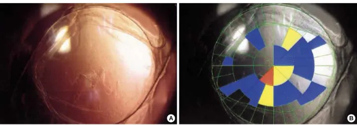
Fig. 1.The severity grades of posterior capsular opacification with the POCOman software. A retroilluminated picture (A) is transformed into a black and white picture (B). The margin of the anterior capsule should be marked then the grid is allocated automatically. Using the standard photographies (grade 1-3), the severity score is calculated with the formula as follows: severity score=(3*Red [grade3]+2*Yel- low [grade2]+1*Blue [grade1]/total area).
A B
To nullify the effects of the anterior capsule in the formation of PCO, we excluded those rabbits with implanted lens optic margins not covered completely by the anterior capsule or extruding into the anterior chamber. The severity of PCO was graded using semi-objective calculation software (POCOman�) from St. Thomas’s Hospital and the King’s College London imaging process group (Fig. 1). Results were compared using the nonparametric rank summation technique and the Mann- Whitney U test, and significance was accepted at the 5%
confidence level. At the end of our study, 8 weeks after im- plantation, all animals were sacrificed and implanted IOLs were carefully removed. The IOLs were again fixed in glu- taraldehyde (2.5%), washed with 0.1 M/L PBS, dehydrated in graded ethanol, critical-point-dried in liquid CO2(CPD 7501), and coated with gold (Giko-IB-3) for scanning elec- tron microscopy (JSM-300, Joel, Tokyo, Japan). Adherent cells were counted in 5 or more fields at 200× and statisti- cal significances between study groups were determined using the Student’s t-test.
RESULTS
In vitro human lens epithelial cell adhesion assay The mean number of adherent LEC’s harvested from each
lens surface was 3.6±1.9×10³for the control and 3.2±1.1
×103for the PEG-IOL, which was not significant (p=0.73, Fig. 2).
In vivo PCO formation assay
No intraoperative complications developed, e.g., capsular tear. However, during follow-up, some parts of IOLs were
No. of cells
8,000 7,000 6,000 5,000 4,000 3,000 2,000
1,000
Control PEG-IOL
Fig. 2.In vitro human lens epithelial cell adhesion after 24-hr cul- ture. The averaged number was slightly higher in the control than that of PEG grafted lens without statistical significance. p=0.73 by Mann whitney-U test.
Fig. 3.The formation of posterior capsular opacification (PCO) after implantation of intraocular lens in rabbits. (A) Polyethylene glycol (PEG), (B) Control. At the 3rd week, the fine fibrotic materials were found on the posterior capsule with minimal proliferation of lens epithelial cells (LECs) in comparison with the control, where the conglomerated LEC’s were dominant. (C) The severity of PCO was significantly lower from the third week in the PEG group until the sixth week of examination. *, p<0.05 by Mann Whitney-U test.
A
C
B
Score
2.4 2 1.6 1.2 0.8 0.4
0
1 2 3 4 5 6 7 8
Week
PEG Control
found to have extruded outside the capsular bag in 7 (4 in the control, 3 in the PEG-IOL group), and thus complete anterior capsular envelopment over the optic was impossible.
These rabbits were excluded from the study. Minimal ante- rior chamber reactions and mild corneal edema were resolved during the first week. The formation of PCO was evident from the third week after surgery and grew weekly until the last follow up. In the PEG-IOL group, PCO began to take on a fine fibrotic appearance rather than a conglomerated cellular proliferation like the Elschnig’s pearl in the control
group, and became a similar figure later on (Fig. 3A, B). The PCO formation was significantly lower during the third to sixth weeks in the PEG-IOL group (Table 1, Fig. 3C, Mann- Whitney-U test). However, during the final two weeks (weeks 7 and 8), no significant differences were found, and some rabbits showed marked LEC proliferation starting from the optic-haptic junction, regardless of group (Fig. 4). Scanning electron microscopic examinations of IOL surfaces revealed fewer adherent LECs on PEG-IOLs. Mean numbers were 26.4
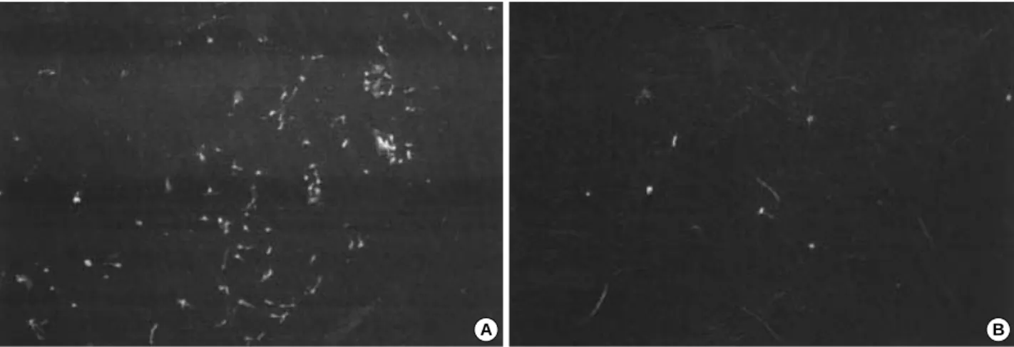
±13.7 for the control and 16.2±11.2 for the PEG -IOL, which were significantly different (p=0.041, Mann-Whitney- U test). Regarding the appearance, more cells were found to be firmly attached to control lens surfaces with a patch-like figure (Fig. 5).
DISCUSSION
The formation of PCO is believed to be a multifactorial process. The surgical technique, lens material, and configu- ration including optic edge design and haptic angulations are thought to play important roles in PCO formation, though patient condition and the postoperative administration of drugs may also have an influence (4, 6, 13, 14). However, there are many different opinions concerning the pathogen- esis of PCO (2, 3). In general, the optic material is not regard-
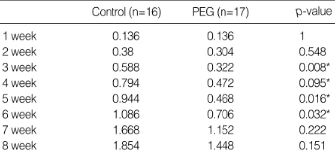
*, p<0.05 by Mann Whitney-U test.
At all time periods, the posterior capsular opacification in PEG-IOL tend- ed to be less severe compared to the control, but the statistical signifi- cance was lost again 7th week of examination.
Control (n=16) PEG (n=17) p-value
1 week 0.136 0.136 1
2 week 0.38 0.304 0.548
3 week 0.588 0.322 0.008*
4 week 0.794 0.472 0.095*
5 week 0.944 0.468 0.016*
6 week 1.086 0.706 0.032*
7 week 1.668 1.152 0.222
8 week 1.854 1.448 0.151
Table 1.The changes of severity score calculated from POCO- man
Fig. 4.An enormous proliferation of lens epithelial cells through the optic-haptic junction. (A) 4th week. Few cells were seen except small cell buds at the optic-haptic junction (arrowhead). (B) 6th week. The cell nests were rapidly growing and threatening the visual axis. (C) 8th week. The visual axis was completely obliter- ated by enormously proliferating cells.
A
C
B
ed so important as optic configuration in the pathogenesis of PCO, although the adhesion of various inflammatory cells and fibronectin is clearly optic material and surface-depen- dent (15, 16). Thus, surface modifications, including heparin incorporation, have been examined, and some reports have shown favorable results under selective conditions (8-11). We undertook this study to investigate the effect of surface mod- ifying acrylic lenses with PEG, which is a high molecular weight hydrophilic polymer and is tethered on the surface, and thus the remainder of the molecule is free to move and can prevent the deposition of protein materials and inflamma- tory cells (17-19). In spite of some debates, hydrophobic acry- lic lenses are known to have an affinity for fibronectin, and thus, we consider that PEG grafting might have a beneficial effect (20). Moreover, although acrylic lenses are better at pre- venting PCO than other type of IOL, PCO formation can also be influenced profoundly by other physical characteris- tics of IOL, and therefore, in the present study we compared the effect of PEG surface grafting on optic materials of same design (7).
In our previous study, PEG-grafted PMMA had a marked- ly lower contact angle and reduced cellular adhesion (12).
However, in the present study, we found no significant dif- ferences between the two study groups in vitro, though a ten- dency for cells to adhere less was observed for the PEG-acry- late. We consider this is due to the characteristics of materi- als that compose the IOL. Since acrylates are soft and their hardness can change markedly with the sudden drop in tem- perature during washing, their adhesiveness might also be changed accordingly.
When PCO formations were compared, it was found to be significantly less severe in the PEG-IOL group than the in control group from the third week postoperatively, when suf- ficient amount of time passed for the cells to migrate and pro- liferate themselves. Moreover, the opacification appeared as a fine fibrosis in the PEG-IOL group, suggesting reduced
visual impairment for the PEG-IOL. However, this PCO lowering effect did not last for long, showing no differences from seventh week of examination. And some rabbits in both groups revealed enormous LEC proliferation starting from the optic-haptic junction, and soon covering throughout the optic surface. The acrylic lens used in our study is a single piece and considered to be better at stretching the posterior capsule because it exerts even forces on the posterior capsule after insertion into the capsular bag, and at the same time avoids early posterior capsular striae, which is believed to be the route of early LEC migration (21, 22). However, the junc- tion between optic and haptic is large and there is no definite barrier, and thus, it can act as a gate for LEC migration. In this study, LECs entered the junction at around 1 month and spread very rapidly to the whole optic surface (Fig. 3). The amount and rapidity of this migration were so great that after six weeks our results might have lost statistical significance.
This demonstrates that such modifications of IOL surface could work during the early phase of PCO formation and that the lens configuration might have a greater effect after a certain time period, as PCO formation is a multifactorial process.
The limitation of our study is that it involved a relatively small number, and thus, a further study using a larger sample number is needed to confirm the findings of this study. Ano- ther limitation is that as we used only one type of IOL, our results cannot be generalized to other lenses with different contours. So it is also needed to study various lens materials and configurations to elucidate their efficacies in terms of preventing PCO formation. Although the formation of PCO is multifactorial and it seems to be impossible to consider its all causative factors in one study, the inflammation induced by the surgery is sure to have significant influence. To inves- tigate its role, more frequent examinations in early postoper- ative phases are also needed.
In conclusion, the surface modification of acrylic IOL with PEG may reduce the formation of PCO during the early stage.
Fig. 5.The scanning electron microscope of adherent cells (×200 fold). (A) In the control group, spindle shape, patch-like cells, together with a few small round cells, were firmly attached to lens surface. (B) In the polyethylence glycol (PEG) group similar cells were found but seemed less adherent.
A B
However, the multifactorial nature of PCO means that we should always consider that other contributors are likely to be important.
REFERENCES
1. Peng Q, Apple DJ, Visessook N, Werner L, Pandey SK, Escobar- Gomez M, Schoderbek R, Guindi A. Surgical prevention of posteri- or capsule opacification. Part 2: enhancement of cortical cleanup by focusing on hydrodissection. J Cataract Refract Surg 2000; 26:
188-97.
2. Linnola RJ, Werner L, Pandey SK, Escobar-Gomez M, Znoiko SL, Apple DJ. Adhesion of fibronectin, vitronectin, laminin, and colla- gen type IV to intraocular lens materials in pseudophakic human autopsy eyes. Part 2: explanted intraocular lenses. J Cataract Refract Surg 2000; 26: 1807-18.
3. Johnston RL, Spalton DJ, Hussain A, Marshall J. In vitro protein adsorption to 2 intraocular lens materials. J Cataract Refract Surg 1999; 25: 1109-15.
4. Nishi O, Nishi K. Preventing posterior capsule opacification by cre- ating a discontinuous sharp bend in the capsule. J Cataract Refract Surg 1999; 25: 521-6.
5. Nishi O, Nishi K, Menapace R. Capsule-bending ring for the preven- tion of capsular opacification: a preliminary report. Ophthalmic Surg Lasers 1998; 29: 749-53.
6. Nishi O, Nishi K, Sakanishi K. Inhibition of migrating lens epithelial cells at the capsular bend created by the rectangular optic edge of a posterior chamber intraocular lens. Ophthalmic Surg Lasers 1998;
29: 587-94.
7. Tanaka T, Yamakawa N, Mizusawa T, Usui M. Interaction between inflammatory cells and heparin-surface-modified intraocular lens. J Cataract Refract Surg 2000; 26: 1409-12.
8. Trocme SD, Li H. Effect of heparin-surface-modified intraocular lenses on postoperative inflammation after phacoemulsification: a randomized trial in a United States patient population. Heparin-Sur- face-Modified Lens Study Group. Ophthalmology 2000; 107: 1031-7.
9. Yuan Z, Sun H, Yuan J. A 1-year study on carbon, titanium surface- modified intraocular lens in rabbit eyes. Graefes Arch Clin Exp Oph- thalmol 2004; 242: 1008-13.
10. Lundvall A, Zetterstrom C, Lundgren B, Kugelberg U. Effect of 3- piece AcrySof and downsized heparin-surface-modified poly (methyl-
methacrylate) intraocular lenses in infant rabbit eyes. J Cataract Refract Surg 2003; 29: 159-63.
11. Gatinel D, Lebrun T, Le Toumelin P, Chaine G. Aqueous flare in- duced by heparin-surface-modified poly(methyl methacrylate) and acrylic lenses implanted through the same-size incision in patients with diabetes. J Cataract Refract Surg 2001; 27: 855-60.
12. Kim MK, Park IS, Park HD, Wee WR, Lee JH, Park KD, Kim SH, Kim YH. Effect of poly (ethylene glycol) graft polymerization of poly (methyl methacrylate) on cell adhesion. In vitro and in vivo study. J Cataract Refract Surg 2001; 27: 766-74.
13. Hollick EJ, Spalton DJ, Meacock WR. The effect of capsulorhexis size on posterior capsular opacification: one-year results of a ran- domized prospective trial. Am J Ophthalmol 1999; 128: 271-9.
14. Fernandez V, Fragoso MA, Billotte C, Lamar P, Orozco MA, Dubovy S, Willcox M, Parel JM. Efficacy of various drugs in the prevention of posterior capsule opacification: experimental study of rabbit eyes.
J Cataract Refract Surg 2004; 30: 2598-605.
15. Smith SR, Daynes T, Hinckley M, Wallin TR, Olson RJ. The effect of lens edge design versus anterior capsule overlap on posterior cap- sule opacification. Am J Ophthalmol 2004; 138: 521-6.
16. Nishi O, Nishi K, Osakabe Y. Effect of intraocular lenses on prevent- ing posterior capsule opacification: design versus material. J Cataract Refract Surg 2004; 30: 2170-6.
17. Desai NP, Hubbell JA. Biological responses to polyethylene oxide modified polyethylene terephthalate surfaces. J Biomed Mater Res 1991; 25: 829-43.
18. Lee JH, Kopecek J, Andrade JD. Protein-resistant surfaces prepared by PEO-containing block copolymer surfactants. J Biomed Mater Res 1989; 23: 351-68.
19. Han DK, Park KD, Ryu GH, Kim UY, Min BG, Kim YH. Plasma protein adsorption to sulfonated poly (ethylene oxide)-grafted poly- urethane surface. J Biomed Mater Res 1996; 30: 23-30.
20. Kim CK, Lee JA, Tchah HW. Adhesion and morphologic change of lens epithelial cell according to materials of intraocular lens. J Kore- an Ophthalmol Soc 2003; 44: 445-53.
21. Lane SS, Burgi P, Milios GS, Orchowski MW, Vaughan M, Schwarte E. Comparison of the biomechanical behavior of foldable intraocu- lar lenses. J Cataract Refract Surg 2004; 30: 2397-402.
22. Kim TW, Kwon JW, Lee JH. Long-term follow-up of the insertion of capsular tension ring in cataract surgery. J Korean Ophthalmol Soc 2005; 46: 1624-9.