INTRODUCTION
Traditional therapy for iliofemoral deep venous thrombo- sis (DVT) has been systemic heparin followed by oral war- farin. Despite widespread use of this method, the results have been largely inadequate in terms of rapid resolution of symp- toms, recanalization of long-segment venous occlusions, and long-term disability from chronic venous insufficiency (1-4).
Thrombolysis for DVT, if performed soon after the onset of symptoms, has the potential to prevent damage to the deep valves, thus maintaining the integrity and preventing post- thrombotic complications (4-9). During the past several years, catheter-directed thrombolysis and endovascular stents have been used to treat extensive lower extremity DVT, as an alter- native to conservative anticoagulation therapy or surgical thrombectomy, in selected cases.
Thrombosis due to compression of the left common iliac vein by the overlying right common iliac artery was first char- acterized by May and Thurner in 1957 (10). With the advent of catheter-directed thrombolysis, iliac vein compression syn- drome (IVCS) is emerging as a frequent finding at venogra-
phy after iliofemoral vein thrombolysis. The term “hyperco- agulable state” is generally used to denote any condition in which the normal balance between clotting and anticlotting mechanisms becomes altered in such a way that the patient is predisposed to thrombus formation. There are a number of conditions that can lead to a hypercoagulable state. Proteins C and S deficiencies are frequently described as causes of the hypercoagulable states (9, 11-19). Although instances of arte- rial thromboses have been reported, proteins C and S defi- ciencies have frequently been associated with venous throm- boembolic events (9, 11-17).
The purpose of this study was to evaluate the early outcome of endovascular management in patients with iliofemoral DVT due to IVCS and protein C and/or S deficiency.
MATERIALS AND METHODS Patients
A total of consecutive 47 patients with DVT were seen in Yong Pil Cho, Je-Hong Ahn*,
Soo-Jung Choi*, Myoung Sik Han, Hyuk Jai Jang, Yong Ho Kim, Hee Jeong Kim�, Tae-Won Kwon�, Sung Gyu Lee�
Department of Surgery and Radiology*, University of Ulsan College of Medicine, Gangneung Asan Hospital, Gangneung; Department of Surgery�, University of Ulsan College of Medicine, Seoul Asan Hospital, Seoul, Korea
Address for correspondence Yong Pil Cho, M.D.
Department of Surgery, Gangneung Asan Hospital, 415 Bangdong-ri, Sacheon-myeon, Gangneung 210-711, Korea
Tel : +82.33-610-3229, Fax : +82.33-641-8120 E-mail : [email protected]
*This work was supported by a research grant from University of Ulsan College of Medicine.
729
Endovascular Management of Iliofemoral Deep Venous Thrombosis due to Iliac Vein Compression Syndrome in Patients with Protein C and/or S Deficiency
The purpose of this study was to evaluate the early outcome of endovascular man- agement in patients with iliofemoral deep venous thrombosis (DVT) due to iliac vein compression syndrome (IVCS) and protein C and/or S deficiency. Between September 2000 and January 2003, catheter-directed thrombolysis was performed in 11 patients with a diagnosis of acute iliofemoral DVT: 7 with protein C and/or S deficiency and 4 without protein C and/or S deficiency. After thrombolysis, the diagnosis of IVCS was confirmed in 6 patients: 4 with protein C and/or S deficien- cy and 2 without protein C and/or S deficiency. Further intervention consisted of angioplasty and stent placement was performed. Four patients with IVCS and pro- tein C and/or S deficiency were included in this study. The immediate technical and clinical success rates were 100% in all 4 patients. There were no complica- tions or clinically detectable pulmonary emboli. This initial experience suggests that endovascular management of iliofemoral DVT due to IVCS in patients with protein C and/or S deficiency is safe and effective.
Key Words : Thrombolytic Therapy; Venous Thrombosis; Protein C Deficiency; Protein S Deficiency
Received : 23 March 2004 Accepted : 1 June 2004
730 Y.P. Cho, J.-H. Ahn, S.-J. Choi, et al.
our department between September 2000 and January 2003.
The diagnosis of DVT was confirmed by characteristic clin- ical, color Doppler ultrasound and/or conventional venogra- phic findings. Of these, 10 patients (21.3%) were diagnosed with DVT associated with protein C and/or S deficiency.
Hypercoagulability studies were done before thrombolysis and systemic anticoagulation therapy. Antigenic protein C and S levels were measured using human protein C/S ‘NL’
NANORIDTMradial immunodiffusion kit (the Binding Site Ltd, Birmingham, U.K.), and the diagnosis of protein C and S deficiencies was confirmed if antigenic protein C and S lev- els were less than 60% of normal. In this study, 3 of the 10 patients with protein C and/or S deficiency did not receive catheter-directed thrombolysis: due to an isolated infrainguinal DVT (1) and a chronic DVT (2). Seven patients with acute iliofemoral DVT and protein C and/or S deficiency were treat- ed with catheter-directed thrombolytic therapy, and 4 with the diagnosis of IVCS (compression of the left iliac vein by the right iliac artery) after thrombolysis were included in this study. There was no family history of DVT in all 4 patients.
We did not performed family studies for protein C and/or S deficiency, because not all patients with these deficiencies will experience episodes of thrombosis and low levels of either factor by itself in an asymptomatic patient are not an indi- cation for anticoagulation. Informed consent was obtained in all patients after the risks and benefits of treatment were fully explained.
Procedure
Local catheter-directed thrombolytic therapy with uroki- nase (Green Cross Co., Yong In, Korea) was performed under the fluoroscopic guidance. Although this drug has recently (mid-1999) been withdrawn from clinical use in the U.S.A.
and U.K. because of problems with purity in its manufacture and on the order of the Food and Drug Administration, we used urokinase as the thrombolytic agent in this study. With the patient in prone position, venous access was through the ipsilateral popliteal vein under the ultrasound guidance. Eight or 9-Fr vascular sheath (Check-flo; Cook, Bloomington, IN, U.S.A.) was placed and direct venography was obtained to estimate the severity and extent of the thrombosis above the popliteal vein. Before the insertion of infusion catheter for the thrombolysis, we aspirated part of the filling defect to curtail the duration and the amount of the thrombolytics with use of another long, smaller-bore vascular sheath. And then, a 5-Fr multi-side-hole infusion catheter was inserted through the sheath. Urokinase was reconstituted by using 500,000 IU in 100 mL of 0.9% NaCl and delivered in continuous low dose of 500-2,000 IU/min via thrombolysis catheter. Patients were systemically treated with heparin during the procedure through the side arm of the venous sheath after a loading bolus of 5,000 IU. Partial thromboplastin time values were obtained at every 4 hr after the start of thrombolytic thera-
py, and the heparin infusion rate was adjusted to maintain the partial thromboplastin time range with 50-90 sec. Fol- low-up venography was performed within 12 hr from the start of thrombolytic therapy. The position of the catheter was changed to facilitate effective thrombolysis. After throm- bolysis, further intervention was performed if there was an underlying venous stenosis of greater than 50%. Intervention consisted of angioplasty and stent placement. The venous stenoses were predilated with an 8- or 10-mm-diameter bal- loon (Ultra-Thin Diamond; Boston Scientific Co., Watertown, MA, U.S.A.) before stent placement (Wallstent; Boston Sci- entific Co.). All stents were placed above the inguinal liga- ment.
Outcome variables
Immediate technical success was defined as complete reca- nalization of the vein with less than 30% residual luminal (area) narrowing (4-6, 9). Immediate clinical success was de- fined as complete or partial resolution of low extremity pain and edema. After the procedure, all patients were given anti- coagulants with a regimen of heparin and, later, orally admin- istered life-long warfarin, adjusted to maintain a prothrom- bin time of 2.0 and followed up both clinically and by color Doppler ultrasound within one month.
RESULTS
Of the 47 patients, catheter-directed thrombolysis was per- formed in 11 patients (23.4%) with symptoms of less than 10 days’ duration and a diagnosis of iliofemoral DVT con- firmed by color Doppler ultrasound: 7 with protein C and/or S deficiency (2 right lower limbs and 5 left lower limbs) and 4 without protein C and/or S deficiency (4 left lower limbs).
After thrombolysis, the diagnosis of IVCS was confirmed in 6 patients: 4 with protein C and/or S deficiency and 2 with- out protein C and/or S deficiency. Further intervention con- sisted of angioplasty and stent placement was performed.
The 4 consecutive patients with IVCS and protein C and/or S deficiency included in this study were 1 man and 3 women, aged 43-75 yr (mean, 62.3 yr). Clinical characteristics are sum-
*Location of thrombus means the highest extent of the thrombus.
�CIV, common iliac vein.
Case Sex/
Age (yr)
Duration of symptom Antigenic protein
C/S (%) Location of
thrombus*
1 F/70 Left CIV� 146/32 5 days
2 M/61 Left CIV 59/27 5 days
3 F/43 Left CIV 37/22 7 days
4 F/75 Left CIV 122/39 10 days
Table 1.Clinical characteristics of 4 patients with iliac vein com- pression syndrome and protein C and/or S deficiency
marized in Table 1. All patients underwent catheter-direct- ed thrombolysis and endovascular stents without antecedent heparin therapy. The mean length of symptoms prior to the initiation of thrombolysis was 6.8 days, with a range of 5- 10 days.
Overall, there were immediate technical and clinical success rates of 100% for treated limbs (Fig. 1). The average dura- tion of therapy was 39.9 hr (range, 16.5-66.5 hr), and the average total urokinase dose was 2.8 million IU (range, 1.0 million to 4.7 million IU). There were no complications or clinically detectable pulmonary emboli. All 4 patients were asymptomatic at mean clinical follow-up of 11.5 months (range, 6-20 months). Serial color Doppler ultrasound in all 4 patients showed no evidence of recurrence of DVT and val-
vular insufficiency in the femoral and popliteal vein segments, using both proximal compression and Valsalva maneuver.
DISCUSSION
Conventional treatment of iliofemoral DVT has typically consisted with systemic heparin therapy followed by antico- agulation with oral warfarin. However, with anticoagulation, eventual recanalization of the occluded vessel depends solely on the effectiveness of the patient’s own fibrinolytic system.
Anticoagulation does not protect the patient from the man- ifestations of post-thrombotic syndrome, which can appear months to years after the acute episode of DVT (3-8). Long-
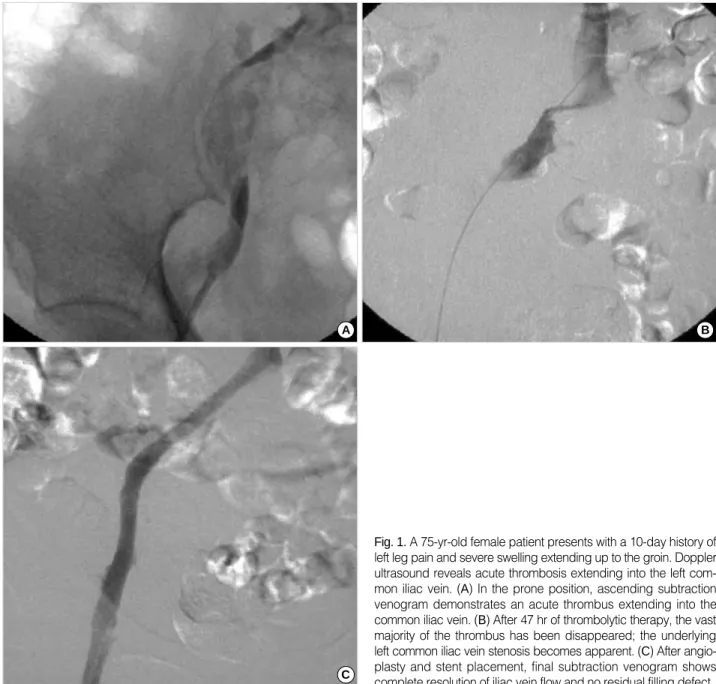
Fig. 1.A 75-yr-old female patient presents with a 10-day history of left leg pain and severe swelling extending up to the groin. Doppler ultrasound reveals acute thrombosis extending into the left com- mon iliac vein. (A) In the prone position, ascending subtraction venogram demonstrates an acute thrombus extending into the common iliac vein. (B) After 47 hr of thrombolytic therapy, the vast majority of the thrombus has been disappeared; the underlying left common iliac vein stenosis becomes apparent. (C) After angio- plasty and stent placement, final subtraction venogram shows complete resolution of iliac vein flow and no residual filling defect.
A B
C
732 Y.P. Cho, J.-H. Ahn, S.-J. Choi, et al.
term studies in patients with iliofemoral DVT treated with anticoagulation alone have demonstrated that muscle pump function and valvular competency are severely compromised in approximately 95% of patients at 5-yr follow-up despite improvement in venous outflow (20). The therapeutic goals for treating the patient with acute DVT include prevention of pulmonary embolism, restoration of unobstructed blood flow through the thrombosed segment, prevention of recur- rent thrombosis, and preservation of venous valve function (4). Considering that success in the achievement of these clini- cal goals will minimize the morbidity and mortality associ- ated with DVT, catheter-directed thrombolysis is a poten- tially attractive form of therapy: (a) it delivers high concen- trations of thrombolytic agents directly into the thrombus while minimizing the potential for a systemic fibrinolytic effect, (b) it provides the opportunity for the prompt restora- tion of venous patency and preservation of venous valve func- tion. This therapy can potentially help prevent long-term sequelae of DVT with acceptable complication rates.
Reported clinical series show that lower extremity DVT occurs three to eight times more frequently on the left when compared to the right (10, 21-23). Virchow first described the anatomic relationship of the right common iliac artery to the left common iliac vein and postulated that the rela- tionship can produce venous stasis, thus accounting for the increased frequency of left leg DVT. Thrombosis due to com- pression of the left common iliac vein by the overlying right common iliac artery was first characterized by May and Thurn- er in 1957 (10). May and Thurner examined 430 cadavers and found 19% to have changes at the junction of the left common iliac vein with the inferior vena cava, which they termed the “spur”. In 1967, Cockett et al. reported the first clinical series of 57 patients presenting with acute iliofemoral DVT secondary to iliac vein compression and coined it the
“IVCS” (24). However, it is well known that compression of the left common iliac vein by the right common iliac artery is a common normal finding during iliocaval venography and all patients with iliac vein compression do not suffer from DVT. With the growing interest in catheter-directed throm- bolysis for extensive lower extremity DVT, IVCS as an anato- mic risk factor for left-sided iliofemoral thrombosis is being identified with increasing frequency (25). Prior to the use of endovascular techniques, the treatment for iliofemoral DVT due to IVCS remained anticoagulation therapy and/or surgery.
During the past several years, there have been several reports of favorable results in managing iliofemoral DVT due to IVCS by endovascular techniques (25, 26). O’Sullivan et al. sug- gest that endovascular techniques are advantageous for sev- eral reasons: (a) diagnostic venography allows for direct assess- ment of the degree of venous obstruction and collateraliza- tion; (b) large acute thrombus burdens can be cleared with use of catheter-directed thrombolysis, thus preserving valve function; (c) obstructive intravenous synechiae and webs (spurs) can be disrupted by means of angioplasty and stent place-
ment; and (d) the integrity of the compressed iliac vein can be restored without apparent long-term harm to the over- riding artery, with acceptable results (25).
The term “hypercoagulable state” is generally used to denote any conditions in which the normal balance between clot- ting and anticlotting mechanisms becomes altered in such a way that the patient is predisposed to thrombus formation.
There are a number of conditions that can lead to a hyperco- agulable state. Proteins C and S deficiencies are frequently described as causes of the hypercoagulable states (11-19). Pro- teins C and S are two of the vitamin K-dependent proteins.
Activated protein C (protein Ca) inactivates factors Va and VIIIa. Protein C is activated to protein Ca 20,000 times faster than by thrombin alone through the interaction of throm- bomodulin and thrombin on the endothelial cell surface (27).
In addition, protein C proteolytically inactivates the inhibitor to tissue plasminogen activator, thus increasing the natural fibrinolytic activity of plasma. Protein S is a cofactor for pro- tein C. The activity of protein Ca is increased several orders of magnitude by its nonenzymatic cofactor protein S. Pro- teins C and S deficiencies may be seen in both congenital and acquired forms. They are inherited in an autosomal domi- nant manner. In the congenital conditions, those homozy- gous for protein C deficiency usually die in infancy, while those heterozygous have antigenic protein C levels less than 60% of normal and present with recurrent venous throm- bosis. Thrombotic events are often precipitated by another factor, such as trauma, surgery, or childbirth. Although the only established treatment for patients with thrombotic events is heparin therapy followed by life-long warfarin therapy, catheter-directed thrombolysis for acute iliofemoral DVT may be safe and effective in patients with protein C and/or S deficiency (9). Furthermore, in patients undergoing coronary thrombolysis, Gruber et al. showed an 11-fold increase in protein Ca during thrombolysis (28). They concluded that thrombolytic therapy generates at least two potent antithrom- botic factors in the circulation, namely the fibrinolytic enzyme, plasmin, and the anticoagulation enzyme, protein Ca. There- fore, we speculate that endogenous protein Ca generated dur- ing thrombolysis has more potent antithrombotic effects in patients with DVT and protein C and/or S deficiency. Pro- tein Ca may help prevent rethrombosis during or after throm- bolysis.
In this small study, 4 with protein C and/or S deficiency and 2 without protein C and/or S deficiency showed IVCS after thrombolysis. Further intervention consisted of angio- plasty and stent placement was performed successfully in all treated limbs without complications or clinically detectable pulmonary emboli. During catheter-directed thrombolysis, major bleeding complications are primarily related to the catheter insertion site (4). Cautious needle access under the ultrasound guidance is mandatory to avoid inadvertent punc- ture of adjacent vessels such as the popliteal artery. A theo- retical consideration with catheter-directed thrombolysis is
dislodgement of significant thrombotic material, leading to a pulmonary embolus. There is a relatively high incidence of asymptomatic pulmonary emboli in iliac venous throm- bosis, as shown by scintigraphy (29). Although the true preva- lence of pulmonary emboli is unknown in our study, there were no clinically significant cases of pulmonary emboli result- ing in increasing dyspnea or arterial oxygen desaturation.
We did not perform scintigraphy and insert caval filters.
In summary, the presence of protein C and/or S deficiency may influence clinical management. In addition to younger patients, other patients who presented with acute, massive iliofemoral DVT not precipitated by other risk factors might benefit from hypercoagulability testing. If there are no con- traindications, patients with DVT associated with this hyper- coagulability should be treated with full anticoagulation in- definitely. Considering that compression of the left common iliac vein by the right common iliac artery is a common nor- mal finding during iliocaval venography, protein C and/or S deficiency may be an important predisposing factor for acute iliofemoral DVT secondary to IVCS. Also, endovascular tech- niques for the treatment of these patients require further eval- uation with full clinical study and long-term follow up, and this presentation shows only the initial experience of this therapeutic modality as a safe and effective method for treat- ing acute iliofemoral DVT with IVCS and protein C and/or S deficiency.
REFERENCES
1. Strandness DE Jr, Langlois Y, Cramer M, Randlett A, Thiele BL.
Long-term sequelae of acute venous thrombosis. JAMA 1983; 250:
1289-92.
2. O’Donnell TF Jr, Browse NL, Burnand KG, Thomas ML. The socio- economic effects of an iliofemoral venous thrombosis. J Surg Res 1977; 22: 483-8.
3. Prandoni P, Lensing AW, Cogo A, Cuppini S, Villalta S, Carta M, Cattelan AM, Polistena P, Bernardi E, Prins MH. The long-term clini- cal course of acute deep venous thrombosis. Ann Intern Med 1996;
125: 1-7.
4. Mewissen MW, Seabrook GR, Meissner MH, Cynamon J, Labropou- los N, Haughton SH. Catheter-directed thrombolysis for lower extrem- ity deep venous thrombosis: report of a National Multicenter Registry.
Radiology 1999; 211: 39-49.
5. Bjarnason H, Kruse JR, Asinger DA, Nazarian GK, Dietz CA Jr, Caldwell MD, Key NS, Hirsch AT, Hunter DW. Iliofemoral deep venous thrombosis: safety and efficacy outcome during 5 years of catheter-directed thrombolytic therapy. J Vasc Interv Radiol 1997;
8: 405-18.
6. Semba CP, Dake MD. Iliofemoral deep venous thrombosis: aggres- sive therapy with catheter-directed thrombolysis. Radiology 1994;
191: 487-94.
7. Sherry S. Thrombolytic therapy for noncoronary diseases. Ann Emerg Med 1991; 20: 396-404.
8. Semba CP, Dake MD. Catheter-directed thrombolysis for iliofemoral venous thrombosis. Semin Vasc Surg 1996; 9: 26-33.
9. Cho YP, Jang HJ, Lee DH, Ahn J, Han MS, Kim JS, Kim YH, Lee SG. Deep venous thrombosis associated with protein C and/or S deficiency: management with catheter-directed thrombolysis. Br J Radiol 2003; 76: 380-4.
10. May R, Thurner J. The cause of the predominantly sinistral occur- rence of thrombosis of the pelvic veins. Angiology 1957; 8: 419-27.
11. Cho YP, Lee DH, Jang HJ, Kim JS, Han MS, Lee SG. Peripheral arterial insufficiency associated with protein C deficiency. Br J Radi- ol 2002; 75: 843-6.
12. Eldrup-Jorgensen J, Flanigan DP, Brace L, Sawchuk AP, Mulder SG, Anderson CP, Schuler JJ, Meyer JR, Durham JR, Schwarcz TH.
Hypercoagulable states and lower limb ischemia in young adults. J Vasc Surg 1989; 9: 334-41.
13. Lane DA, Mannucci PM, Bauer KA, Bertina RM, Bochkov NP, Boulyjenkov V, Chandy M, Dahlback B, Ginter EK, Miletich JP, Rosendaal FR, Seligsohn U. Inherited thrombophilia: Part 1. Thromb Haemost 1996; 76: 651-62.
14. Lane DA, Mannucci PM, Bauer KA, Bertina RM, Bochkov NP, Boulyjenkov V, Chandy M, Dahlback B, Ginter EK, Miletich JP, Rosendaal FR, Seligsohn U. Inherited thrombophilia: Part 2. Thromb Haemost 1996; 76: 824-34.
15. De Stefano V, Finazzi G, Mannucci PM. Inherited thrombophilia:
pathogenesis, clinical syndromes, and management. Blood 1996;
87: 3531-44.
16. Allaart CF, Poort SR, Rosendaal FR, Reitsma PH, Bertina RM, Briet E. Increased risk of venous thrombosis in carriers of hereditary pro- tein C deficiency defect. Lancet 1993; 341: 134-8.
17. De Stefano V, Leone G, Mastrangelo S, Tripodi A, Rodeghiero F, Castaman G, Barbui T, Finazzi G, Bizzi B, Mannucci PM. Clinical manifestations and management of inherited thrombophilia: retro- spective analysis and follow-up after diagnosis of 238 patients with congenital deficiency of antithrombin III, protein C, protein S. Thromb Haemost 1994; 72: 352-8.
18. Sakata T, Kario K, Katayama Y, Matsuyama T, Kato H, Miyata T.
Studies on congenital protein C deficiency in Japanese: prevalence, genetic analysis, and relevance to the onset of arterial occlusive dis- eases. Seminars Thromb Haemost 2000; 26: 11-6.
19. Sakata T, Kario K, Katayama Y, Matsuyama T, Kato H, Miyata T.
Analysis of 45 episodes of arterial occlusive disease in Japanese patients with congenital protein C deficiency. Thromb Res 1999;
94: 69-78.
20. Akesson H, Brudin L, Dahlstrom JA, Eklof B, Ohlin P, Plate G.
Venous function assessed during a five year period after acute ilio- femoral venous thrombosis treated with anticoagulation. Eur J Vasc Surg 1990; 4: 43-8.
21. Cockett FB, Thomas ML. The iliac compression syndrome. Br J Surg 1965; 52: 816-21.
22. Okrent D, Messersmith R, Buckman J. Transcatheter fibrinolytic therapy and angioplasty for left iliofemoral venous thrombosis. J Vasc Interv Radiol 1991; 2: 195-7.
23. Berger A, Jaffe JW, York TN. Iliac compression syndrome treated with stent placement. J Vasc Surg 1995; 21: 510-4.
24. Cockett FB, Lea Thomas M, Negus D. Iliac vein compression: its relation to iliofemoral thrombosis and the post-thrombotic syndrome.
Br Med J 1967; 2: 14-9.
25. O’Sullivan GJ, Semba CP, Bittner CA, Kee ST, Razavi MK, Sze DY, Dake MD. Endovascular management of iliac vein compression (May-Thurner) syndrome. J Vasc Interv Radiol 2000; 11: 823-36.
26. Patel NH, Stookey KR, Ketcham DB, Cragg AH. Endovascular management of acute extensive iliofemoral deep venous thrombosis
caused by May-Thurner syndrome. J Vasc Interv Radiol 2000; 11:
1297-302.
27. Clouse LH, Comp PC. The regulation of hemostasis: the protein C system. N Engl J Med 1986; 314: 1298-304.
28. Gruber A, Pal A, Kiss RG, Sas G, Griffin JH. Generation of activat- ed protein C during thrombolysis. Lancet 1993; 342: 1275-6.
29. Plate G, Ohlin P, Eklof B. Pulmonary embolism in acute ilio-femoral venous thrombosis. Br J Surg 1985; 72: 912-5.
734 Y.P. Cho, J.-H. Ahn, S.-J. Choi, et al.
′