© 2018 Korean Breast Cancer Society. All rights reserved. http://ejbc.kr | pISSN 1738-6756 eISSN 2092-9900
This is an Open Access article distributed under the terms of the Creative Commons Attribution Non-Commercial License (http://creativecommons.org/
licenses/by-nc/4.0) which permits unrestricted non-commercial use, distribution, and reproduction in any medium, provided the original work is properly cited.
INTRODUCTION
The 4th edition of the World Health Organization (WHO) classification of breast tumors defines encapsulated papillary carcinoma (EPC) as a variant of papillary carcinoma that is surrounded by a thick fibrous capsule and lacks myoepithelial cells (MECs) within the papillae and around the periphery of the tumor, as demonstrated by a panel of immunohistochem- istry (IHC) markers [1]. According to the classical breast pa- thology textbooks, the lack of MECs suggests malignant clin- ical behavior breast neoplasms; however, EPC is reported to be a relatively indolent tumor, and very few cases have shown axillary lymph node metastases [2]. Several hypotheses about the biological nature of EPC exist; for example, a tumor in transition from an in situ to an invasive phase and an indolent low-grade carcinoma with pushing rounded invasion [3]. The WHO Working Group advocates that EPC should be staged as an in situ lesion (Tis) [1,4]. EPC is a well-circumscribed tu- mor of the elderly and can be accompanied by a conventional
in situ and/or invasive carcinoma component, most frequently in the form of invasive no special type (NST) carcinoma [5].
The present case perfectly matches these characteristics.
Apocrine breast neoplasms are characterized by a specific histologic appearance, which resembles apocrine metaplasia on hematoxylin and eosin (H&E) stained slides. Such tumors consist of abundant granular eosinophilic cytoplasm and large nuclei, with prominent nucleoli [1]. This subset of breast tu- mors is generally estrogen receptor (ER) negative, progester- one receptor (PR) negative, and androgen receptor (AR) posi- tive [6]. Our previous results suggest that these tumors are also almost uniformly positive for growth hormone-releasing hormone-receptor (GHRH-R) [7,8]. The majority of apocrine carcinomas can be considered special variants of NST carci- nomas, although apocrine differentiation has also been re- ported in several special-type cancers, including EPC [9-12].
CASE REPORT
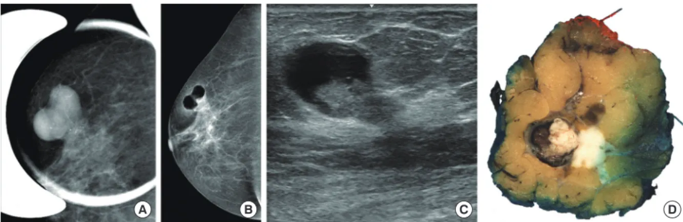
On a breast screening mammography, a 70-year-old woman was diagnosed with a complex breast lesion in the upper-outer quadrant of her right breast. The lesion contained a cystic part and a neighboring spiculated area, which was suggestive of an infiltrative component (Figure 1A-1C). Fine needle aspiration cytology of the lump showed atypical apocrine epithelial cells, but confirmation of the suspected malignancy was not possible.
The subsequent core needle biopsy disclosed an invasive car-
Apocrine Encapsulated Papillary Carcinoma of the Breast: The First Reported Case with an Infiltrative Component
Bence Kővári1, Katalin Ormándi2,3, Zsolt Simonka4, András Vörös1, Gábor Cserni1,5
Departments of 1Pathology and 2Radiology, University of Szeged, Szeged; 3Affidea Diagnostics–Szeged, Szeged; 4Department of Surgery, University of Szeged, Szeged; 5Department of Pathology, Bács-Kiskun County Teaching Hospital, Kecskemét, Hungary
CASE REPORT
J Breast Cancer 2018 June; 21(2): 227-230 https://doi.org/10.4048/jbc.2018.21.2.227
Apocrine encapsulated papillary carcinoma (EPC) of the breast is a rare neoplasm, and only 10 cases have been reported in the literature to date. Although EPC by definition lacks a peripheral myoepithelial layer, all previously published apocrine EPC cases were clinically indolent and lacked a conventional invasive com- ponent. Herein, we report the 11th case of apocrine EPC, which had a conventional invasive carcinoma component and provides
evidence of the malignant potential of this entity. We postulate that apocrine EPC is most likely a morphological variant of con- ventional EPC, with the same unpredictable malignant potential as non-apocrine cases.
Key Words: Apocrine glands, Breast neoplasms, Cysts, Papillary carcinoma
Correspondence to: Bence Kővári
Department of Pathology, University of Szeged, Állomás u. 1, 6720 Szeged, Hungary
Tel: +36-304069859, Fax: +36-62545868 E-mail: [email protected]
This study was supported by the National Research, Development and Innovation Office (grant number: GINOP-2.3.2-15-2016-00020).
Received: November 30, 2017 Accepted: April 18, 2018
Journal of
Breast
Cancer
228 Bence Kővári, et al.
http://ejbc.kr https://doi.org/10.4048/jbc.2018.21.2.227 Figure 1. Imaging and gross findings. Architecture of the dual tumor with mammography (A), pneumocystography (B), ultrasound (C), and gross mor- phology (D). Note the sharply outlined component, showing a clear cystic nature (B, C) with an intraluminal mass. In connection with the intracystic tumor an ill-defined lesion with coarse microcalcifications (A) suggestive of an infiltrative tumor component is also present.
B
A C D
Figure 2. Histological characteristics and immunoprofile. (A) An intracystic papillary component (right) is surrounded by a thick fibrous capsule and an invasive carcinoma (left) component on the low-power view of the lesion (H&E stain, ×1.2). (B) The intracystic part shows a prominent papillary archi- tecture and apocrine cytomorphology (H&E stain, ×5). (C) The invasive carcinoma part also demonstrates apocrine cytomorphology (H&E stain,
×20). The p63 (D, H&E stain, ×5), cytokeratin 5 (E, H&E stain, ×5), and CD10 (F, H&E stain, ×5) immunohistochemistry markers demonstrate the absence of myoepithelial cells in the intracystic papillary component; note the positive internal control and the focal luminal CD10 expression (insert) frequently observed in apocrine lesions [13]. As an evidence for apocrine differentiation, both the encapsulated papillary carcinoma component (G-I,
×15) and the invasive component (J-L, ×20) express androgen receptor (G and J, respectively), gross cystic disease fluid protein 15 (H and K, re- spectively), and growth hormone-releasing hormone-receptor (I and L, respectively).
B
E
H
K A
D
G
J
C
F
I
L
Apocrine Encapsulated Papillary Breast Carcinoma with Infiltrative Component 229
https://doi.org/10.4048/jbc.2018.21.2.227 http://ejbc.kr
cinoma with apocrine differentiation. The patient underwent breast-conserving surgery with a sentinel lymph node biopsy.
The gross examination discovered a correlating dual lesion, which consisted of an intracystic proliferation and a tumor mass that was ill-defined and infiltrative in appearance (Figure 1D). Microscopically, the intracystic portion showed a thick fibrous capsule, an intraluminal dominantly papillary, partial- ly solid proliferation, and a total absence of MEC, under H&E staining and p63, cytokeratin 5, smooth muscle actin, and CD10 IHC (Figure 2) [13]. The ill-defined component repre- sented a NST invasive carcinoma with desmoplasia. Both the intracystic and infiltrative parts showed cytomorphologies that agreed with apocrine differentiation, with eosinophilic granular cytoplasm, large nuclei, and prominent nucleoli.
Small apocrine ductal carcinoma in situ foci were also present.
The invasive tumor was categorized as grade 2 (2+3+ 2=7 points for tubule formation, pleomorphism, and mitotic activity, respectively), according to the Elston-Ellis modification of the Scarff-Bloom-Richardson system. With respect to prognostic and predictive IHC markers, all tumor components were neg- ative for ER, PR, and human epithelial growth factor receptor 2 (HER2), and showed a Ki-67 labeling index of 15%. The tu- mor was positive for AR, with an Allred score of 8, gross cys- tic disease fluid protein 15 (GCDFP-15) and GHRH-R were both observed in the intracystic papillary carcinoma, and the tumor contained an infiltrative NST carcinoma component (Figure 2). Based on our findings, the diagnosis of apocrine EPC of the breast, associated with an invasive NST (ductal) carcinoma with apocrine differentiation, was established.
DISCUSSION
EPC of the breast, formerly known as intracystic and en- cysted papillary carcinomas, is a rare tumor type that repre- sents approximately 1% of all breast carcinomas. The previ- ously published 10 cases of apocrine EPC (A-EPC) all lacked a conventional invasive component and have been described to have indolent clinical behaviors (Table 1) [9-12]. MECs are sometimes very hard or even impossible to identify around benign cystic apocrine glands [14,15], and they may virtually, partially, or completely lack benign cystic apocrine lesions with papillary proliferations [15]. Therefore, the definition that was used for classic EPCs should be applied to apocrine variants with caution. This doubt was also formulated in the first series of such lesions, where the term encapsulated apo- crine papillary carcinoma was used, based on compliance with the WHO terminology: a papillary lesion without MECs in the papillary area and at the periphery. The authors also stressed that the malignant potential of A-EPC has not yet been proven [9]. A characteristic feature of EPCs, the presence of a thick fibrous capsule, was lacking in at least some of the five cases, as shown in Figure 8 of the paper [9]. The present A-EPC case, on the other hand, displayed a thick fibrous cap- sule and a threefold variation in nuclear caliber. This case is unique in the sense that it represents a two-component tumor, with an EPC and a conventional invasive NST component, both of which demonstrate apocrine differentiation. The re- ported case, therefore, may provide proof that A-EPC has the potential to display the same disease spectrum as convention- al EPC, including an association with ductal carcinoma in situ and invasive carcinoma [5]. We therefore propose that A-EPCs Table 1. Patient information, treatment, and outcome of previously published apocrine EPCs and present case
Case Age (yr) Clinical presentation Surgical procedure Radiation Systemic therapy
Follow-up
(mo) Recurrence Status
1 [9] 44 Palpable mass Left partial mastectomy No No 36 None Alive
2 [9] 44 Recurrent cyst Left partial mastectomy+sentinel node biopsy
42.5 Gy in 16 fractions
No 17 None Alive
3 [9] 84 Bilateral recurrent cysts Left partial mastectomy No No 41 None Alive
4 [9] 50 Bilateral recurrent cysts Left partial mastectomy+re-excision
+sentinel node biopsy No No 7 None Alive
5 [9] 50 Recurrent cyst Left partial mastectomy+sentinel node
biopsy No No 3 None Alive
6 [11] 49 Screening-detected Breast-conserving surgery No No 22 None Alive
7 [10] 50 Palpable mass Right partial mastectomy NA NA NA None Alive
8 [12] 68 Palpable mass Left partial mastectomy+sentinel node
biopsy No No 11 None Alive
Present 70 Palpable mass Right partial mastectomy+sentinel
node biopsy 50 Gy Denied 8 None Alive
Two previously published apocrine EPC cases are not shown, as no clinical information was available in the report.
EPC=encapsulated papillary carcinoma; NA=not applicable.
230 Bence Kővári, et al.
http://ejbc.kr https://doi.org/10.4048/jbc.2018.21.2.227
are not necessarily as indolent as previously suggested by the nine pure A-EPC lesions. Our case implies that the clinical behavior of A-EPCs falls into the same unpredictable malignant potential category as the behavior of classical EPCs, rather than into a completely indolent category, as put forward by the first reported cases.
CONFLICT OF INTEREST
The authors declare that they have no competing interests.
ACKNOWLEDGMENTS
The authors thank photographer Mihály Dezső for his as- sistance with the figures.
REFERENCES
1. Lakhani SR, Ellis IO, Schnitt SJ, Tan PH, van de Vijver MJ. WHO Classification of Tumours of the Breast. 4th ed. Lyon: International Agency for Research on Cancer; 2012.
2. Esposito NN, Dabbs DJ, Bhargava R. Are encapsulated papillary carci- nomas of the breast in situ or invasive? A basement membrane study of 27 cases. Am J Clin Pathol 2009;131:228-42.
3. Rakha EA, Ahmed MA, Ellis IO. Papillary carcinoma of the breast:
diagnostic agreement and management implications. Histopathology 2016;69:862-70.
4. Tan PH, Schnitt SJ, van de Vijver MJ, Ellis IO, Lakhani SR. Papillary and neuroendocrine breast lesions: the WHO stance. Histopathology 2015;66:761-70.
5. Leal C, Costa I, Fonseca D, Lopes P, Bento MJ, Lopes C. Intracystic
(encysted) papillary carcinoma of the breast: a clinical, pathological, and immunohistochemical study. Hum Pathol 1998;29:1097-104.
6. Vranic S, Schmitt F, Sapino A, Costa JL, Reddy S, Castro M, et al.
Apocrine carcinoma of the breast: a comprehensive review. Histol Histopathol 2013;28:1393-409.
7. Kővári B, Rusz O, Schally AV, Kahán Z, Cserni G. Differential immu- nostaining of various types of breast carcinomas for growth hormone- releasing hormone receptor: apocrine epithelium and carcinomas emerging as uniformly positive. APMIS 2014;122:824-31.
8. Kővári B, Vranic S, Marchio C, Sapino A, Cserni G. The expression of GHRH and its receptors in breast carcinomas with apocrine differenti- ation-further evidence of the presence of a GHRH pathway in these tu- mors. Hum Pathol 2017;64:164-70.
9. Seal M, Wilson C, Naus GJ, Chia S, Bainbridge TC, Hayes MM.
Encapsulated apocrine papillary carcinoma of the breast: a tumour of uncertain malignant potential: report of five cases. Virchows Arch 2009;455:477-83.
10. Laforga JB, Gasent JM, Sánchez I. Encapsulated apocrine papillary carcinoma of the breast: case report with clinicopathologic and immu- nohistochemical study. Diagn Cytopathol 2011;39:288-93.
11. Hayashi H, Ohtani H, Yamaguchi J, Shimokawa I. A case of intracystic apocrine papillary tumor: diagnostic pitfalls for malignancy. Pathol Res Pract 2013;209:808-11.
12. Kuroda N, Fujishima N, Hayes MM, Moritani S, Ichihara S. Encapsu- lated papillary carcinoma, apocrine type, of the breast. Malays J Pathol 2014;36:139-43.
13. Kővári B, Báthori Á, Cserni G. CD10 immunohistochemical expres- sion in apocrine lesions of the breast. Pathobiology 2015;82:259-63.
14. Cserni G. Lack of myoepithelium in apocrine glands of the breast does not necessarily imply malignancy. Histopathology 2008;52:253-5.
15. Cserni G. Benign apocrine papillary lesions of the breast lacking or virtually lacking myoepithelial cells: potential pitfalls in diagnosing ma- lignancy. APMIS 2012;120:249-52.