Original Article
The utility of the human papillomavirus DNA load for the diagnosis and prediction of persistent vaginal intraepithelial
neoplasia
Kyeong A So, Jin-Hwa Hong, Jong Ha Hwang, Seung-Hun Song, Jae-Kwan Lee, Nak Woo Lee, Kyu Wan Lee
Department of Obstetrics and Gynecology, Korea University College of Medicine, Seoul, Korea
Objective: We evaluated the human papillomavirus (HPV) DNA load for the diagnosis and prediction of persistent vaginal intraepithelial neoplasia (VAIN).
Methods: A retrospective review of the medical records of patients with a pathological diagnosis of VAIN was performed. Eligible women (N=48) were followed for cytology and HPV DNA test, and colposcopic biopsies were taken at 3- to 6-month intervals. Thirty-seven patients were followed for more than 6 months; their HPV DNA test results were compared to the cytology results for the prediction of disease prognosis.
Results: The degree of VAIN was more severe in patients with a high initial HPV DNA load (p=0.009). Patients with VAIN 2 and VAIN 3 were older than those with VAIN 1 (p=0.005 and 0.008, respectively). In 26 out of 37 patients (70.3%), the VAIN resolved. The other patients had persistent lesions with no progression to invasive vaginal carcinoma. The last follow-up HPV DNA load was significantly higher in the group with persistent VAIN compared to the group with resolved VAIN (p<0.0001). Negative cytology was observed in 25 out of 26 patients in the VAIN resolved group and in nine out of 11 patients in the VAIN persistent group (p=0.205).
Conclusion: These results suggest that the HPV DNA test, especially for viral load, was more effective for the diagnosis and prediction of persistent VAIN than cytology.
Key Words: Vaginal intraepithelial neoplasia, Human papillomavirus, Viral load
Received September 7, 2009, Revised November 15, 2009, Accepted November 24, 2009
Correspondence to Jae-Kwan Lee
Department of Obstetrics and Gynecology, Korea University Guro Hospital, Korea University College of Medicine, 80, Guro-dong, Guro-gu, Seoul 152-703, Korea
Tel: 82-2-2626-1831, Fax: 82-2-838-1560 E-mail: joyfulplace@hanmail.net
INTRODUCTION
Vaginal intraepithelial neoplasia (VAIN) is thought to be a precursor lesion of invasive vaginal carcinoma.1 VAIN is much less common than cervical intraepithelial neoplasia (CIN); it is estimated to account for about 1% of all lower gen- ital tract intraepithelial neoplasias.2 However, the incidence of VAIN is expected to increase due to the wider application of cytological screening and colposcopy as well as the in- creased awareness of the condition. There are several different treatment options available for VAIN such as laser vapor- ization, radiation therapy, vaginectomy, immunotherapy, and watchful expectancy.
The risk factors for VAIN are similar to the risk factors asso-
ciated with neoplasia of the cervix and vulva. A prior hyster- ectomy for cervical neoplasia3 is the main risk factor for VAIN.
In addition, the human papillomavirus (HPV) has been im- plicated as a causative agent.4,5 Other risk factors include an- tecedent or coexistent neoplasia of the lower genital tract, a history of pelvic radiation,6,7 and immunosuppression.6 VAIN often accompanies CIN, and is thought to have a similar etiology.8 Such lesions may be extensions into the vagina from the CIN, or they may be satellite lesions occurring mainly in the upper vagina. High-grade VAIN usually occurs in associa- tion with high-grade CIN lesions, which, in approximately 3%
of cases, extend into the vaginal fornices. However, primary foci of high-grade VAIN does occur.9 Because the vagina does not have a transformation zone with immature epithelial cells that can be infected by HPV, the mechanism of entry of HPV is by way of skin abrasions from coitus or tampon use. As these abrasions heal with metaplastic squamous cells, HPV may begin its growth in a manner similar to that occurring in the cervical transformation zone.
For the detection of VAIN, abnormal cytology usually ini- tiates a diagnostic survey for cervical pathology in patients with a history of a previous hysterectomy; in the past, many cases of VAIN were detected incidentally. The utility of HPV
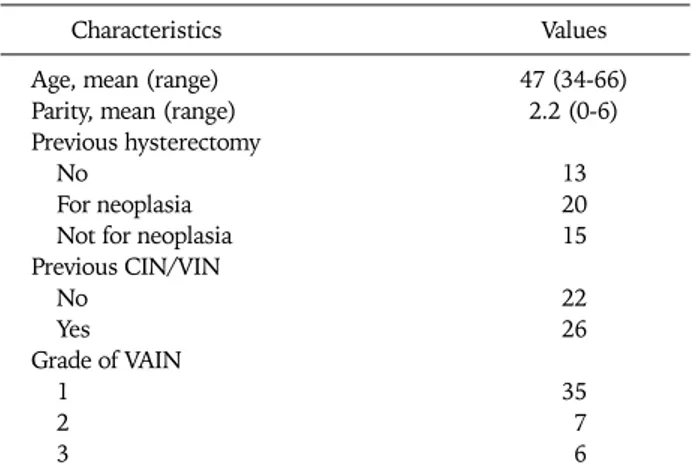
Table 1. Patient and disease characteristics
Characteristics Values
Age, mean (range) 47 (34-66)
Parity, mean (range) 2.2 (0-6)
Previous hysterectomy
No 13
For neoplasia 20
Not for neoplasia 15
Previous CIN/VIN
No 22
Yes 26
Grade of VAIN
1 35
2 7
3 6
CIN: cervical intraepithelial neoplasia, VIN: vulvar intraepithelial neoplasia, VAIN: vaginal intraepithelial neoplasia.
DNA test for the diagnosis and follow-up of VAIN has not been widely used, and its value for this purpose has not been previously analyzed. The aim of this study was to evaluate the efficacy of the HPV DNA test, especially for viral load, for the diagnosis and prediction of regression or persistence of VAIN.
MATERIALS AND METHODS
A retrospective review of the medical records of 48 patients that presented with VAIN lesions between March 2001 and February 2008 was performed. The records were obtained from the database at the Department of Obstetrics and Gynecology, Guro Hospital, at the Korea University. All VAIN lesions were diagnosed by histopathological examination of specimens ob- tained by colposcopy guided biopsies. The grade of the histo- logical diagnosis was determined according to the WHO accepted criteria. All patient specimens and data were obtained with Institutional Review Board approval and informed consent.
All 48 eligible women were followed for cytology, HPV DNA test, and colposcopy guided biopsies at 3- to 6-month inter- vals. The grade of the cytological diagnosis was determined according to the Bethesda system10 for classification of Papanicolaou smear reports. The hybrid capture 2 (HC2) as- say was used to identify patients as HPV DNA positive or neg- ative, and the viral load was determined.
Data regarding the history of previous CIN or vulvar intra- epithelial neoplasia (VIN), a prior hysterectomy, and previous treatment for cervical dysplasia were confirmed by review of the medical records. Women that were previously treated for VAIN were excluded. Cytology results, HPV DNA test results and viral load, colposcopy findings, and treatment modality (vaginectomy, radiation therapy, immunotherapy, laser ther- apy or no treatment) were also recorded.
Among the 48 women, 11 patients were excluded from the study due to loss to follow-up. Thus, there were 37 patients studied with an average follow-up of 30 months (range, 12 to 72 months). The association of HPV DNA test results with VAIN, according to grade, was compared to the cervical cytology. In addition, the efficacy of the HPV DNA test was compared to cytology for the prediction of disease prognosis.
The persistence or regression of lesions was confirmed by histopathology.
1. Liquid-based cytology
A Cervex brush (Rovers Medical Devices, Oss, Netherlands) was used to obtain samples from the cervical os and vaginal wall. The brush was immediately rinsed in a vial of PreservCyt solution (Cytyc Corporation, Marlborough, MA, USA) and the vial was placed in the ThinPrep (Cytyc Corporation) Processor.
2. HC2 assay
Vaginal scrape samples from the high end of the vaginal wall (vaginal fornix) were collected for the HC2 assay using Dacron swabs, specifically targeting areas not touched by the
speculum, and the samples were placed in UCM (Digene Corporation, Gaithersburg, MD, USA).
HPV DNA test by the HC2 assay was performed according to the manufacturer’s protocol (Digene Corporation); signal amplification was performed based on the production of DNA/RNA hybrids using a chemiluminescent reporter sys- tem. Results are reported as the relative light unit (RLU) ra- tio, a semi quantitative estimate of the viral load in the samples. This test has been validated to detect approximately 4,700 genome equivalents (or 1 pg/ml) of target HPV repre- sented by an RLU greater than or equal to the cutoff value cal- culated in each run by a series of standards. Measurements be- low the cutoff were scored as negative. Samples were analyzed for the presence of 13 high-risk (HR) HPV types: 16, 18, 31, 33, 35, 39, 45, 51, 52, 56, 58, 59, and 68. Positive and negative controls were included in each run.
3. Statistical analysis
The data were computerized and analyzed with the SPSS ver.
12.0 (SPSS Inc., Chicago, IL, USA) statistical package. Statisti- cal analyses were performed using the Kruskal-Wallis test, the Chi-Square test, and the Fisher Exact test. The Kruskal- Wallis test was used to assess the association between the grades of VAIN and age, parity and the HPV viral load. The Chi-Square test and the Fisher Exact test were used to evaluate the associa- tion between the grades of VAIN and the cytological results, HC2 results, previous CIN/VIN, and previous hysterectomy.
In addition, these two tests were used to evaluate differ- entiation of the HPV DNA viral load between the regressed and persistent VAIN groups. The p-values for all tests were 2-sided and considered statistically significant when less than 0.05.
RESULTS
The clinical characteristics of the initial 48 patients with VAIN are shown in Table 1. The mean age at the time of diag-
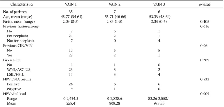
Table 2. Association of clinical characteristics with VAIN according to grade
Characteristics VAIN 1 VAIN 2 VAIN 3 p-value
No. of patients 35 7 6
Age, mean (range) 45.77 (34-61) 55.71 (46-66) 53.33 (48-64)
Parity, mean (range) 2.09 (0-5) 2.86 (1-5) 2.33 (0-5) 0.405
Previous hysterectomy 0.016
No 7 5 1
For neoplasia 21 2 1
Not for neoplasia 7 0 4
Previous CIN/VIN 0.06
No 12 5 5
Yes 23 2 1
Pap results 0.289
No 1 1 0
WNL/ASC-US 23 3 2
LSIL/HSIL 11 3 4
HPV DNA results 0.533
Positive 26 6 6
Negative 9 1 0
HPV viral load 0.009
Range 0-2,494.8 0-2,828.6 83.26-2,550.1
Mean 258.4 909.28 983.55
VAIN: vaginal intraepithelial neoplasia, CIN: cervical intraepithelial neoplasia, VIN: vulvar intraepithelial neoplasia, WNL: within normal lim- its, ASC-US: atypical squamous cells of undetermined significance, LSIL: low-grade squamous intraepithelial lesion, HSIL: high-grade squ- amous intraepithelial lesion, HPV: human papillomavirus.
Table 3. Follow-up period and the disease prognosis
Characteristics No. (%)
Mean follow-up period, mon (range) 30.97 (12-72) Follow-up results
Regression 26 (70.3)
Persistence 11 (29.7)
Progression 0 (0)
nosis was 47 years (range, 34 to 66 years). Among the 48 pa- tients with VAIN, 35 had VAIN 1 (72.9%), seven VAIN 2 (14.6%), and six VAIN 3 (12.5%) at the mean ages of 45.8, 55.7, and 53.3 years, respectively (Table 2). Patients with VAIN 2 and VAIN 3 were older than those with VAIN 1 (p=0.005 and 0.008, respectively).
The patients with VAIN more frequently had a history of a previous hysterectomy (p=0.016). Among 35 VAIN patients with a previous hysterectomy, a hysterectomy for neoplasia was significantly higher in the VAIN 1 group compared to the VAIN 3 group (p=0.035) and vice versa for patients with a history of hysterectomy for reasons other than neoplasia.
We also analyzed the association of the HC2 results and viral load according to the grade of VAIN. The HPV DNA load was significantly higher in patients with high grade VAIN com- pared to the patients with low grade VAIN (p=0.009). The pa- tient’s parity, history of CIN or VIN and Pap smear results did not show any significant difference according to the grade of VAIN.
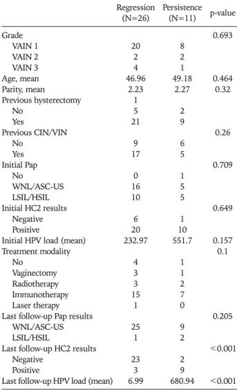
Among the 48 patients, 37 were considered to have adequate follow-up (a mean of 30 months). Among 26 out of the 37 pa- tients (70.3%), the VAIN lesions resolved during the fol- low-up visits. For the other patients (11/37) the lesions per- sisted. However, there was no patient with progression to in- vasive vaginal carcinoma (Table 3).
In addition, there was no difference in the grade of VAIN be- tween the regressed and persistent groups (Table 4). The mean age of the patients in the VAIN regressed and persistent groups was 47.0 and 49.2 years, respectively; this difference was not
statistically significant. The mean parity also was not sig- nificantly different between the two groups. In addition, a his- tory of previous hysterectomy and of previous CIN or VIN did not affect the prognosis of the patients with VAIN (Table 4).
Once VAIN was diagnosed, we initiated treatment of the le- sions using various modalities, for example, vaginectomy, ra- diation therapy, immunotherapy, laser therapy or observa- tion. There was no difference in the disease prognosis be- tween the two groups among the various treatment mod- alities. We evaluated the initial HPV DNA results and the viral load in the regressed and persistent groups and found no stat- istically significant difference. As previously mentioned, dur- ing the 3- to 6-months of patient follow-up, we performed both cytology and HC2 assays. The last follow-up HC2 and vi- ral load results of the regressed and persistent groups were analyzed and compared. The results showed that the de- tection rate for HR HPV DNA was significantly higher in the persistent group than in the regressed group (p<0.001).
Furthermore, the mean HPV DNA viral load in the regressed group was 6.99, whereas that in the persistent group it was
Table 4. Comparison between the regressed and persistent groups Regression
(N=26) Persistence (N=11) p-value Grade
VAIN 1 VAIN 2 VAIN 3 Age, mean Parity, mean
Previous hysterectomy No
Yes
Previous CIN/VIN No
Yes Initial Pap
No
WNL/ASC-US LSIL/HSIL Initial HC2 results
Negative Positive
Initial HPV load (mean) Treatment modality
No Vaginectomy Radiotherapy Immunotherapy Laser therapy Last follow-up Pap results
WNL/ASC-US LSIL/HSIL
Last follow-up HC2 results Negative
Positive
Last follow-up HPV load (mean) 20
2 4 46.96
2.23 1 5 21
9 17
0 16 10
6 20 232.97
4 3 3 15
1
25 1
23 3 6.99
8 2 1 49.18
2.27
2 9
6 5
1 5 5
1 10 551.7
1 1 2 7 0
9 2
2 9 680.94
0.693
0.464 0.32
0.26
0.709
0.649
0.157 0.1
0.205
<0.001
<0.001 VAIN: vaginal intraepithelial neoplasia, CIN: cervical intraepithelial neoplasia, VIN: vulvar intraepithelial neoplasia, WNL: within nor- mal limits, ASC-US: atypical squamous cells of undetermined sig- nificance, LSIL: low-grade squamous intraepithelial lesion, HSIL:
high-grade squamous intraepithelial lesion, HC2: hybrid capture 2, HPV: human papillomavirus.
680.94. These findings demonstrate that the viral load was significantly higher in the persistent group compared to the regressed group (p<0.001).
In the normal or atypical squamous cells of undetermined significance (ASC-US) cytology group, 23 VAIN 1, three VAIN 2 and two VAIN 3 patients were identified. In the low-grade squamous intraepithelial lesion (LSIL) and high- grade squamous intraepithelial lesion (HSIL) cytology group, 11 VAIN 1, three VAIN 2 and four VAIN 3 patients were present. Among the 21 patients that initially presented with within normal limits (WNL) or ASC-US cytology, 16 were in the regressed group and 5 were in the persistent group; while among the 15 patients with the initial diagnosis of LSIL or HSIL, 10 regressed and 5 persisted. At the last follow-up cy-
tology, a negative cytology was observed in 25 out of 26 pa- tients that had regressed lesions and in 9 out of 11 patients that had persistent lesions; this difference was not statisti- cally significant (p=0.205).
During the follow-up of the VAIN lesions, the diagnostic ac- curacy for the HPV DNA positive results as predictive of VAIN persistence or regression was as follows: the sensitivity was 81.8%, specificity 88.5%, positive predictive value 75.0%
and negative predictive value 92.0%. By contrast, the diag- nostic accuracy for the Pap smear had a sensitivity of 18.2%, specificity of 96.2%, positive predictive value of 66.7%, and a negative predictive value of 73.5%.
DISCUSSION
VAIN in almost all cases is asymptomatic, and in many pa- tients there is no lesion visible in the vagina. Previously, the possibility of the presence of VAIN was suggested on the basis of abnormal cytology in patients that had a hysterectomy.
Why has VAIN been overlooked so far? Is it because of the difficulty of colposcopic assessment of the lesions? Perhaps because the so-called flat condyloma might have been ne- glected? The true reason may well be a cytological screening artifact since cytological smears are usually taken from the cervix but not from the vagina. To recognize VAIN with cer- tainty, it is useful to look for HPV DNA in vaginal smears. The natural history of VAIN 1-3 and the risk of progression to in- vasive vaginal carcinoma remain poorly defined. The immedi- ate importance of VAIN might be its role as a viral reservoir for potential sexual transmission. Therefore, it is important to recognize and monitor VAIN to control lower genital tact neoplasia.
HPV has proven to be an important etiologic agent for the de- velopment of CIN and invasive cervical cancer. In a population based study, Castle et al. found a similar distribution of HR HPV genotypes in vaginal and cervical specimens in patients without a history of gynecological malignancy.11 Although far less common than cervical neoplasia, VAIN and invasive vagi- nal carcinoma are also caused by HPV in the majority of cases.5,12 Infection by HR HPV genotypes in both CIN and VAIN may manifest as both high and low-grade lesions. In fact, it was due to the high percentage of LSIL lesions asso- ciated with HR HPV genotypes, that the ALTS group modified the triage algorithm in these patients.13
The purpose of follow up after treatment of VAIN was to de- tect regression or progression in both the short term and the long term. The rationale of adding HPV DNA test to the cytol- ogy was to increase the sensitivity of detection of HR HPV positive disease, and accordingly continue to follow these patients. Similarly, a significant number of vaginal low-grade lesions were associated with HR HPV infections. A previous study reported that 84% of VAIN 1 lesions were associated with HR HPV DNA.14 In this study, we found that 74.3% of VAIN 1, 85.7% of VAIN 2 and 100% of VAIN 3 lesions were
associated with HR HPV DNA.
Previously, it was reported that malignant pathology of the vagina (intraepithelial neoplasia or vaginal cancer)3,15-17 may be more frequently identified in women that had a hyster- ectomy for benign disease. Hence, it was suggested that vagi- nal smears should continue to be performed at regular inter- vals, even after a hysterectomy. In this study, there were 35 patients that had a hysterectomy and also had VAIN; among this group 24 (68.6%) had neoplasia, and the others (11/35, 31.4%) were did not have neoplasia. Although the sample size was quite small, the proportion of VAIN after benign pathol- ogy cannot be ignored; we recommend that vaginal vault cy- tology be continued in women that previously had a hyster- ectomy with benign pathology.
Age was a strong demographic predictor of HR HPV infec- tion. There was a significant association between patients with a higher degree of VAIN and older age (p=0.001). VAIN 1 was diagnosed at around 45 years of age compared to VAIN 2 and VAIN 3, which are most frequently diagnosed after 50 years of age. These results are consistent with the findings of Aho et al.18 (41 years), Benedet and Sanders8 (55 years), and Lenehan et al.19 (49 years). Audet-Lapointe et al.7 reported that patients with VAIN were on average older than those di- agnosed with CIN. Other authors reported an increasing mean patient age with increasing grade of VAIN.6
As shown in Table 2, among the 28 patients with VAIN 1 that had had a hysterectomy, 21 (75.0%) had the hyster- ectomy for neoplasia and seven for other reasons. By contrast, among the VAIN 3 patients, those that had a hysterectomy for reasons other than neoplasia were 4-times more common than patients that had a hysterectomy for neoplasia. A possi- ble explanation for this finding is that more attention may be paid to the individuals that had a hysterectomy for neoplasia due to the possibility of the development of VAIN, and there- fore VAIN may be detected earlier.
As mentioned above, the relationship between HPV in- fection and the development of VAIN is well recognized. In this study, positive HPV DNA was superior to negative HPV DNA regardless of the grade of VAIN; reflecting the close rela- tionship between HPV infection and the development of VAIN. Furthermore, this is the first study to demonstrate a positive correlation between the HPV DNA load and the se- verity of the VAIN grade. In contrast to HPV DNA testing, the Pap smear results did not show a positive correlation with the VAIN grade. Therefore, the results of this study showed that the HPV DNA test was a more useful predictor of the VAIN grade.
During the follow-up of patients with VAIN after treatment, we evaluated the Pap smear and HPV DNA test results as pre- dictors of disease prognosis. As in the prediction of the VAIN grade, the HPV DNA test, by the HC2 assay, showed a sig- nificantly higher viral clearance in the regressed group than in the persistent group (p<0.001). More importantly, the HPV DNA viral load was significantly higher in the persistent
group than in the regressed group (p<0.0001). These find- ings suggest that decrease of the HPV DNA viral load might be a marker for disease regression. Our results indicate that the HR HPV viral load was a better predictor of VAIN regression or persistence than abnormal cytology.
One study recommended including HR HPV test to monitor women initially treated for CIN 2 or 3.20 They suggested that all women should be tested at 6 and 24 months after treat- ment, and should only be referred for routine cervical cancer screening when these tests are negative on both visits. They evaluated the predictive value of the follow-up HPV test in pa- tients treated for CIN 2 or 3. After monitoring patients by cy- tology and HR HPV testing at 3, 6, 9, 12, and 24 months after treatment for CIN, they found that a positive HR HPV DNA test performed 6 months after treatment was more predictive than abnormal cervical cytology, with sensitivities of 90% and 62%, respectively, and with similar specificity.
In this study, we did not evaluate the best treatment among all modalities. Because of the retrospective nature of this study, it was not possible to identify all of the factors that in- fluenced the choice of treatment for all patients. Some of the factors that were considered included the location of the le- sion and multifocality, as well as the patient’s and physician’s preference. This study was performed based on patient data in a retrospective fashion, and the sample size was small; these are significant limitations of this study. Future studies should include prospective data and a larger sample size.
In conclusion, HPV DNA test, especially the HPV DNA load was more effective than cytology for the diagnosis and pre- diction of regression or persistence of VAIN. Thus, HPV DNA test should be performed in addition to cytology during the follow-up of patients with VAIN after treatment.
REFERENCES
1. Rutledge F. Cancer of the vagina. Am J Obstet Gynecol 1967;
97: 635-55.
2. Sillman FH, Sedlis A, Boyce JG. A review of lower genital intra- epithelial neoplasia and the use of topical 5-fluorouracil. Obstet Gynecol Surv 1985; 40: 190-220.
3. Gallup DG, Morley GW. Carcinoma in situ of the vagina: a study and review. Obstet Gynecol 1975; 46: 334-40.
4. Minucci D, Cinel A, Insacco E, Oselladore M. Epidemiological aspects of vaginal intraepithelial neoplasia (VAIN). Clin Exp Obstet Gynecol 1995; 22: 36-42.
5. Sugase M, Matsukura T. Distinct manifestations of human pap- illomaviruses in the vagina. Int J Cancer 1997; 72: 412-5.
6. Sillman FH, Fruchter RG, Chen YS, Camilien L, Sedlis A, McTigue E. Vaginal intraepithelial neoplasia: risk factors for persistence, recurrence, and invasion and its management. Am J Obstet Gynecol 1997; 176: 93-9.
7. Audet-Lapointe P, Body G, Vauclair R, Drouin P, Ayoub J.
Vaginal intraepithelial neoplasia. Gynecol Oncol 1990; 36:
232-9.
8. Benedet JL, Sanders BH. Carcinoma in situ of the vagina. Am J Obstet Gynecol 1984; 148: 695-700.
9. Dodge JA, Eltabbakh GH, Mount SL, Walker RP, Morgan A.
Clinical features and risk of recurrence among patients with vaginal intraepithelial neoplasia. Gynecol Oncol 2001; 83:
363-9.
10. Solomon D, Davey D, Kurman R, Moriarty A, O'Connor D, Prey M, et al. The 2001 Bethesda System: terminology for re- porting results of cervical cytology. JAMA 2002; 287: 2114-9.
11. Castle PE, Schiffman M, Bratti MC, Hildesheim A, Herrero R, Hutchinson ML, et al. A population-based study of vaginal hu- man papillomavirus infection in hysterectomized women. J Infect Dis 2004; 190: 458-67.
12. Daling JR, Madeleine MM, Schwartz SM, Shera KA, Carter JJ, McKnight B, et al. A population-based study of squamous cell vaginal cancer: HPV and cofactors. Gynecol Oncol 2002; 84:
263-70.
13. The Atypical Squamous Cells of Undetermined Significance/
Low-Grade Squamous Intraepithelial Lesions Triage Study (ALTS) Group. Human papillomavirus testing for triage of women with cytologic evidence of low-grade squamous intra- epithelial lesions: baseline data from a randomized trial. J Natl Cancer Inst 2000; 92: 397-402.
14. Logani S, Lu D, Quint WG, Ellenson LH, Pirog EC. Low-grade vulvar and vaginal intraepithelial neoplasia: correlation of his-
tologic features with human papillomavirus DNA detection and MIB-1 immunostaining. Mod Pathol 2003; 16: 735-41.
15. Bell J, Sevin BU, Averette H, Nadji M. Vaginal cancer after hys- terectomy for benign disease: value of cytologic screening.
Obstet Gynecol 1984; 64: 699-702.
16. Ruiz-Moreno JA, Garcia-Gomez R, Vargas-Solano A, Alonso P.
Vaginal intraepithelial neoplasia: report of 14 cases. Int J Gynaecol Obstet 1987; 25: 359-62.
17. Murta EF, Neves Junior MA, Sempionato LR, Costa MC, Maluf PJ. Vaginal intraepithelial neoplasia: clinical-therapeutic analy- sis of 33 cases. Arch Gynecol Obstet 2005; 272: 261-4.
18. Aho M, Vesterinen E, Meyer B, Purola E, Paavonen J. Natural history of vaginal intraepithelial neoplasia. Cancer 1991; 68:
195-7.
19. Lenehan PM, Meffe F, Lickrish GM. Vaginal intraepithelial neo- plasia: biologic aspects and management. Obstet Gynecol 1986;
68: 333-7.
20. Nobbenhuis MA, Meijer CJ, van den Brule AJ, Rozendaal L, Voorhorst FJ, Risse EK, et al. Addition of high-risk HPV testing improves the current guidelines on follow-up after treatment for cervical intraepithelial neoplasia. Br J Cancer 2001; 84:
796-801.