서 론
Human epidermal growth factor receptor 2 (HER2)/neu 원종양 유전자(proto-oncogene)의 증폭 또는 HER2 단백질 과 발현은 침습성 유방 암종의 예후적, 예측적 요인들 중 하나이다.
나쁜 예후, 더 침습적인 질병 경과, 높은 재발 가능성, 전체 생존 기간의 단축 등과 연관되어 있으며, 항암 화학요법에 대한 저항성 과 상관관계가 있다.(1)
Purpose: The purpose of this study was to evaluate silver in situ hybridization (SISH) as an effective test to identify HER2 gene amplification in patients with breast cancer.
Methods: A systematic literature review was used to eval- uate the effectiveness of SISH. The literature review covered from October 27, 2009 to December 1, 2009, and eight domestic databases including KoreaMed and foreign data- bases including Ovid-MEDLINE, EMBASE, and Cochrane Library were used. Keywords, such as ‘silver in situ hybridiza- tion’ and ‘SISH’, were used to search 63 documents. Ten studies regarding the evaluation of diagnostics were included in the final evaluation. The Scottish Intercollegiate Guidelines Network (SIGN) tool was used by two evaluators to inde- pendently evaluate the quality of the ten studies. Results:
A total of ten studies (nine diagnostic evaluation studies and one correlation study) were identified to evaluate SISH. The
effectiveness of this test was evaluated based on diagnostic accuracy, concordance rate, and correlation with fluores- cence in situ hybridization (FISH) results. The sensitivity of SISH was 0.81-1.00, and the specificity was 0.82-1.00. The positive predictive value was 0.95-1.00, negative predictive value was 0.81-1.00, and the test accuracy was 0.90-1.00.
The concordance rate of SISH was 87.0-100% and two stud- ies reported a correlation with FISH results. The body of evi- dence as a whole suggests a Grade D for SISH. Conclusion:
SISH is a safe and useful test in patients with breast cancer and at least grade D evidence based on existing positive studies.
Key Words: Breast neoplasms, Fluorescence in situ hybridization, Meta-analysis 중심단어: 유방암, 형광제자리부합법, 체계적 문헌고찰
The Effectiveness of Silver In Situ Hybridization in Patients with Breast Cancer:
A Systematic Review
Sunyoung Jang, Seon-Heui Lee, Soojin Kim, You-Kyoung Lee
1, Young-Hyuck Im
2, Wonshik Han
3, Hee-Sook Park
4Health Technology Assessment Department, National Evidence-Based Healthcare Collaborating Agency, Seoul; 1Department of Laboratory Medicine, Soonchunhyang University College of Medicine, Seoul; 2Department of Internal Medicine, Sungkyunkwan University School of Medicine, Seoul; 3Department of Surgery, Seoul National University College of Medicine, Seoul; 4Department of Internal Medicine, Soonchunhyang University College of Medicine, Seoul, Korea
Breast Cancer
O R I G I N A L A R T I C L E
장선영ㆍ이선희ㆍ김수진ㆍ이유경1ㆍ임영혁2ㆍ한원식3ㆍ박희숙4
한국보건의료연구원신의료기술평가사업본부, 1순천향대학교 의과대학진단검사의학과, 2성균관대학교의과대학내과학교실 혈액종양내과, 3서울대학교의과대학 외과학교실, 4순천향대학교의과대학내과학교실 혈액종양내과
유방암 환자에서의 은제자리부합법의 유효성:
체계적 문헌 고찰
책임저자: 박희숙
140-743 서울시 용산구 한남동 657-58, 순천향대학교 의과대학 내과학교실 혈액종양내과
Tel: 02-709-9185, Fax: 02-709-9200 E-mail: parkhs@schmc.ac.kr
접수일: 2010년 12월 3일 게재승인일: 2011년 2월 11일 본 논문은 2009년 보건복지부의 연구기금에서 연구비를 지원받음.
S1
HER2 유전자의 증폭 또는 과발현은 유방암 환자에게 있어 나 쁜 예후와 관련이 있고, 타목시펜(tamoxifen)을 이용한 호르몬 치료에 저항성을 나타내지만 anthracycline 계열의 항암제나 paclitaxel의 치료에 비교적 좋은 반응을 나타낸다. 또한 HER2 표적 치료제인 trastuzumab (Herceptin�; Genentech, South San Francisco, USA)이나 lapatinib (Tykerb�; GlaxoSmith- Kline, Philadelphia, USA)이 개발됨으로써 HER2 상태는 유 방암 환자 치료방침 결정에 필수적인 요소가 되었다.(2)
2007년 American Society of Clinical Oncology (ASCO) tumor marker guideline에서는 새로 진단받은 원발 유방암 환 자와 재발 유방암 환자 및 전이 유방암 환자 등에서 HER2 검사 시 행을 권장하였고, 2007년 발표된 ASCO/College of American Pathologist (CAP) guidelines에서는 HER2 상태를 평가하기 위한 방법으로 면역조직화학검사(immunohistochemistry, IHC) 와 형광제자리부합법(Fluorescence in situ hybridization, FISH)을 포함하는 두 가지 검사 체계를 제안하고 있다. IHC 염 색은 HER2 단백의 과발현을 관찰하는 방법으로서, 사용되는 항 체의 종류가 매우 다양하며 민감도와 특이도, 판정 기준에 따라 다른 결과를 보여 표준화에 대한 지속적인 노력이 필요한 검사법 이다. FISH법은 HER2 유전자의 증폭 여부를 직접 검사하기 때 문에 항암제에 대한 효과와 예후에 대한 보다 정확한 예측이 가능 하며, IHC 염색보다 민감도와 특이도 및 정확도가 우수하고 안정 성이 높으며 소량의 조직만으로도 검사가 가능하다는 장점을 가지 고 있다.(3)
최근 유전자 증폭을 직접 평가하는 검사법으로 은제자리부합법 (silver-enhanced in situ hybridization, SISH)이 소개되었 는데, 이 방법은 탐색자(probe)가 해당 DNA 부위에 부착하면 침 착을 유도하여 조직 내에서 오랫동안 유지되는 검은색 점으로 반 응 결과를 나타내어 광학현미경으로 판독이 가능한 검사 방법이 다.(2) 은제자리부합법은 유방암 환자의 진단 및 적절한 치료제 선 택을 위하여 HER2 유전자의 증폭 여부를 검사하는 기술로, 기존
의 형광제자리부합법과 비교 시 검사시간이 짧고 자동화가 가능하 며, 검사 후 결과물 보관이 가능하다는 장점이 있다. 따라서 현존 하는 HER2 유전자의 증폭 여부를 확인하는 표준검사인 형광제자 리부합법과 은제자리부합법을 비교한 보고된 문헌들을 데이터베 이스 검색을 통하여 선택한 후 HER2 양성 진단의 정확성을 확인 하여 은제자리부합법 검사의 유효성을 판단하고자 한다.
방 법
은제자리부합법의 유효성은 유방암 환자를 대상으로 은제자리 부합법과 형광제자리부합법과의 비교를 통해 검사의 진단 정확성, 비교검사와의 일치율 및 상관성을 평가하였다.
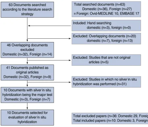
은제자리부합법은 KoreaMed를 포함한 8개 국내 데이터베이스 와 Ovid-MEDLINE, EMBASE 및 Cochrane Library의 국외 데이터베이스를 이용하여 2009년 12월 1일 최종 검색을 완료하였 다. 국내 문헌은‘silverin situ hybridizaion’, ‘SISH’, ‘은제 자리부합법’과 같은 관련 검색어를 위주로 한 검색전략을 통해 8개 의 인터넷 검색 데이터베이스를 이용하여 검색하였다. 국외 문헌 은MEDLINE과EMBASE를이용하여Tables 1, 2와같은검색전 략을 도출하였다. 총 63개의 문헌을 검색하였고, 동물 실험 및 전 임상시험, 원저가 아닌 연구, 초록만 발표된 경우 및 한국어와 영 어로 기술되지 않은 문헌은 배제하였다. 중복검색을 포함하여 총 36개 문헌이 제외되었고 진단법 평가연구 9편, 기타 연구(상관성 연구) 1편, 총 10개의 연구가 최종 평가에 포함되었다(Figure 1).
문헌선택기준
선택기준(Inclusion criteria)
�
�은제자리부합법이 수행된 연구
�
�적절한 의료결과(outcome)가 하나 이상 보고된 연구
�
�한국어나 영어로 출판된 연구
배제기준(Exclusion criteria)
�
�동물 실험(non-human)
�
�원저(original article)가 아닌 연구나 초록만 발표된 경우 혹은 회색문헌 등(non-systematic reviews, editorial,
PICO=Patients, Index test, Comparators, Outcomes.
Patients 1 Breast Neoplasms/ 170,188
Index test 2 In Situ Hybridization/ 42,451
3 Silver/ 9,461
4 silver in situ hybridization.mp. 5
5 sish.mp. 18
6 And/2-3 27
Index test total 7 or/4-6 43
Total 9 1 and 7 10
Table 1. Ovid-MEDLINE search strategy
PICO No. Search term Searched no.
PICO=Patients, Index test, Comparators, Outcomes.
Patients 1 ‘breast tumor’/exp 232,309
Index test 2 ‘silver in situ hybridization’ 8
Total 3 1 and 2 7
Table 2. EMBASE search strategy
PICO No. Search term Searched no.
letter, comment, opinion pieces, review, congress or conference material, guideline, note, news article, abstract, etc.)
문헌의질평가
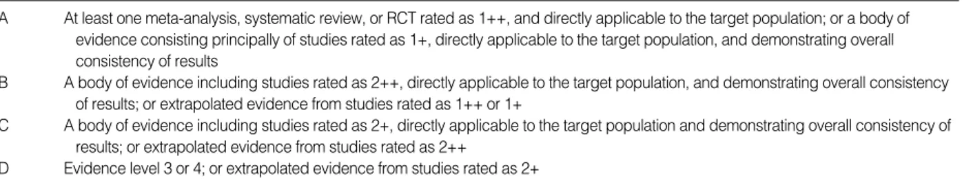
선택된 문헌의 질 평가 도구는 영국 Scottish Intercollegiate Guidelines (SIGN)의‘Methodology Checklist’(2004년 3월) 를 채택하였다. SIGN의 질 평가 도구는 원래 체계적 문헌 고찰, 무작위 임상시험, 코호트 연구, 환자-대조군 연구, 진단법 평가 및 경제성 평가연구로 구분되어 있으나, 의료기술평가에서 대부분의 문헌이 비무작위 임상시험(quasi-experimental studies)과 비 교관찰 연구(observational study)가 많아 SIGN과 협의 하에
일부 도구를 수정하였고, 연구 유형별 질 평가 도구는 Table 3과 같다.(5) 문헌 검색부터 선택기준 적용 및 자료 추출까지 각 단계 는 2명의 평가자가 각 과정을 독립적으로 수행하였으며 이에 따라 근거의 수준과 권고의 등급을 선정하였다(Table 4).
자료추출
평가에 포함된 문헌이 한 유형으로만 한정되지 않아 자료의 추 출은 여러 차례 반복되었다. 자료 추출은 2명의 평가자가 각각 중 복하여 분석하였으며, 연구를 진행하면서 나타난 문제점을 토의하 는 과정이 여러 차례 반복되었다. 동 과정에서 문헌에 기술된 내 용과 결과에 영향을 주는 연구대상의 특성 및 필수적으로 기술되 어야 하는 유효성에 대한 자료를 구분하였다. 1차 자료 추출은 2명 Total searched documents (n=63)
Domestic (n=36), Foreign (n=27)
※Foreign: Ovid-MEDLINE 10, EMBASE 17 Induded: Hand searching
domestic (n=3), foreign (n=0) Excluded: Overlapping documents (n=20)
domestic (n=7), foreign (n=13)
Excluded: Studies that are not original articles (n=5)
Excluded: Studies in which no silver in situ hybridization was performed (n=31)
Total excluded papers (n=36: Domestic 29, Foreign 7) Total included papers (n=10: Domestic 3, Foreign 7) 63 Documents searched
according to the literature search strategy
46 Overlapping documents excluded
Domestic (n=32), Foreign (n=14)
41 Documents published as original articles Domestic (n=32), Foreign (n=9)
10 Documents with silver in situ hybridization being the major test
Domestic (n=3), Foreign (n=7)
10 Documents selected for evaluation of silver in situ
hybridization
Figure 1. Documents selected for evaluation of silver in situ hybridization according to the literature search strategy.
Table 3. Levels of evidence (From Scottish Intercollegiate Guidelines Network. SIGN 50: a guideline developer’s handbook) (4)
1++ High quality meta-analyses, systematic reviews of RCTs, or RCTs with a very low risk of bias 1+ Well-conducted meta-analyses, systematic reviews, or RCTs with a low risk of bias 1- Meta-analyses, systematic reviews, or RCTs with a high risk of bias
2++ High quality systematic reviews of case control or cohort or studies
High quality case control or cohort studies with a very low risk of confounding or bias and a high probability that the relationship is causal
2+ Well-conducted case control or cohort studies with a low risk of confounding or bias and a moderate probability that the relationship is causal
2- Case control or cohort studies with a high risk of confounding or bias and a significant risk that the relationship is not causal 3 Non-analytic studies, e.g. case reports, case series
4 Expert opinion
RCT=randomized controlled trial.
의 평가자가 각각 중복하여 분석하였으며, 이후 실무 회의를 통해 나타난 문제점을 토의하고 재차 자료 추출 과정을 수행하였다.
통계분석
본 연구에 사용된 문헌들의 진단 정확성을 확인하기 위하여 부 정을 제외한 후 고정효과모형(fixed effect model)에 기초하여 메타분석을 실시하였고, 전반적인 은제자리부합법의 유용성을 receiver operating characteristics (ROC) curve와 area under the curve (AUC)를 통해 평가하였다. 추가적으로 출판 편향 정도를 확인하기 위하여 funnel plot을 확인하였다. 본 연 구의메타분석을위해서, STATA 통계프로그램11.1 MP version (Stata Corp., College Station, USA)와 MetaDiSc 1.4 version (Hospital Universtario Ramon y Cajal, Madrid, Spain)을 사용하였다.
결 과
은제자리부합법은 총10편(진단법평가연구 10편)의 문헌적 근 거로 평가하였으며, 본 평가에 포함된 문헌의 주요 자료추출 목록 은 Table 5와 같다.
은제자리부합법의 유효성은 진단의 정확성, 비교검사와의 일치 율, 비교검사와의 상관성에 대하여 평가하였다. 은제자리부합법은 HER2 유전자 과발현 여부를 확인하고 그 결과에 따라 예후 예측 및 치료 방향을 선택하는데 도움을 주는 검사로 현재 HER2 유전 자를 확인하는 검사 중 표지자만 상이하고 원리는 동일한 표준검 사인 형광제자리부합법을 비교자로 하여 진단정확성 및 일치율과 상관성에 초점을 두어 문헌의 결과를 해석하기로 결정하였다.
은제자리부합법의 진단 정확성 평가는 7편의 문헌(2,8-13)을 근거로 평가하였다. 은제자리부합법의 경우 보통 양성, 부정, 음 성으로 나누어 결과를 보고하여, 진단의 정확성을 명확하게 평가 Table 4. Grades of recommendations (From Scottish Intercollegiate Guidelines Network. SIGN 50: a guideline developer’s handbook) (4)
A At least one meta-analysis, systematic review, or RCT rated as 1++, and directly applicable to the target population; or a body of evidence consisting principally of studies rated as 1+, directly applicable to the target population, and demonstrating overall consistency of results
B A body of evidence including studies rated as 2++, directly applicable to the target population, and demonstrating overall consistency of results; or extrapolated evidence from studies rated as 1++ or 1+
C A body of evidence including studies rated as 2+, directly applicable to the target population and demonstrating overall consistency of results; or extrapolated evidence from studies rated as 2++
D Evidence level 3 or 4; or extrapolated evidence from studies rated as 2+
RCT=randomized controlled trial.
Table 5. Documents selected for evaluation of silver in situ hybridization
No. Author Research
location
Research
type Subjects Comparators Level of
evidence Publication
yr
1 2010 Sung et al.(2) Korea Diagnostic 144 invasive IHC, FISH 2+
evaluation study breast cancer tissue
2 2009 Bartlett et al.(6) England Diagnostic 45 breast cancer tissue FISH 2-
evaluation study
3 2009 Francis et al.(7) Australia Diagnostic 593 cancer patients IHC 2-
evaluation study
4 2009 Kang et al.(8) Korea Diagnostic 165 invasive breast cancer FISH 2-
evaluation study
5 2009 Kim et al.(9) Korea Diagnostic 58 breast cancer patients FISH 2-
evaluation study
6 2009 Shousha et al.(10) England Diagnostic 56 breast cancer tissue IHC, FISH 2-
evaluation study
7 2008 Capizzi et al.(11) Italy Diagnostic 83 women breast cancer IHC 2-
evaluation study patients
8 2008 Carbone et al.(12) Italy Diagnostic 89 breast cancer tissue IHC, FISH 2-
evaluation study
9 2007 Dietel et al.(13) Germany Diagnostic 99 breast cancer tissue IHC, FISH 2+
evaluation study
10 2007 Si´nczak-Kuta et al.(1) Poland Correlation study 63 breast cancer tissue IHC, FISH 3 IHC=immunohistochemistry; FISH=fluorescence in situ hybridization.
하기 어려웠으나 부정을 제외한 양성 및 음성을 대상으로 재 구성 하여 진단 정확성을 산출하였다(Table 6).
Sung 등(2)의 한국에서 유방암으로 진단받은 144명의 유방암 환자의 조직을 대상으로 한 연구에서는 1.8 미만일 경우 음성, 1.8 이상 2.2 이하인 경우 부정(equivocal), 2.2를 초과하면 양성으로 판정하였다. 부정인 경우를 제외하고 형광제자리부합법과 비교한 결과민감도는0.92 (95% confidence interval [CI], 0.82-0.95), 특이도는 0.99 (95% CI, 0.97-1.00), 양성예측도는 0.96 (95%
CI, 0.85-0.99), 음성예측도는 0.98 (95% CI, 0.96-0.99), 양 성우도비는 103.39 (95% CI, 25.14-542.13), 음성우도비는 0.08 (95% CI, 0.05-0.19), 검사의 정확도는 0.98 (95% CI, 0.94- 0.99), 진단교차비는1,332.00 (95% CI, 135.81-1,175.15)로보고하 였다. Kang 등(8)의 한국의 165개 유방암 조직을 대상으로 한 연 구에서는 1.8 미만일 경우 음성, 1.8 이상 2.2 이하인 경우 부정, 2.2를 초과하면 양성으로 판정하였다. 부정인 경우를 제외하고 형 광제자리부합법과 비교한 결과 민감도는 0.95 (95% CI, 0.89- 0.95), 특이도는 1.00 (95% CI, 0.98-1.00), 양성예측도는 1.00 (0.94-1.00), 음성예측도는 0.98 (95% CI, 0.97-0.98), 음성우 도비는 0.05 (95% CI, 0.05-0.11), 검사의 정확도는 0.98 (95%
CI, 0.96-0.99)로 보고하였다. Kim 등(9)의 한국에서 유방암으 로 진단받은 58명의 유방암 환자의 조직을 대상으로 한 연구에서 는 1.8 미만일 경우 음성, 1.8 이상 2.2 이하인 경우 부정, 2.2를 초과하면 양성으로 판정하였다. 형광제자리부합법과 비교한 결 과 민감도는 1.00 (95% CI, 0.90-1.00), 특이도는 1.00 (95%
CI, 0.96-1.00), 양성예측도는 1.00 (0.90-1.00), 음성예측도는 1.00 (95% CI, 0.96-1.00), 음성우도비는 0.00 (95% CI, 0.00-
0.11), 검사의 정확도는 1.00 (95% CI, 0.94-1.00)로 보고하였 다. Shousha 등(10)의 영국에서 총 65개의 유방암 조직과 동일한 환자의 56개의 중심부 생검조직을 대상으로 한 연구에서는 1.8 미 만일경우음성, 2.2를초과하면 양성으로 판정하였다. 형광제자리 부합법과 비교한 결과 민감도는 0.85 (95% CI, 0.66-0.92), 특이 도는0.98 (95% CI, 0.92-1.00), 양성예측도는 0.92 (0.72-0.98), 음성예측도는 0.96 (95% CI, 0.89-0.97), 양성우도비는 33.85 (95% CI, 7.77-182.88), 음성우도비는 0.16 (95% CI, 0.09- 0.37), 검사의 정확도는 0.94 (95% CI, 0.85-0.97), 진단교차비 는 214.50 (95% CI, 20.98-1973.78)로 보고하였다. Capizzi 등(11)의 이탈리아에서 유방암으로 치료받은 83명 여성의 조직을 대상으로 한 연구에서는 1.8 미만일 경우 음성, 1.8 이상 2.2 이하 인 경우 부정, 2.2를 초과하면 양성으로 판정하였다. 부정인 경우 를 제외하고 형광제자리부합법과 비교한 결과 민감도는 0.81 (95%
CI, 0.70-0.81), 특이도는 1.00 (95% CI, 0.70-1.00), 양성예측 도는 1.00 (95% CI, 0.87-1.00), 음성예측도는 0.81 (95% CI, 0.70-0.81), 음성우도비는 0.19 (95% CI, 0.19-0.35), 검사의 정확도는 0.90 (95% CI, 0.77-0.90)로 보고하였다. Carbone 등(12)의 이탈리아에서 유방암으로 치료받은 89명 여성의 조직을 대상으로 한 연구에서는 1.5 미만일 경우 음성, 1.5 이상 3.0 이하 인 경우 부정, 3.0를 초과하면 양성으로 판정하였다. 부정인 경우 를제외하고형광제자리부합법과비교한 결과 민감도는 0.95 (95%
CI, 0.85-0.95), 특이도는 1.00 (95% CI, 0.96-1.00), 양성예측 도는 1.00 (0.90-1.00), 음성예측도는 0.98 (95% CI, 0.95-0.98), 음성우도비는 0.05 (95% CI, 0.05-0.15), 검사의 정확도는 0.99 (95% CI, 0.93-0.99)로 보고하였다. Dietel 등(13)의 독일에서 Table 6. Summary of diagnostic accuracy
Author Critical value No. of
cases
Negative likelihood
ratio Positive likelihood
ratio Negative predictive
value Positive
predictive value Speci-
ficity Sensi-
tivity Accuracy
Diagnostic odds ration
Sung et al.(2) Positive >2.2 144 0.92 0.99 0.96 0.98 103.39 0.08 0.98 1,332.00
Negative <1.8
Kang et al.(8) Positive >2.2 165 0.95 1.00 1.00 0.98 - 0.05 0.99 -
Negative <1.8
Kim et al.(9) Positive >2.2 58 1.00 1.00 1.00 1.00 - 0.00 1.00 -
Negative <1.8
Shousha et al.(10) Positive >2.2 56 0.85 0.98 0.95 0.95 33.85 0.16 0.94 214.50
Negative <1.8
Capizzi et al.(11) Positive >2.2 83 0.81 1.00 1.00 0.81 - 0.19 0.90 -
Negative <1.8
Carbone et al.(12) Positive >3.0 89 0.95 1.00 1.00 0.98 - 0.05 0.99 -
Negative <1.5
Dietel et al.(13) Positive >2.2 99 0.98 0.82 0.98 0.82 5.37 0.03 0.96 186.75
Negative <1.8
유방암으로 치료받은 99명 여성의 조직을 대상으로 한 연구에서 는 1.8 미만일 경우 음성, 1.8 이상 2.2 이하인 경우 부정, 2.2를 초과하면 양성으로 판정하였다. 부정인 경우를 제외하고 형광제 자리부합법과 비교한 결과 민감도는 0.98 (95% CI, 0.95-0.99), 특이도는 0.82 (95% CI, 0.59-0.93), 양성예측도는 0.98 (95%
CI, 0.95-0.99), 음성예측도는 0.82 (95% CI, 0.59-0.93), 양 성우도비는 5.37 (95% CI, 0.31-13.75), 음성우도비는0.03 (95%
CI, 0.01-0.09), 검사의 정확도는 0.96 (95% CI, 0.91-0.98), 진 단교차비는186.75 (95% CI, 25.69-1369.13)로 보고하였다.
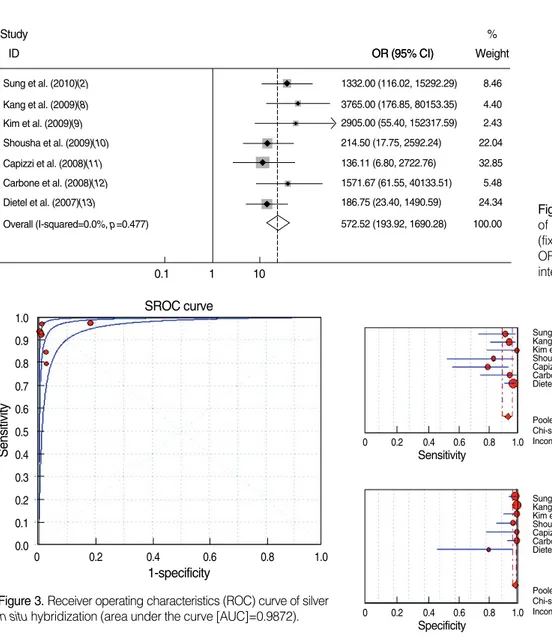
이에 부정을 제외한 진단정확성을 산출할 수 있는 문헌들을 고 정효과모형에 기초하여 메타분석을 실시하였다. 은제자리부합법 의진단적의미를분석한연구들간의이질성을검정한결과(Figure 2), 통합교차비는 572.52 (95% CI, 193.92-1690.28)로 통계적 으로 유의하였으며(
χ
2=500.29, df=1, p<0.001), 병합된 연구간의 동질성을 보였다(
χ
2=5.53, df=6, p=0.477). 또한 이 문헌 들을 대상으로 ROC curve를 확인한 결과는 Figure 3과 같으며, AUC가 0.99로 나타났다.유방암에서의 은제자리부합법의 진단적 의미를 분석한 연구들 Figure 4. Forest plot of sensitivities and specificities.
CI=confidence interval.
0 0.2 0.4 0.6 0.8 1.0
Sensitivity
Sensitivity (95% CI) Sung et al. (2010)(2)
Kang et al. (2009)(8) Kim et al. (2009)(9) Shousha et al. (2009)(10) Capizzi et al. (2008)(11) Carbone et al. (2008)(12) Dietel et al. (2007)(13)
Pooled Sensitivity=0.94 (0.90 to 0.97) Chi-square=10.52; df=6 (p=0.1043) Inconsistency (I-square)=43.0%
0.92 (0.75-0.99) 0.95 (0.83-0.99) 1.00 (0.80-1.00) 0.85 (0.55-0.98) 0.81 (0.58-0.95) 0.95 (0.76-1.00) 0.98 (0.92-1.00)
0 0.2 0.4 0.6 0.8 1.0
Specificity
Specificity (95% CI) Sung et al. (2010)(2)
Kang et al. (2009)(8) Kim et al. (2009)(9) Shousha et al. (2008)(10) Capizzi et al. (2008)(11) Carbone et al. (2008)(12)
Pooled Specificity=0.99 (0.98 to 1.00) Chi-square=4.32; df=5 (p=0.5041) Inconsistency (I-square)=0.0%
0.99 (0.95-1.00) 1.00 (0.97-1.00) 1.00 (0.91-1.00) 0.98 (0.87-1.00) 1.00 (0.80-1.00) 1.00 (0.94-1.00)
0 0.2 0.4 0.6 0.8 1.0
Specificity
Specificity (95% CI) Sung et al. (2010)(2)
Kang et al. (2009)(8) Kim et al. (2009)(9) Shousha et al. (2009)(10) Capizzi et al. (2008)(11) Carbone et al. (2008)(12) Dietel et al. (2007)(13)
Pooled Specificity=0.99 (0.97 to 1.00) Chi-square=13.65; df=6 (p=0.0338) Inconsistency (I-square)=56.0%
0.99 (0.95-1.00) 1.00 (0.97-1.00) 1.00 (0.91-1.00) 0.98 (0.87-1.00) 1.00 (0.80-1.00) 1.00 (0.94-1.00) 0.82 (0.48-0.98)
Figure 2. Diagnostic meaning of silver in situ hybridization (fixed-effect model).
OR=odds ratio; CI=confidence interval.
0.1
Sung et al. (2010)(2) 1332.00 (116.02, 15292.29) 8.46
% Weight OR (95% CI)
Study
ID OR (95% CI)
3765.00 (176.85, 80153.35) 4.40 2905.00 (55.40, 152317.59) 2.43 214.50 (17.75, 2592.24) 22.04 136.11 (6.80, 2722.76) 32.85 1571.67 (61.55, 40133.51) 5.48 186.75 (23.40, 1490.59) 24.34 572.52 (193.92, 1690.28) 100.00 Kang et al. (2009)(8)
Kim et al. (2009)(9) Shousha et al. (2009)(10) Capizzi et al. (2008)(11) Carbone et al. (2008)(12) Dietel et al. (2007)(13)
Overall (I-squared=0.0%, p=0.477)
1 10
Sensitivity
1.0 0.9 0.8 0.7 0.6 0.5 0.4 0.3 0.2 0.1 0.0
0 0.2 0.4 0.6 0.8 1.0
1-specificity SROC curve
Figure 3. Receiver operating characteristics (ROC) curve of silver in situ hybridization (area under the curve [AUC]=0.9872).
간의 이질성을 검정한 결과 통합민감도는 0.94 (95% CI, 0.90- 0.97)이었으며, 병합된 연구 간 이질성이 없음을 보였고(
χ
2= 10.52, df=6, p=0.104), 통합특이도는 0.99 (95% CI, 0.97-1.00)이었으며, 병합된 연구 간 이질성을 보였다(
χ
2=13.65, df=6, p=0.034). 통합특이도에서 이질성을 보인 Dietel 등(12)의 연 구를 제외한 연구들 간의 병합을 살펴본 결과 이질성을 보이지 않 았다(
χ
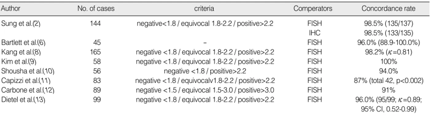
2=4.32, df=5, p=0.504) (Figure 4). 문헌의 이질성 검 정을 위해 funnel plot을 확인한 결과 출판편향은 없는 것으로 나타났다(p=0.349) (Figure 5).형광제자리부합법과의 일치율은 총 8편(2,5,7-12)의 문헌에 의해 평가되었다(Table 7).
Sung 등(2)은 형광제자리부합법에서 양성 혹은 음성으로 판독 된 경우와 은제자리부합법과의 일치율은 98.5% (135/137건)로, 면역조직화학검사에서 양성 혹은 음성으로 판독된 경우와 은제자 리부합법과의 일치율은 98.5% (133/135건)로 보고하였고, 두 병 리의사 간 판독 결과는 96.4% (134/139건; κ=0.89; p<0.001)로 보고하였다. Bartlett 등(6)의 연구에서는 형광제자리부합법과 의 전반적인 일치율을 96.0% (88.9-100%)로 보고하였고, 검사 자간 변이 8.0%, 검사실간 변이를 12.66%로 보고하였다. Kang
Table 8. Correlation with comparators
Author No. of cases Comperators Methods Values p-value
Kim et al.(9) 58 FISH HER2 copy enumeration Correlation coefficient (Pearson) R=0.83 <0.001 Linear regression R2=0.69
FISH CEP17copy enumeration Correlation coefficient (Pearson) R=0.23 <0.078 Linear regression R2=0.04
FISH HER2/CEP17 ratio Correlation coefficient (Pearson) R=0.75 <0.001 Linear regression R2=0.56
Si´nczak-Kuta et al.(1) 63 IHC Pearson χ2test 13.27 <0.01
Gamma correlation coefficient 0.69 -
Spearman rank correlation coefficient 0.45
Kappa coefficient 0.58
FISH Pearson χ2test 39.91 <0.01
Gamma correlation coefficient 0.96 -
Spearman rank correlation coefficient 0.72
Kappa coefficient 0.38
IHC=immunohistochemistry; FISH=fluorescence in situ hybridization.
Table 7. Summary table of diagnostic concordance rate
Author No. of cases criteria Comperators Concordance rate
Sung et al.(2) 144 negative<1.8 / equivocal 1.8-2.2 / positive>2.2 FISH 98.5% (135/137)
IHC 98.5% (133/135)
Bartlett et al.(6) 45 - FISH 96.0% (88.9-100.0%)
Kang et al.(8) 165 negative <1.8 / equivocal 1.8-2.2 / positive>2.2 FISH 98.2% (κ=0.81)
Kim et al.(9) 58 negative <1.8 / equivocal 1.8-2.2 / positive>2.2 FISH 100%
Shousha et al.(10) 56 negative <1.8 / positive>2.2 FISH 94.0%
Capizzi et al.(11) 83 negative <1.8 / equivocalv1.8-2.2 / positive>2.2 FISH 87% (total 42, p<0.002) Carbone et al.(12) 89 negative <1.5 / equivocal 1.5-3.0 / positive>3.0 FISH 91%
Dietel et al.(13) 99 negative <1.8 / equivocal 1.8-2.2 / positive>2.2 FISH 96.0% (95/99; κ=0.89;
95% CI, 0.52-0.99) IHC=immunohistochemistry; FISH=fluorescence in situ hybridization; CI=confidence interval.
S.E. of InOR
0
0.5
1
1.5
2
2 4 6 8 10
InOR
Funnel plot with pseudo 95% confidence limits
Figure 5. Funnel plot of selected studies.
S.E.=standard error; InOR=natural log of odds ratio.
등(8)은 형광제자리부합법과 비교한 결과, 총 165조직 중 162조 직이 일치하여 98.2% (κ=0.94)의 일치율을 보고하였다. 또한 면 역조직화학검사와의 일치율은 총 157조직 중 147조직이 일치하 여 92.5% (κ=0.81)로 보고하였다. Kim 등(9)은 형광제자리부합 법과 100%의 일치율을 보였다. Shousha 등(10)은 형광제자리부 합법과의 전반적인 일치율은 94.0%로 보고하였다. Capizzi 등(11) 의 전반적인 형광제자리부합법과 은제자리부합법과의 일치율은 87% (총 42건, p<0.002)로 보고하였다. Carbone 등(12)의 전 반적인 형광제자리부합법과 은제자리부합법과의 일치율은 91%
로 보고하였다. Dietel 등(13)의 전반적인 형광제자리부합법과 은 제자리부합법과의 일치율은 96.0% (95/99건; κ=0.89; 95% CI, 0.52-0.99)로 보고하였다.
비교검사와의 상관성은 2편(1,8)의 문헌을 근거로 평가되었으 며 형광제자리부합법과의 HER2/CEP17의 비율의 상관관계를 확인한 결과 두 편 모두 통계적으로 유의한 상관관계를 나타내는 것으로 보고하였다(Table 8).
이러한 결과를 토대로 유방암 환자에서의 은제자리부합법은 기 존 임상에서 사용되는 형광제자리부합법 결과와의 일치율 및 상 관성이 높아 유효성의 근거가 있는 검사로 평가하였다.
근거의등급
은제자리부합법은 진단법평가연구(1,2,6-13) 총 10편을 근거 로 평가하였고, 질(level of evidence) 평가 결과 증례연구를 제 외한 문헌 중‘2+’가 2편, ‘2-’가 7편이었다.
따라서 현존하는 문헌을 근거로 은제자리부합법은 권고등급 D 의 유효성이 있는 기술로 판단하였다.
고 찰
현재까지 유방암의 발병과 직접적이고도 확실한 인과관계를 갖 는 인자가 정확히 밝혀져 있지는 않지만 몇몇 위험 인자들이 발병 에 관여하는 것으로 알려져 있다. 이러한 위험인자로는 여성, 고령, 유방암 가족력, 임신 및 월경력, 장기간의 호르몬 요법, 유전자 이 상 등이 거론되고 있다.
최근 HER2 유전자의 증폭 여부가 임상적으로 크게 주목을 받 기 시작한 것은 tratuzumab, lapatinib와 같은 HER2에 대한 표 적치료제의 치료가 밝혀지면서부터이다. 현재 가장 정확한 HER2 상태 판단 기준은 FISH로 여겨지나 비용과 시간이 많이 들고, 검 사 여력을 갖춘 기관이 부족하다는 점 등 제약이 있어 널리 보급되 지는 못한 상태이다.(9) 그 대안으로 국내에서는 일부 병원에서 표 지자로 형광물질 대신 은을 사용하는 은제자리부합법을 도입하게 되었는데, 이는 기존의 형광제자리부합법으로 시행하던 검사를
자동으로 시행할 수 있고, 그 결과물을 보관할 수 있어 추후 결과 확인이 가능하다는 장점을 가지고 있다. 현재 국내 문헌을 포함하 여 여러 문헌에서 형광제자리부합법과 은제자리부합법의 일치율 및 진단 정확성을 확인하였는데, 검사실마다 차이가 있으나 높은 진단 정확성을 보고하고 있다. 하지만 이에 대한 명확한 근거가 제시되고 있지 않아 체계적 문헌고찰 방법을 통해 유방암 환자에 서의 은제자리부합법의 유효성을 평가하였다.
형광제자리부합법과 비교한 7편에서 은제자리부합법의 진단정 확성은 민감도 0.81-1.00, 특이도 0.82-1.00, 양성예측도 0.95- 1.00, 음성예측도 0.81-1.00, 검사정확도 0.90-1.00으로 보고 되었다. 형광제자리부합법과의 일치율은 총 8편의 문헌에 의해 평가되었는데, 87.0-100.0%로 보고되었다. 비교검사와의 상관 성은 2편의 문헌을 근거로 평가되었으며 형광제자리부합법과의 HER2/CEP17의 비율의 상관관계를 확인한 결과 두 편 모두 통계 적으로 유의한 상관관계를 나타내는 것으로 보고하였다.
따라서 은제자리부합법은 기존 임상에서 사용되는 형광제자리 부합법 결과와의 일치율 및 상관성이 높고, 기존검사에 비해 간편 하고 신속한 방법으로, 유방암환자의 HER2 유전자 과발현 여부 를 확인하고 그 결과에 따라 예후 예측 및 치료 방향을 선택하는 데 있어 유효성의 근거가 있는 검사로 평가하였다(권고등급, D).
본 연구의 제한점은 연구들 간의 이질성의 검증 없이 은제자리 부합법을 평가하였다는 점이다. 즉, 결과에 영향을 줄 수 있는 연 구에 사용된 검체의 특성, 채취자 및 검사자의 변이등이 보정되지 않았고, 진단 정확성의 경우 문헌에서 제시한 부정의 단계를 제외 하고 음성과 양성만으로 산출하였다. 따라서 은제자리부합법을 실제 임상에 적용할 시에는 이러한 점을 고려하여 검사를 시행하 여야 할 것이다.
그럼에도 불구하고, 본 연구는 현존하는 문헌을 근거로 은제자 리부합법은 기존 임상에서 사용되는 형광제자리부합법 결과와의 일치율 및 상관성이 높고, 기존검사에 비해 간편하고 신속한 방법 으로, 유방암환자의 HER2 유전자 증폭 여부를 확인하고 그 결과 에 따라 예후 예측 및 치료 방향을 선택하는 데 있어 유효성의 근 거가 있는 검사로 평가하였다.
참고문헌
1. Si´nczak-Kuta A, Tomaszewska R, Rudnicka-Sosin L, Oko´n K, Stachura J. Evaluation of HER2/neu gene amplification in patients with invasive breast carcinoma. Comparison of in situ hybridization methods. Pol J Pathol 2007;58:41-50.
2. Sung WJ, Park SJ, Gu MJ, Bae YK. Automated silver-enhanced in situ hybridization for evaluation of HER2 gene status in breast carci-
noma: comparison with fluorescence in situ hybridization and immu- nohistochemistry. Korean J Pathol 2010;44:28-34.
3. Lee JW, Noh WC, Kim MS, Kim HA, Chang YH, Hong YJ, et al.
Availability of fine needle aspirates for the assessment of HER2 gene amplification in invasive breast cancer patients. Korean J Lab Med 2008;28:392-9.
4. SIGN 50: a guideline developer’s handbook. Scottish Intercollegiate Guidelines Network. http://www.sign.ac.uk/pdf/sign50.pdf. Accessed January 22nd, 2011.
5. Health Insurance Review Agency. A Study on the Construction and Management of Health Technology Assessment System. Seoul: Health Insurance Review Agency; 2005. p.227-8.
6. Bartlett JM, Campbell FM, Ibrahim M, Wencyk P, Ellis I, Kay E, et al. Chromogenic in situ hybridization: a multicenter study comparing silver in situ hybridization with FISH. Am J Clin Pathol 2009;132:
514-20.
7. Francis GD, Jones MA, Beadle GF, Stein SR. Bright-field in situ hybridization for HER2 gene amplification in breast cancer using tissue microarrays: correlation between chromogenic (CISH) and automated silver-enhanced (SISH) methods with patient outcome.
Diagn Mol Pathol 2009;18:88-95.
8. Kang J, Kwon GY, Lee YH, Gong G. Comparison of silver-enhanced in situ hybridization and fluorescence in situ hybridization for HER2 gene status in breast carcinomas. J Breast Cancer 2009;12:235-40.
9. Kim TJ, Kim TE, Jung ES, Yim HW, Song BJ, Jung SS, et al. The comparison of automated silver in situ hybridization and fluorescence in situ hybridization for evaluating HER2 gene amplification in breast carcinoma. J Breast Cancer 2009;12:295-301.
10. Shousha S, Peston D, Amo-Takyi B, Morgan M, Jasani B. Evaluation of automated silver-enhanced in situ hybridization (SISH) for detec- tion of HER2 gene amplification in breast carcinoma excision and core biopsy specimens. Histopathology 2009;54:248-53.
11. Capizzi E, Gruppioni E, Grigioni AD, Gabusi E, Grassigli A, Grigioni WF, et al. Real time RT-PCR approach for the evaluation of ERBB2 overexpression in breast cancer archival samples: a comparative study with FISH, SISH, and immunohistochemistry. Diagn Mol Pathol 2008;17:220-6.
12. Carbone A, Botti G, Gloghini A, Simone G, Truini M, Curcio MP, et al. Delineation of HER2 gene status in breast carcinoma by silver in situ hybridization is reproducible among laboratories and pathol- ogists. J Mol Diagn 2008;10:527-36.
13. Dietel M, Ellis IO, Höfler H, Kreipe H, Moch H, Dankof A, et al.
Comparison of automated silver enhanced in situ hybridisation (SISH) and fluorescence ISH (FISH) for the validation of HER2 gene status in breast carcinoma according to the guidelines of the American Society of Clinical Oncology and the College of American Pathol- ogists. Virchows Arch 2007;451:19-25.