INTRODUCTION
Immunophenotype is one of the most useful tool for the identification of leukemia subtypes[1-5] and can be assi- gned within a few hours after bone marrow sampling.
The typical immunophenotypic feature of acute promyelo- cytic leukemia (APL) has been reported as CD34-, HLA- DR-, CD13+, and CD33+[6-8]. It has been reported that HLA-DR antigens are consistently negative in APL, whe- reas the majority of non-APL cases are positive for HLA- DR expression[9, 10]; therefore, HLA-DR negativity is known to be useful for distinguishing APL from other AML subtypes, even before the PML-RARA gene rear- rangement is identified by cytogenetic or molecular stud- ies[4, 6-10]. However, recent studies have reported that HLA-DR is not expressed in some AML without PML- 313
DOI 10.3343/kjlm.2007.27.5.313
313
HLA-DR 음성 급성골수성백혈병의 혈액학적 소견
Characteristics of Acute Myeloid Leukemia without HLA-DR Expression
Heewon Moon, M.D., Sookyoung Lee, M.D., Jungwon Huh, M.D., and Wha Soon Chung, M.D.
Department of Laboratory Medicine, School of Medicine, Ewha Womans University, Seoul, Korea
문희원∙이숙영∙허정원∙정화순
이화여자대학교 의학전문대학원 진단검사의학교실
313 313
Background : HLA-DR negativity is known to be useful for distinguishing acute promyelocytic leu- kemia (APL) from other subtypes of AML, but non-APL cases without HLA-DR antigen expression have been reported. The purpose of this study was to evaluate and compare the characteristics of APL, HLA-DR negative non-APL, and HLA-DR positive non-APL cases.
Methods :A total of 114 cases of AML admitted at Ewha Womans University, Mokdong Hospital between March 1997 and June 2006 were included in this study. A diagnosis of AML was made based on the results of morphology, cytochemistry, immunophenotype, cytogenetics, and/or fluo- rescence in situ hybridization.
Results :Among the 114 AML patients, HLA-DR antigen was not expressed in 39 (34%), includ- ing 24 non-APL (62%) and 15 APL patients (38%). The HLA-DR negative non-APL group showed higher leukocyte counts and positive rate of CD19 expression than did APL group (P<0.05). The remaining laboratory findings were not statistically different between the HLA-DR negative non-APL and APL groups. CD34 expression was more frequent in the HLA-DR positive non-APL group than in the HLA-DR negative non-APL group and APL group. Of the 24 patients with HLA-DR negative non-APL, 7 patients had disseminated intravascular coagulation and 2 patients showed morpho- logic features similar to those of APL.
Conclusions :CD19 expression and leukocyte count may be helpful for differentiating HLA-DR negative non-APL from APL. However, the final diagnosis and classification should be confirmed by cytogenetic or molecular studies. (Korean J Lab Med 2007;27:313-7)
Key Words : HLA-DR negative, Immunophenotype, AML, Acute promyelocytic leukemia
접 수: 2007년 7월 25일 접수번호:KJLM2058 수정본접수: 2007년 8월 14일
게재승인일: 2007년 8월 29일 교 신저 자: 허 정 원
우 158-710 서울시 양천구 목동 911-1
이화여자대학교 의학전문대학원 진단검사의학교실 전화: 02-2650-5320, Fax: 02-2650-5091 E-mail: [email protected]
RARA gene rearrangement[10-13], and clinical characte- ristics of HLA-DR negative non-APL patients have yet to be clarified.
The purpose of this study was to evaluate and com- pare the characteristics of HLA-DR negative non-APL cases, APL, and HLA-DR positive non-APL cases.
MATERIALS AND MTHODS
1. Patients
The records and laboratory findings of 114 AML cases admitted at Ewha Womans University Mokdong Hospi- tal between March 1997 and June 2006 were reviewed.
The median age of these patients was 46 yr, ranging from 1 day to 83 yr (mean 46.1, SD 20.1 yr), and 58 were male and 56 female. The diagnosis of AML were made based on the results of morphology, cytochemistry, immunophenotype, cytogenetics, and/or fluorescence in situ hybridization[14, 15]. The diagnosis of disseminated intravascular coagulation (DIC) was made by finding a prolonged prothrombin time, activated partial thrombo- plastin time, and positive fibrinogen degradation product/
D-dimer with reduced fibrinogen and platelet count. The patients with conditions other than AML that could cause DIC were not included.
2. Immunophenotyping
Samples were analyzed using FACScan (Becton Dick- inson Biosciences, San Diego, CA, USA); Cytomics FC500 (Beckman Coulter, CA, USA); and three color combina- tions of FITC, PE, and PerCP-conjugated monoclonal antibodies (Becton Dickinson Biosciences and DakoCy- tomation, Glostrup, Denmark) for: CD13, CD33, CD117, CD3, CD7, CD5, CD16/56, CD19, CD20, CD22, CD10, CD34, HLA-DR, cytoplasmic myeloperoxidase, cytoplas- mic CD79a, cytoplasmic CD22, cytoplasmic CD3, and nu- clear TdT. Leukemic cells were gated based on their side and forward scatter characteristics or CD 45 expression and side scatter. Negative controls included a mouse iso- type matched nonrelevant immunoglobulin. A sample was considered positive for an antigen when more than 20%
of leukemic cells reacted with the monoclonal antibody, except for TdT (10% cutoff) according to the European
Group for the Immunological characterization of Leuke- mias (EGIL) criteria[2].
3. Statistics
Statistic analysis was performed using SPSS software, version 11.0 (SPSS Inc., Chicago, IL, USA). Relations between variables were analyzed using the Fisher exact test for categorical variables and the Student’s t-test for continuous variables. P values of less than 0.05 were con- sidered statistically significant.
RESULT
1. Incidence of HLA-DR negative AML
Among the 114 AML patients, HLA-DR antigen was not expressed in 39 patients (34%), including 24 non- APL (62%) and 15 APL patients (38%). Among the non-APL patients, 24% of the patients didn’t show HLA- DR expression.
2. Comparison of HLA-DR negative non-APL and APL groups
(Table 1)The laboratory findings at diagnosis were not statisti- cally different between HLA-DR negative non-APL and APL cases, except leukocyte counts and CD19 expression.
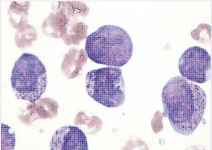
The HLA-DR negative non-APL group showed higher leukocyte counts than did the APL group. CD19 was ex- pressed more frequently in HLA-DR negative non-APL than in APL (P<0.05). Among the HLA-DR negative non-APL, 7 patients had DIC and 2 patients had mor- phologic features similar to those of APL, i.e., indented nuclei and heavy coarse cytoplasmic granules (a repre- sentative example was shown in Fig. 1). These 2 cases did not express CD34 and CD19 as well as HLA-DR and showed normal karyotype without PML-RARA rearran- gement by a molecular study.
3. Comparison of HLA-DR positive and negative non-APL patients
(Table 1)In HLA-DR positive non-APL group, the leukemic cell counts of bone marrow and DIC rates were lower, where-
as platelet counts were higher than in HLA-DR negative non-APL group (P<0.05). CD34 was more frequently expressed in the HLA-DR positive group than in HLA-
DR negative group.
DISUSSION
In this study, 60% of the HLA-DR negative AML patients were non-APL and 24% of the non-APL pati- ents did not show HLA-DR expression. Other studies reported that the incidence of HLA-DR negative non- APL was 9 to 24%[4, 10, 11]. Taken together, the ab- sence of HLA-DR antigen expression cannot be consid- ered sufficient for establishing a diagnosis of APL.
We found that the laboratory findings at diagnosis were not significantly different between HLA-DR negative non- APL and APL, except for leukocyte counts and CD19 expression. Worthy of note was the finding that 46% of the HLA-DR negative non-APL cases expressed CD19, whereas CD19 was rarely expressed in APL. The Liter- ature review, showed that CD19 antigen was not expre- ssed in any of the 250 APL patients (including micro- granular variant) using a 20% cutoff[4, 7, 8, 10], whe- reas one study reported CD19 expression in 11% of APL patients[16]. The absence of CD19 may be a useful mar- ker for APL.
A previous study reported that CD34 was expressed in 62% of non-APL and 17% of APL (all microgranular variants) cases[17]. In the present study, CD34 expres- sion was significantly less frequent in APL cases than in non-APL. Furthermore, HLA-DR negative non-APL showed a lower incidence of CD34 expression than did HLA-DR
negative non APL (N=24)
HLA-DR positive non APL (N=74) APL (N=16)
Age (yr), 36 (16-80) 46 (4-68) 49 (5-81) median (range)
Sex (M:F ) 9:7 11:13 38:36
WBC (×109/L)*, 2.2 (0.4-76.2) 47.4 (1-497.6) 13.1 (0.3-554.4) median (range)
BM leukemic cell (%)�, 74 (47.6-97.8) 89.8 (21.4-99.2) 67.1 (22.0-98.8) median (range)
Hb (g/dL), 7.7 (4.7-11.2) 8.5 (5.7-12.0) 8.1 (4.6-14.9) median (range)
PLT (×109/L)�, 32 (10-104) 37 (1-177) 50 (4-538) median(range)
DIC (%)� 7/15 (46.7) 7/18 (38.9) 5/44 (11.3)
FAB classification (%) 2 (8) 5 (7)
M0
M1 16 (67) 20 (27)
M2 4 (17) 36 (49)
M3 16 (100)
M4 1 (4) 11 (15)
M5 0 (0) 0 (0)
M6 1 (1)
Others� 1 (4) 1 (1)
Positive of antigen expression (%)
CD 13 15 (94) 18 (75) 66 (89)
CD 33 16 (100) 22 (92) 71 (96)
HLA-DR� 1 (6) 0 (0) 74 (100)
CD 34� 1 (6) 5 (21) 52 (70)
CD 7 1 (6) 1 (4) 13 (18)
CD 5 0 (0) 0 (0) 1 (1)
CD 3 0 (0) 0 (0) 2 (3)
CD 19* 1 (6) 11 (46) 23 (31)
CD 10 0 (0) 0 (0) 1 (1)
CD 22 0 (0) 1 (4) 6 (8)
CD 20 0 (0) 0 (0) 2 (3)
CD 14 0 (0) 1 (4) 10 (14)
CD 16/56 0 (0) 3 (13) 7 (9)
Karyotype (%)
t(8;21)(q22;q22) 0 (0) 8 (12)
t(15;17)(q22;q21) 16 (100) 0 (0) 0 (0)
inv(16) 0 (0) 2 (3)
t(9;22)(q34;q11) 0 (0) 3 (5)
Other abnormalities 2 (12) 14 (22)
Normal karyotype 14 (88) 38 (58)
Not available 8 9
Table 1.Comparison of the characteristics of non-APL and APL according to HLA-DR expression
*p<0.05 between APL vs HLA-DR negative non APL; �p<0.05 between HLA-DR negative vs positive non APL; �This group included acute ba- sophilic leukemia.
Abbreviations: APL, acute promyelocytic leukemia; WBC, white blood cell; BM, bone marrow; PLT, platelet count; DIC, disseminated intravas- cular coagulation; FAB, French-American-British.
Fig. 1.A case of 21 yr-old female patient diagnosed as HLA-DR negative AML. Immature cells showed indented nuclear margins and coarse cytoplasmic granules (BM aspiration, Wright stain,
×1,000).
HLA-DR positive non-APL (21% & 70%, respectively).
Another study also demonstrated that the incidence of CD34 expression was higher in HLA-DR positive non- APL (79%) than in HLA-DR negative non-APL pati- ents (17%)[18]. Especially, 10% of non-APL patients were negative for both CD34 and HLA-DR[17]. One author reported that invaginated nuclear morphology was associated with loss of HLA-DR and CD34 expressions in non-APL[19]. These results suggested that absence of HLA-DR antigen was accompanied by absence of CD34 antigen in AML. In the present study, positive predictive value was found to be further enhanced when the exp- ressions of CD19 and CD34 were taken into account with HLA-DR negativity for predicting APL (40% for only HLA-DR negativity; 46% for HLA-DR and CD34 neg- ativity; and 57% for HLA-DR, CD34, and CD19 nega- tivity).
We found that the leukemic cell counts of bone mar- row and DIC rates were higher in HLA-DR negative non- APL than in HLA-DR positive non-APL group, where- as platelet counts were higher in HLA-DR positive non- APL group (P<0.05). It has been reported that treatment response of HLA-DR negative patients is similar to those of HLA-DR positive non-APL[10].
Furthermore, we found that two cases among 24 HLA- DR negative non-APL cases showed morphologies simi- lar to those of APL, and these cases were negative for both CD19 and CD34. Thus, cytogenetic and molecular studies were found necessary in such cases for an accu- rate diagnosis. Other authors have also reported that cells from HLA-DR negative non-APL patients resemble those of hypogranular variant APL, whereas morphologic fea- tures resembling APL are not present in any HLA-DR positive AML patients[10]. Moreover, some investigators have described that HLA-DR negative AML patients who seem to have APL variants based on morphology and immunophenotype, are re-classified as non-APL after cytogenetic and molecular analyses[12, 13]. Taken toge- ther, HLA-DR negative non-APL may show the similar characteristics to APL, which may make it more difficult to differentiate non-APL, especially, those resembling APL, from APL.
In conclusion, AML without HLA-DR expression in- cludes both non-APL and APL. The leukocyte count and CD19 expression may be helpful for differentiating HLA-DR negative non-APL from APL. However, the
final diagnosis and classification should be confirmed by cytogenetic or molecular studies.
요 약
배경 : HLA-DR 음성소견은 급성전골수성백혈병(acute pro- myelocytic leukemia, APL)과 다른 급성골수성백혈병(AML)을 구별하는데 도움을 준다고 알려져 있으나, HLA-DR이 음성이나 APL이 아닌 증례들도 보고되었다. 본 연구에서는 APL, APL이 아닌 HLA-DR 음성 및 양성 AML의 특징을 비교, 분석하였다.
방법 : 1997년 3월부터 2006년 6월까지 이대목동병원에 입원한 AML 114증례가 본 연구에 포함되었다. 급성골수성백혈병의 진 단은 형태학적 소견, 세포화학적 소견, 면역표현형, 염색체 검사 혹은 형광제자리부합법 등을 통해 이루어졌다.
결과 : 114예의 AML 중에 HLA-DR은 39예(34%)에서 표현 되지 않았으며 그 중 24예(62%)는 APL이 아니었고 15예(38%) 는 APL이었다. HLA-DR 음성이며 APL이 아닌 증례들은 APL 에 비해 더 높은 백혈구 수치와 CD19 양성률을 보였다(P<0.05).
그 외의 소견은 통계학적으로 유의하지 않았다. APL이 아닌 증 례에서 CD34 양성률은 HLA-DR 양성인 경우가 HLA-DR 음 성인 군보다 높았다. 24예의 HLA-DR 음성이며 APL이 아닌 증 례 중 7예에서 파종혈관내응고가 있었으며 2예에서는 형태학적으 로 APL과 유사하였다.
결론 : CD19 양성 여부와 백혈구 수치가 HLA-DR 음성이며 APL이 아닌 증례와 APL을 감별하는데 도움을 줄 수 있으나 최 종진단은 반드시 세포유전학적 혹은 분자유전학적 결과로 확인되 어야 할 것이다.
REFERENCES
1. Bene MC. Immunophenotyping of acute leukaemias. Immunol Lett 2005;98:9-21.
2. Casasnovas RO, Slimane FK, Garand R, Faure GC, Campos L, De- neys V, et al. Immunological classification of acute myeloblastic leukemias: relevance to patient outcome. Leukemia 2003;17:515-27.
3. Bene MC, Bernier M, Castoldi G, Faure GC, Knapp W, Ludwig WD, et al. Impact of immunophenotyping on management of acute leu- kemias. Haematologica 1999;84:1024-34.
4. Kaleem Z, Crawford E, Pathan MH, Jasper L, Covinsky MA, John- son LR, et al. Flow cytometric analysis of acute leukemias. Diagno- stic utility and critical analysis of data. Arch Pathol Lab Med 2003;
127:42-8.
5. Jennings CD and Foon KA. Recent advances in flow cytometry:
application to the diagnosis of hematologic malignancy. Blood 1997;
90:2863-92.
6. Paietta E, Andersen J, Gallagher R, Bennett J, Yunis J, Cassileth P, et al. The immunophenotype of acute promyelocytic leukemia (APL):
an ECOG study. Leukemia 1994;8:1108-12.
7. Oelschlagel U, Nowak R, Mohr B, Thiede C, Ehninger G, Schaub A, et al. Specificity of immunophenotyping in acute promyelocytic leukemia. Cytometry 2000;42:396-7.
8. Exner M, Thalhammer R, Kapiotis S, Mitterbauer G, Knobl P, Haas OA, et al. The ‘‘typical’’ immunophenotype of acute promyelocytic leukemia (APL-M3): does it prove true for the M3-variant? Cytom- etry 2000;42:106-9.
9. Baer MR, Stewart CC, Dodge RK, Leget G, Sule N, Mrozek K, et al.
High frequency of immunophenotype changes in acute myeloid leukemia at relapse: implications for residual disease detection (Can- cer and Leukemia Group B Study 8361). Blood 2001;97:3574-80.
10. Wetzler M, McElwain BK, Stewart CC, Blumenson L, Mortazavi A, Ford LA, et al. HLA-DR antigen-negative acute myeloid leukemia.
Leukemia 2003;17:707-15.
11. Orfao A, Chillon MC, Bortoluci AM, Lopez-Berges MC, Garcia-Sanz R, Gonzalez M, et al. The flow cytometric pattern of CD34, CD15 and CD13 expression in acute myeloblastic leukemia is highly char- acteristic of the presence of PML-RARalpha gene rearrangements.
Haematologica 1999;84:405-12.
12. Fenu S, Carmini D, Mancini F, Guglielmi C, Alimena G, Riccioni R, et al. Acute myeloid leukemias M2 potentially misdiagnosed as M3 variant French-American-Britain (FAB) subtype: a transitional form?
Leuk Lymphoma 1995;18(S1):49-55.
13. Lazarchick J and Hopkins M. HLA-Dr negative acute non-lympho- cytic leukemia. Ann Clin Lab Sci 1998;28:150-2.
14. Jaffe ES HN, Stein H, et al, eds. WHO classification of tumours: Tu- mours of Hematopoietic and Lymphoid tissue. Lyon: IARC Press, 2001.
15. Bene MC, Castoldi G, Knapp W, Ludwig WD, Matutes E, Orfao A, et al. Proposals for the immunological classification of acute leuke- mias. European Group for the Immunological Characterization of Leukemias (EGIL). Leukemia 1995;9:1783-6.
16. Guglielmi C, Martelli MP, Diverio D, Fenu S, Vegna ML, Cantu- Rajnoldi A, et al. Immunophenotype of adult and childhood acute promyelocytic leukaemia: correlation with morphology, type of PML gene breakpoint and clinical outcome. A cooperative Italian study on 196 cases. Br J Haematol 1998;102:1035-41.
17. Khoury H, Dalal BI, Nantel SH, Horsman DE, Lavoie JC, Shepherd JD, et al. Correlation between karyotype and quantitative immuno- phenotype in acute myelogenous leukemia with t(8;21). Mod Pathol 2004;17:1211-6.
18. Syampurnawati M, Tatsumi E, Furuta K, Takenokuchi M, Naka- machi Y, Kawano S, et al. HLA-DR-negative AML (M1 and M2):
FLT3 mutations (ITD and D835) and cell-surface antigen expression.
Leuk Res 2007;31:921-9.
19. Kussick SJ, Stirewalt DL, Yi HS, Sheets KM, Pogosova-Agadjanyan E, Braswell S, et al. A distinctive nuclear morphology in acute mye- loid leukemia is strongly associated with loss of HLA-DR expression and FLT3 internal tandem duplication. Leukemia 2004;18:1591-8.