ISSN 2234-3806 • eISSN 2234-3814
Ann Lab Med 2017;37:438-442
https://doi.org/10.3343/alm.2017.37.5.438
Identification of Pathogenic Variants in the CHM Gene in Two Korean Patients With Choroideremia
Kunho Bae, M.D.1,*, Ju Sun Song, M.D.2,*, Chung Lee, B.S.3,4, Nayoung K.D. Kim, Ph.D.3, Woong-Yang Park, M.D.3,5, Byoung Joon Kim, M.D.6, Chang-Seok Ki, M.D.2, and Sang Jin Kim, M.D.1
Departments of Ophthalmology1, Laboratory Medicine and Genetics2, and Neurology6, Samsung Medical Center, Sungkyunkwan University School of Medicine, Seoul; Samsung Genome Institute3, Samsung Medical Center, Sungkyunkwan University School of Medicine, Seoul; Department of Health Sciences and Technology4, SAIHST, Sungkyunkwan University, Seoul; Department of Molecular Cell Biology5, Sungkyunkwan University School of Medicine, Seoul, Korea
Choroideremia is a rare X-linked disorder causing progressive chorioretinal atrophy. Af- fected patients develop night blindness with progressive peripheral vision loss and even- tual blindness. Herein, we report two Korean families with choroideremia. Multimodal im- aging studies showed that the probands had progressive loss of visual field with character- istic chorioretinal atrophy, while electroretinography demonstrated nearly extinguished cone and rod responses compatible with choroideremia. Sanger sequencing of all coding exons and flanking intronic regions of the CHM gene revealed a novel small deletion at a splice site (c.184_189+3delTACCAGGTA) in one patient and a deletion of the entire exon 9 in the other. This is the first report on a molecular genetic diagnosis of choroideremia in Korean individuals. Molecular diagnosis of choroideremia should be widely adopted for proper diagnosis and the development of new treatment modalities including gene therapy.
Key Words: Choroideremia, CHM, Inherited retinal degeneration
Received: December 15, 2016 Revision received: January 22, 2017 Accepted: May 31, 2017
Corresponding author: Sang Jin Kim Department of Ophthalmology, Samsung Medical Center, Sungkyunkwan University School of Medicine, 81 Irwon-ro, Gangnam-gu, Seoul 06351, Korea Tel: +82-2-3410-6775
Fax: +82-2-3410-0074 E-mail: [email protected]
Co-corresponding author: Chang-Seok Ki Department of Laboratory Medicine and Genetics, Samsung Medical Center, Sungkyunkwan University School of Medicine, 81 Irwon-ro, Gangnam-gu, Seoul 06351, Korea
Tel: +82-2-3410-2709 Fax: +82-2-3410-0022 E-mail: [email protected]
*Contributed equally as first authors.
© Korean Society for Laboratory Medicine This is an Open Access article distributed under the terms of the Creative Commons Attribution Non-Commercial License (http://creativecom- mons.org/licenses/by-nc/4.0) which permits unrestricted non-commercial use, distribution, and reproduction in any medium, provided the original work is properly cited.
INTRODUCTION
Choroideremia is a rare X-linked disorder causing progressive degeneration of the retina, retinal pigment epithelium (RPE), and choroid [1-3]. Affected male patients develop night blindness with progressive peripheral vision loss and central vision loss, usually observed later in their lives [4]. Female carriers may be
asymptomatic but can demonstrate patchy chorioretinal atrophy [5]. CHM was identified in the 1990s as a gene responsible for choroideremia. CHM encodes Rab Escort Protein-1 (REP-1), which facilitates posttranslational modification of Rab proteins regulating intracellular trafficking [6-8]. Various types of muta- tions in CHM have been identified including small deletions, non- sense mutations, missense mutations, frameshift mutations, splice
2017-03-16 https://crossmark-cdn.crossref.org/widget/v2.0/logos/CROSSMARK_Color_square.svg
Fig. 1. Ocular phenotypes exhibited by the probands (indicated by arrows) and the pedigrees of family A (A-D) and family B (E-H). (A, E) Fundus photograph. (B, F) Spectral-domain optical coherence tomography. (C, G) Fundus autofluorescence photograph. (D, H) Family pedigree.
A B
C
F
G E
D
H
site defects, and deletion of the entire gene [9]. These mutations cause truncation, loss of functional domain, or absence of REP-1 [10]. In Korea, there has been no report on genetic diagnosis of choroideremia, although a few cases of clinical diagnosis of cho- roideremia have been reported [11, 12]. The purpose of this study is to report the first molecular diagnosis of choroideremia in two Korean families, one of which had a novel CHM mutation.
CASE REPORT
In family A, the proband was a 45-yr-old man complaining of night blindness and visual field defect with decreased visual acuity. His uncorrected visual acuity was 20/30 in the right eye and hand motion in the left eye. The proband had been taking immunosuppressant medication subsequent to undergoing kid- ney transplantation because of chronic glomerulonephritis. In addition, he underwent cataract surgery for posterior subcapsu- lar opacity in both eyes eight years ago. The fundus exam showed bilateral chorioretinal atrophy and areas of RPE disruption with sparing of the central macula (Fig. 1A-D). The residual RPE tis- sue appeared as a well-demarcated hyperfluorescent area in fundus autofluorescence (FAF) photographs. Standard electro- retinograms showed almost extinguished cone and rod respon- ses. An automated visual field test showed a severely constricted visual field in both eyes. Spectral domain optical coherence to- mography (SD-OCT) scans showed retinal thinning, choriocapil- lary atrophy, and abrupt transition to atrophic areas. Increased loss of outer nuclear layer and collapse of outer retina were ob- served compared to SD-OCT images taken five years ago. The proband’s elder brother showed similar symptoms with severe vision loss, which was considered as legal blindness; the ocular phenotype was highly suggestive of choroideremia. In family B, the proband was a 41-yr-old man who was referred to the retina clinic with night blindness and visual field defect in both eyes.
His best-corrected visual acuity was 20/40 in the right eye and 20/30 in the left eye. Prior to receiving refractive surgery, his eyes were highly myopic (-10 diopters in both eyes). The find- ings of the fundus exam, FAF, and SD-OCT scans were analo- gous to those of obtained for proband A (Fig. 1E-H).
To confirm choroideremia in the probands of family A and fam- ily B, genetic analysis of CHM was performed after obtaining in- formed consent. Genomic DNA was extracted from peripheral blood leukocytes by using the Wizard Genomic DNA Purification kit (Promega, Madison, WI, USA) according to the manufactur- er’s instructions. Fifteen coding exons and their flanking introns were PCR amplified, and the resulting amplicons were sequenced
by using an ABI 3730xl Genetic Analyzer (Applied Biosystems, Foster City, CA, USA) with primers described in Table 1. Targeted sequencing of candidate retinal degeneration genes (including 98 known genes associated with inherited retinal degeneration) was also performed for proband B by using an Illumina HiSeq 2500 platform (Illumina, San Diego, CA, USA) with 101-bp paired- end reads.
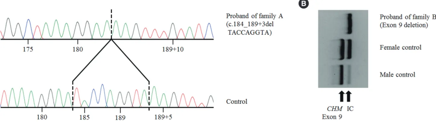
A 9-bp deletion in exon 3 and adjacent intron sequences (c.184_189+3delTACCAGGTA; Fig. 2A) was identified in the proband of family A. The exon 9 PCR product was not detected in the proband of family B, indicating exon 9 deletion (Fig. 2B).
Table 1. Primers used for sequencing analysis of the CHM gene Primer name Primer sequence 5´-3´ Amplicon size (bp)
CHM_e01F agcctggaaaatgagtcgag 414
CHM_e01R ggagttggcagttacaggga
CHM_e02F agcaaggatgggtctctttg 334
CHM_e02R gttagaagaaagatcggagttgttt
CHM_e03F ccacttatgtgagccttcca 339
CHM_e03R gcttcacctgtaacacaggatt
CHM_e04F ttctttggtgactctgaggtga 396
CHM_e04R cgttaatatgctggttttgcc
CHM_e05F tgagtcacataagcaaaacgtaca 578
CHM_e05R tgagatgcagaacatttgttttg
CHM_e06F tcaattctgagcctgtaatagattgt 400 CHM_e06R taaattccagtcctccgtgg
CHM_e07F actgatggacggtgatgtga 396
CHM_e07R tctgcactatcaataggttagcca
CHM_e08F cctttgtgaggtctgtgaaaca 499
CHM_e08R acctacctatctacccacctaagtga
CHM_e09F tgcctctgagagatttttaatactatg 399 CHM_e09R acacacacacatatcccaaaca
CHM_e10F gaaaacatggaattgtaggcaag 395
CHM_e10R ggtctggttttagggaagcc
CHM_e11F tttcatgagccaaggaaaga 378
CHM_e11R tttttgtggtgagaacacttaaga
CHM_e12F tgtttcaaattctgttccaaaa 431
CHM_e12R tcatttcacaccatcccctt
CHM_e13F aacaaatgttgtaaccaccatga 391
CHM_e13R tgtctgcctaaacatgtggg
CHM_e14F acatacgaagctctgatttcct 400
CHM_e14R gcatctctcagtagtaccatttctg
CHM_e15F acggaagttcatgtattctgattaag 491 CHM_e15R tccaaaaggggattttcctt
Fig. 2. CHM variants identified in the probands of family A and family B. (A) Chromatogram of c.184_189+3delTACCAGGTA (p.Tyr62_Gln- 63del) detected in the proband of family A. (B) PCR products of exon 9. Exon 9 deletion was detected in the proband of family B.
A B
Sequencing analysis of other amplified products did not un- cover any pathogenic variants; targeted sequencing of 98 can- didate retinal degeneration genes in proband B did not reveal any suspicious variations. However, exon 9 of CHM was not captured at all, indicating exon 9 deletion (see Supplemental data Figure S1). No genetic study of proband B’s daughter has been performed; however, she is an obligate female carrier of the CHM exon 9 deletion, which is inherited in an X-linked re- cessive manner.
DISCUSSION
We present two Korean families with choroideremia diagnosed by sequencing CHM. One patient showed a novel small deletion at an exon/intron boundary, and the other revealed a full dele- tion of exon 9, a known mutation in choroideremia. The novel small deletion variant involves canonical ±1 or 2 splice sites, which have been predicted to lead to a null effect and hypothe- sized to cause disease in choroideremia because the known CHM disease mechanism is loss-of-function. This variant is not present in the data from 622 Korean control exomes or approxi- mately 8,600 East Asian alleles from the Exome Aggregation Consortium (ExAC) [13, 14]. According to the American College of Medical Genetics and Genomics and the Association for Mo- lecular Pathology (ACMG-AMP) 2015 guidelines [15], this vari- ant is classified as a likely pathogenic variant based on one very strong piece of evidence (PVS1) and one moderate piece of evi- dence (PM2). However, an additional family study verifying cose- gregation or functional analysis by RNA is needed to confirm the pathogenicity of this variant. Exon 9 deletion has been re- ported in several choroideremia families and patients [9, 16].
Deletion of exon 9 encompassing nucleotides 1,167 to 1,244
has been predicted to cause a frameshift, leading to a null vari- ant. This deletion is not present in the data from 622 Korean control exomes or approximately 8,600 East Asian alleles from ExAC. Therefore, exon 9 deletion is classified as a pathogenic variant according to the ACMG-AMP 2015 guidelines.
Patients with choroideremia usually show characteristic retinal and choroidal features. However, at times it is difficult to clini- cally differentiate choroideremia from other inherited retinal de- generations including retinitis pigmentosa and cone-rod dystro- phy [17, 18]. Therefore, a number of researchers have advocated next generation sequencing (NGS)-based approaches and have reported cases of successful diagnosis of choroideremia [17].
However, because choroideremia is often caused by large dele- tions in CHM, NGS alone may not enable a proper molecular di- agnosis for a considerable number of patients with choroidere- mia. Therefore, when there is a clinical suspicion of choroidere- mia, a combined molecular genetics approach including direct CHM sequencing, multiplex ligation-dependent probe amplifica- tion, and RNA (cDNA) sequencing, as well as NGS-based meth- ods should be considered [19].
In summary, we performed molecular diagnosis of choroider- emia in two unrelated Korean families. To the best of our knowl- edge, this is the first report on the molecular genetic diagnosis of choroideremia in Korean individuals. This study will expand the mutational spectrum of CHM and may help in the develop- ment of new treatment modalities such as gene therapy.
Authors’ Disclosures of Potential Conflicts of Interest
No potential conflicts of interest relevant to this article were re- ported.
Acknowledgments
This work was supported by a grant from the Korean Health Tech- nology R&D Project, Ministry of Health & Welfare, Republic of Korea (HI13C1826) and Collaborative Genome Program for Fos- tering New Post-Genome industry through the National Research Foundation of Korea (NRF) funded by the Ministry of Science, ICT and Future Planning (NRF-2014M3C9A 2064620).
REFERENCES
1. Rafuse EV and McCulloch C. Choroideremia. A pathological report. Can J Ophthalmol 1968;3:347-52.
2. Rubin ML, Fishman RS, McKay RA. Choroideremia. Study of a family and literature review. Arch Ophthalmol 1966;76:563-74.
3. MacDonald IM, Hume S, Chan S, Seabra MC. Choroideremia. In: Pragon RA, Adam MP, Ardinger HH, Wallace SE, Amemiya A, Bean LJH, et al.
eds. GeneReviews [Internet], Seattle, WA: University of Washington, 1993- 2015.
4. Roberts MF, Fishman GA, Roberts DK, Heckenlively JR, Weleber RG, Anderson RJ, et al. Retrospective, longitudinal, and cross sectional study of visual acuity impairment in choroideraemia. Br J Ophthalmol 2002;
86:658-62.
5. MacDonald IM, Chen MH, Addison DJ, Mielke BW, Nesslinger NJ. His- topathology of the retinal pigment epithelium of a female carrier of cho- roideremia. Can J Ophthalmol 1997;32:329-33.
6. Seabra MC, Brown MS, Slaughter CA, Südhof TC, Goldstein JL. Purifi- cation of component A of Rab geranylgeranyl transferase: possible iden- tity with the choroideremia gene product. Cell 1992;70:1049-57.
7. van den Hurk JA, Schwartz M, van Bokhoven H, van de Pol TJ, Bogerd L, Pinckers AJ, et al. Molecular basis of choroideremia (CHM): mutations involving the Rab escort protein-1 (REP-1) gene. Hum Mutat 1997;9:
110-7.
8. Pfeffer SR. Rab GTPases: master regulators of membrane trafficking.
Curr Opin Cell Biol 1994;6:522-6.
9. Sanchez-Alcudia R, Garcia-Hoyos M, Lopez-Martinez MA, Sanchez-Bo- livar N, Zurita O, Gimenez A, et al. A comprehensive analysis of choroi- deremia: From genetic characterization to clinical practice. PLoS One 2016;11:e0151943.
10. Sergeev YV, Smaoui N, Sui R, Stiles D, Gordiyenko N, Strunnikova N, et al. The functional effect of pathogenic mutations in Rab escort protein 1. Mutat Res 2009;665:44-50.
11. O SJ, Kim SH, Lee HY. A case of choroideremia with recurrent anterior uveitis. Korean J Ophthalmol 2003;17:55-62.
12. Lee CY and Shin TY. Choroideremia. J Korean Ophthalmol Soc 1981;
22:433-8.
13. Korea National Research Institute of Health. Korean Reference Genome Database (KRGDB). http://152.99.75.168/KRGDB (Accessed on May 2016).
14. Exome Aggregation Consortium (ExAC). http://exac.broadinstitute.org (Accessed on May 2016).
15. Richards S, Aziz N, Bale S, Bick D, Das S, Gastier-Foster J, et al. Stan- dards and guidelines for the interpretation of sequence variants: a joint consensus recommendation of the American College of Medical Genet- ics and Genomics and the Association for Molecular Pathology. Genet Med 2015;17:405-24.
16. Ramsden SC, O’Grady A, Fletcher T, O’Sullivan J, Hart-Holden N, Bar- ton SJ, et al. A clinical molecular genetic service for United Kingdom families with choroideraemia. Eur J Med Genet 2013;56:432-8.
17. Shimizu K, Oishi A, Oishi M, Ogino K, Morooka S, Sugahara M, et al.
Next-Generation Sequencing-based molecular diagnosis of choroidere- mia. Case Rep Ophthalmol 2015;6:246-50.
18. Li S, Guan L, Fang S, Jiang H, Xiao X, Yang J, et al. Exome sequencing reveals CHM mutations in six families with atypical choroideremia ini- tially diagnosed as retinitis pigmentosa. Int J Mol Med 2014;34:573-7.
19. Furgoch MJ, Mewes-Arès J, Radziwon A, Macdonald IM. Molecular ge- netic diagnostic techniques in choroideremia. Mol Vis 2014;20:535-44.
Supplemental Data Fig. S1. BAM file created by using IGV viewer of targeted sequencing of candidate retinal degeneration genes includ- ing CHM. CHM exon 9 is absent in proband B and present in the control patients.