Due to untreated tuberculosis, an increase in the num- ber of patients with acquired immunodeficiency syn- drome (AIDS), drug abuse, immunosuppression, and the development of drug resistant mycobacteria, the in- cidence of abdominal tuberculosis is not declining. The pathogenesis of abdominal tuberculosis is often thought to be from the ingestion of bacilli in infected sputum or contaminated food, or hematogenous spread from a pri- mary pulmonary lesion (1, 2). Although abdominal tu- berculosis usually involves a chronic process of nonspe- cific clinical manifestations including fever, weight loss, abdominal pain, discomfort or mass, acute abdomen can develop through localized foci of recent or remote tuberculous infection. Because of the lack of specific clinical manifestations and radiologic findings, the diag- nosis of acute abdomen due to tuberculosis is not easy.
This pictorial review presents surgically confirmed in- traperitoneal tuberculosis giving rise to clinically signifi- cant acute abdomen; the radiologic findings which can
help diagnose tuberculous lesions are described.
Imaging Findings of Acute Abdomen with Intraperitoneal Tuberculosis
1Ji Seon Joo, M.D., Mi Young Kim, M.D., Jin Hoi Koo, M.D., Soon Gu Cho, M.D., Chang Hae Suh, M.D.
Acute abdomen caused by abdominal tuberculosis is a rare manifestation, and in- cludes bleeding of a gastric or ileal ulcer, obstruction of the small bowel by an adhesive band, perforation of the ileum, ileocolic intussusception and fistula, and mesenteric abscesses caused by necrotic lymph nodes. The clinical and radiologic features of these complicated tuberculosis may mimic other acute abdominal diseases. Although not definitive, careful evaluation of the radiologic findings of the bowel wall, mesen- teric fat infiltration, and lymph node enlargement may provide useful diagnostic clues to the presence of acute abdomen due to tuberculosis.
Index words : Abdomen, acute conditions Tuberculosis, gastrointestinal
1Department of Radiology, Inha University College of Medicine Received March 22, 2000; Accepted September 29, 2000
Address reprint requests to: Mi Young Kim, M.D., Department of Radiology, Inha University College of Medicine, Inha Hospital
Taepyung-dong, Su-jung-gu 7336 Sung-nam City, Kyung-gi-do, 461-712, Korea.
Tel. 82-31-720-5225 Fax. 82-31-755-2812 E-mail: [email protected]
Fig. 1. 24-year-old woman with severe epigastric pain.
Barium study of stomach shows lobulated polypoid mass (open arrows) with central ulcer (arrow) in gastric antrum. At surgery, gastric mass was pathologically confirmed as gastric tuberculosis.
Bleeding of Gastric Ulcer
Because of a paucity of lymphoid tissue, gastric tuber- culosis is rare, presenting as ulcerative or hypertrophic fibrotic encasement and commonly involving the lesser curvature of the antrum and prepylorus (3, 4). The usual manifestations of gastric tuberculosis include ulcers, gastric outlet obstruction, or a gastric mass (5). An ulcer- ative lesion is the most frequent of these: it is usually ir- regular and surrounded by inflamed and necrotic tissue, and similar in endoscopic appearance to a malignant ul- cer (3). In our case, there was severe abdominal pain and upper gastric bleeding, and barium study revealed that in the gastric antrum, a nodular ulcerative mass re- sembling a submucosal tumor of the stomach was pre- sent (Fig. 1). The second most common type of gastric tuberculosis is hypertrophic infiltrative form. The linitis plastica pattern may resemble scirrhous carcinoma, sar- coidosis, syphilis, lymphoma, prior ingestion of a caustic agent, or radiation injury (5).
Jejunal Obstruction with Adhesive Band
Dry type tuberculous peritonitis is characterized by the presence of a caseous nodule, fibrous peritoneal re- action, and obstruction of the small bowel caused by an adhesive band (1). CT demonstrates the presence of such obstruction with abrupt narrowing of the proximal
jejunum, multiple small lymph nodes, increased vascu- larity and thickened strands within the mesentery (Fig.
2). These findings are highly suggestive but not pathog- nomonic of tuberculosis, and are also detected in carci- nomatosis, lymphoma, inflammatory lesions such as pancreatitis, Crohn’s disease, diverticulitis, paniculitis, and mesenteric vascular disease.
Bleeding and Perforation of Ileal Ulcer
The ileocecal region is the most commonly involved site of gastrointestinal tuberculosis, a fact probably relat- ed to the abundance of lymphatics and lymphoid tissue in this area (6). The intestinal mucosa responds with an inflammatory exudate that may progress to an area of ulceration, and though rare, acute bleeding may devel- op. The radiologic findings of small bowel tuberculosis include symmetric annular napkin-ring stenosis caused by an ulcer, shortening and retraction (Fig. 3).
An unusual complication of tuberculous ileal ulcer is penetrating to the peritoneal cavity, or adjacent small bowel or colon. When the ileum is perforated, CT re- veals the presence of free air, ascites, irregular fat infiltra- tion of the omentum and mesentery, and associated mesenteric lymph node enlargement (Fig. 4). In tubercu- losis, however, a CT finding of bowel perforation can not be distinguished from peritonitis secondary to acute ap- pendicitis, diverticulitis, or other inflammatory diseases.
A B
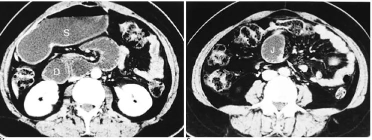
Fig. 2. 40-year-old woman with vomiting and abdominal distension.
A. CT scan shows distended stomach (S) and duodenum (D).
B. CT scan demonstrates abrupt transitional zone (arrow) of dilated proximal jejunum (J), and mesenteric lymph nodes (open ar- rows). At surgery, fibrous band and multiple whitish lymph nodes were detected and tuberculous lymphadenitis was confirmed.
Ileocolic Intussusception
The causes of tuberculous intussusception are an ileal lesion and mesenteric lymph nodes. Mesenteric lymph nodes may be clustered or matted together, adhering to the ileum, the lesion can be cause of a leading point of intussusception. CT demonstrates the outer intussuscip- ience and inner intussusceptum containing mesenteric fat and lymph nodes (Fig. 5). Because mesenteric lymph nodes are frequently associated with primary or metastatic malignant neoplasms, Whipple’s disease or
lymphoma (6), the preoperative diagnosis of the intus- susception caused by tuberculous lymphadenitis is, however, impossible,
Ileocolic Fistula
In abdominal tuberculosis, a colonic fistula is very rare, a fistula or sinus tract can develop between the
A B
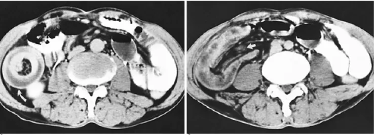
Fig. 5. 62-year-old man with nausea, vomiting and right lower abdominal pain.
A. CT scan shows target like intussusception (arrow) with low attenuated mesenteric fat and hyperattenuated intussusceptum.
B. CT scan (more caudal than A) demonstrates the longitudinal contour of intussusception including enlarged mesenteric lymph node (arrow). At surgery, leading point of intussusception was confirmed as ileal tuberculosis and mesenteric lymph node.
Fig. 3. 24-year-old man with abdominal pain, and massive hematochezia.
Barium study of small bowel shows multiple ileal ulcers (ar- rows). Segmental resection of ileum was performed. Bleeding focus was confirmed as multiple ulcers induced by tubercu- lous enteritis.
Fig. 4. 48-year-old man with abdominal pain and severe ten- derness.
CT scan shows intraperitoneal free air (small arrows), mesen- teric lymph nodes (long arrows), focal thickening of terminal ileum (curved arrow), and loculated peritoneal fluid collection (asterisk). Laparatomy demonstrated the ileal perforation with ulcer at 100 cm proximal to the ileocecal valve, and multiple segmental narrowing and dilatation of small bowel loops.
Pathologic examination confirmed the penetrating ulcer with tuberculous enteritis and mesenteric lymphadenitis.
colon and the abdominal wall, the colon, pancreas, ileum and duodenum, the most common site is the sig- moid colon (7). A fistula or sinus tract results from a mu- cosal ulcer or severe caseous necrosis occurring in mesenteric lymphadenitis (8). The patient complains of abdominal pain, bloody stool and diarrhea; barium study shows that the sigmoid colon has a cobble stone appearance and a fistula to the ileum (Fig. 6A). These le-
sions closely mimic Crohn’s colitis. CT reveals an irreg- ular heterogeneous pelvic mass adhering to adjacent small bowel and mesentery (Fig. 6B).
Mesenteric Abscess
Mesenteric abscess with tuberculosis is believed to be the result of rupture of necrotic lymph nodes, and due
A
B
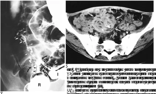
Fig. 6. 21-year-old man with diarrhea, low abdominal pain and guarding.
A. Barium study shows cobble stone appearance of sigmoid colon (S) with seg- mental narrowing (arrowheads). R: rectum. Ileocolic fistula filled with barium (curved arrows) is demonstrated between sigmoid colon to the terminal ileum (ar- rows) and distal ileum (I).
B. CT scan shows a irregular heterogeneous pelvic mass consisted of small bowel loops (arrows). At surgery, severe bowel adhesion and ileocolic fistula with tuber- culous enteritis were found.
Fig. 7. 28-year-old woman with right low abdominal pain.
CT scan shows low attenuated abscess (black arrows) with small calcification (small open arrow), mesenteric lymph node (white arrow) and massive fat infiltration of mesentery (large open arrows). Laparotomy demonstrated the tuberculous ab- scess with caseous necrosis, multiple mesenteric lymph node enlargement, and severe mesenteric adhesion.
Fig. 8. 33-year-old man with right upper quadrant pain and se- vere tenderness.
CT scan shows heterogeneously enhancing mass with periph- eral enhancement (large arrow) and regional lymph node en- largement (small curved arrows). The mass was confirmed as well encapsulated abscess contained caseous materials.
to similar clinical and radiological findings, is often mis- taken for periappendiceal inflammation or diverticulitis.
Tuberculosis commonly involves the mesentery, omen- tum, and peripancreatic lymph nodes, showing a vari- ety of enhancing patterns on CT, even within the same nodal group, and possibly related to the stage of patho- logical process (9). The lymph node exhibits an enhanc- ing rim, due to the presence of a highly vascular inflam- matory capsule and perinodal reaction surrounding an area of central caseating necrosis or liquefaction (2, 9).
CT scanning clearly demonstrates the nodular lesions with a peripheral enhancing rim and low attenuated center, punctuate calcifications and mesenteric fat infil- tration (Fig. 7). The associated mesenteric findings are helpful for the diagnosis of tuberculosis, but in our case, a tuberculous abscess appeared as a single mass at the mesenteric root with no associated peritoneal lesion (Fig. 8). It is not easy to differentiate between a single tu- berculous abscess and mesenchymal mesenteric mass.
Conclusion
Tuberculous complications may mimic the radiologic findings of other causes of acute abdomen. The imaging features that suggest the correct diagnosis are nodular mucosal change, the presence of an ulcer, ileoceal thick- ening, mesenteric fat infiltration and lymphadenitis.
The absence of the bowel-related or mesenteric manifes-
tations dose not exclude the possibility of a tuberculous lesion, however, and by means of appropriate clinical correlation, radiologists should consider the possibility of acute abdomen with tuberculosis.
References
1. Epstein BM, Mann JH. CT of abdominal tuberculosis. AJR Am J Roentgenol 1982;139:861-866
2. Sheikh M, Abu-Zidan F, al-Hilaly M, Behbehani A. Abdominal tu- berculosis: comparison of sonography and computed tomography.
J Clin Ultrasound 1995;23:413-417
3. Subei I, Attar B, Schmitt G, Levendoglu H. Primary gastric tuber- culosis: a case report and literature review. Am J Gastroenterol 1987;82:769-772
4. Brody JM, Miller DK, Zeman RK, et al. Gastric tuberculosis: a manifestation of acquired immunodeficiency syndrome. Radilogy 1986;159:347-348
5. Jadvar H, Mindelzun RE, Olcott EW, Levitt DB. Still the great mimicker: abdominal tuberculosis. AJR Am J Roentgenol 1997;168:
1455-1460
6. Balthazar EJ, Gordon R, Hulnick D. Ileoceal Tuberculosis: CT and radiologic evaluation. AJR Am J Roentgenol 1990;154:499-503 7. Tsukada T, Nishioka T, Ishida N, et al. Colonic and peritoneal tu-
berculosis associated with coloduodenal fistula. J Gastroenterol 1995;30:520-523
8. Hulnick DH, Megibow AJ, Naidich DP, Hilton S, Cho KC, Balthazar EJ. Abdominal tuberculosis: CT evaluation. Radiology 1985;157:199-204
9. Pombo F, Rodriguez E, Mato J, Perez-Fontan J, Rivera E, Valvuena L. Patterns of contrast enhancement of tuberculous lymph nodes demonstrated by computed tomography. Clin Radiol 1992;46:13- 17
대한방사선의학회지 2000;43:745-749
복강내 결핵에 의한 급성 복증의 영상소견1
인하대학교 의과대학 방사선과학교실 주지선・김미영・구진회・조순구・서창해
복부 결핵에 의한 급성 복증은 매우 드문 소견으로서 위궤양 출혈, 장간막 유착에 의한 공장 폐쇄, 회장 궤양의 출혈 및 천공, 회장-결장 장중첩 및 누공, 괴사된 임파절에 의한 장간막 농양이 포함된다. 이러한 결핵 합병증의 임상적 또는 방사선과적 소견들은 다른 급성 복증과 유사하게 나타날 수 있다. 확진적이지는 않지만 장관벽, 장간막의 지방 침윤과 임파절 비대의 방사선학적 소견의 주의 깊은 분석은 결핵에 의한 급성 복증의 진단에 유용한 단서를 시사할 수 있을 것 이다.