449
Copyrights © 2013 The Korean Society of Radiology
INTRODUCTION
Ossifying fibroma is a rare, well-demarcated, fibro-osseous tu- mor that is composed of bone, fibrous tissue and cementum. It usually occurs in the craniofacial bones, with the mandible be- ing the most common site. Ossifying fibroma originating from the middle turbinate is extremely rare (1, 2). Psammomatoid ju- venile ossifying fibroma (PsJOF) represents a unique subtype of fibro-osseous lesion. It usually occurs in the sinonasal area of children and adolescents, and has a tendency toward locally ag- gressive behavior.
We report here a case of PsJOF of the middle turbinate with aggressive imaging features in an 18-year-old adolescent female along with CT, MRI and pathologic features.
CASE REPORT
An 18-year-old adolescent female was presented with propto-
sis of her right eye. She had experienced proptosis and right na- sal obstruction for 3 years. On physical examination, a large mu- cosa-lined mass was identified in the right nasal cavity. No palpable mass elsewhere or cervical lymphadenopathy was iden- tified. Multidetector CT showed that an approximately 35 × 42
× 41 mm-sized, well-demarcated, expansile, heterogeneous mass originated from the right middle turbinate and occupied the right nasal cavity and ethmoid sinus (Fig. 1A-C). The tumor ma- trix consisted of areas of ground-glass attenuation with moder- ate enhancement, non-enhancing cystic spaces and irregular os- sifications (Fig. 1B, C). It was surrounded by a thick peripheral rim ossification (Fig. 1B, C). Focal bony destruction was noted at the right lamina papyracea and cribriform plate (Fig. 1A). MRI was performed for better characterization of the mass. T1-weight- ed images (T1WI) portrayed that the mass was heterogeneous and hypointense compared with the brain (Fig. 1D). T2-weight- ed images (T2WI) with fat saturation revealed heterogeneous mass containing multiple hyperintense cystic spaces (Fig. 1E),
Case Report
pISSN 1738-2637
J Korean Soc Radiol 2013;68(6):449-452 http://dx.doi.org/10.3348/jksr.2013.68.6.449
Received February 9, 2013; Accepted March 22, 2013 Corresponding author: Sang Kwon Lee, MD Department of Radiology, Dongsan Medical Center, Keimyung University School of Medicine, 56 Dalseong-ro, Jung-gu, Daegu 700-712, Korea.
Tel. 82-53-250-7735 Fax. 82-53-250-7766 E-mail: [email protected]
This is an Open Access article distributed under the terms of the Creative Commons Attribution Non-Commercial License (http://creativecommons.org/licenses/by-nc/3.0) which permits unrestricted non-commercial use, distri- bution, and reproduction in any medium, provided the original work is properly cited.
Ossifying fibroma of the middle turbinate is extremely rare. We report a case of psammomatoid juvenile ossifying fibroma (PsJOF) of the middle turbinate in an 18-year-old adolescent female along with its CT, MRI and pathologic features. Ps- JOF of the middle turbinate may present a well-demarcated, expansile, solidly en- hancing mass with focal bony destruction, which may mimic various benign and malignant neoplasms of the sinonasal area. A combination of clinical, imaging and pathologic findings is prerequisite for establishing an accurate diagnosis.
Index terms Ossifying Fibroma Middle Turbinate
Tomography, X-Ray Computed Magnetic Resonance Imaging
CT and Magnetic Resonance Imaging Findings of Psammomatoid Juvenile Ossifying Fibroma of the Middle Turbinate: A Case Report
1 중비갑개의 사종체양 연소성 골화성 섬유종의 전산화단층촬영 및자기공명영상소견: 증례 보고1
Sang Kwon Lee, MD
1, Mi Sun Choe, MD
2Departments of 1Radiology, 2Pathology, Dongsan Medical Center, Keimyung University School of Medicine, Daegu, Korea
CT and MR Imaging Findings of Psammomatoid Juvenile Ossifying Fibroma of the Middle Turbinate
submit.radiology.or.kr
J Korean Soc Radiol 2013;68(6):449-452
450
and metastatic diseases. The histological examination of the specimen obtained by endoscopic sinus surgery (ESS) confirmed the diagnosis of PsJOF. Histologically, the lesion consisted of highly cellular and loose or myxoid areas (Fig. 1G). It was char- acterized by the presence of small mineralized (psammomatoid) which corresponded to the non-enhancing, hypointense areas
within the moderately enhancing tumor matrix on gadolinium (Gd)-enhanced images (Fig. 1F). In view of CT and MR imag- ing features, our tentative diagnosis was ossifying fibroma, and alternative diagnoses included fibrous dysplasia, osteosarcoma
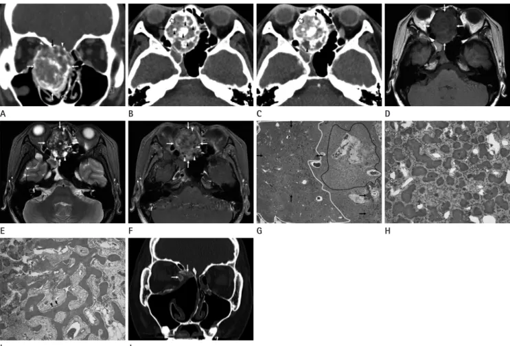
Fig. 1. CT, MRI, and pathologic features of psammomatoid juvenile ossifying fibroma of the right middle turbinate in an 18-year-old adolescent female.
A. Coronal enhanced CT image shows that a well-demarcated, expansile, heterogeneous mass (arrows) originating from the right middle turbi- nate (open arrow) involves the right nasal cavity and ethmoid sinus. Note focal bony destruction of the right lamina papyracea and right cribri- form plate (arrowheads).
B. Non-enhanced CT image reveals that the tumor matrix, which consists of areas of ground-glass attenuation (open arrow), hypoattenuating areas, and irregular ossifications (arrowheads), is surrounded by a thick peripheral rim of ossification (arrows).
C. Enhanced CT image demonstrates moderate enhancement of the areas of ground-glass attenuation (open arrow), while the hypoattenuating areas (arrowheads) are not enhanced.
D. Axial T1-weighted image shows that the mass (arrows) is heterogeneous and hypointense compared with brain.
E. Axial T2-weighted image (T2WI) with fat saturation reveals heterogeneous mass (arrows) containing multiple hyperintense cystic spaces (ar- rowheads). Also noted is a hypointense ossification (asterisk) in the center of the mass.
F. Enhanced axial T1-weighted image demonstrates moderate enhancement of the mass (arrows). Several small non-enhancing, hypointense areas (ar- rowheads), which are corresponded to the hyperintense cystic spaces on fast spin echo T2WI, are seen within the moderately enhancing tumor matrix.
G. Low-power photomicrograph of the specimen reveals a highly cellular area (white circle), a loose or myxoid area (black circle), and scattered psammomatoid bodies (arrows) (hematoxylin & eosin staining, × 40).
H. High-power photomicrograph shows characteristic psammomatoid bodies (asterisks) and intervening fibroblastic stroma (hematoxylin & eosin staining, × 200).
I. High-power photomicrograph demonstrates bony trabeculae lined by osteoblastic rimming (arrowheads).
J. Bone algorithm CT image obtained 13 months after endoscopic sinus surgery reveals residual disease of ground-glass attenuation at the right medial orbital wall and cribriform plate (arrows).
J F B
G C
H D
I E A
*
*
*
Sang Kwon Lee, et al
submit.radiology.or.kr J Korean Soc Radiol 2013;68(6):449-452
451
Accordingly, ossifying fibroma should be resected whenever possible, although “wait and see” with regular clinical and imag- ing follow-up is appropriate for fibrous dysplasia (8, 9). As such, a correct diagnosis is prerequisite for appropriate management.
However, owing to the overlapping clinical and histomorpho- logic features of fibro-osseous lesions, the diagnosis of fibro-os- seous lesions of the head and neck is not always easy. It is often difficult to diagnose them histologically. Thus, the correlation of imaging and pathologic findings is prerequisite for establishing a definitive diagnosis.
Radiographically, the lesion of an ossifying fibroma is well-de- marcated from the surrounding bone by a radio-opaque border, whereas fibrous dysplasia is presented with a diffuse border. The CT features of a typical ossifying fibroma consist of a monostot- ic and well-demarcated lesion, which contains tissues of varying attenuation values, while those of fibrous dysplasia are poorly delineated osseous expansion covered by a thin cortex. Han et al. (10) reported CT and MR imaging findings of five case of si- nonasal psammomatoid ossifying fibroma, four in the spheno- ethmoidal area with extension into the nasal cavity or orbit and one in the perpendicular plate of the ethmoid bone. They no- ticed that the lesion was well-circumscribed, multiloculated, and expansile with a thick wall of bone density on CT, and showed enhancement on postcontrast MR images. Ossifying fibroma of the nasal turbinates is extremely rare. Only four cases have ap- peared in the English literature (1-4). Caylakli et al. (1) reported a case of ossifying fibroma that showed a monostotic, hyper- dense (ground-glass attenuation) lesion confined to the middle turbinate. Galvan et al. (2) reported a case of ossifying fibroma that showed a large, expansile mass arising from the left middle turbinate and extending to the ethmoid sinus with an extensive ground-glass attenuation surrounded by hypoattenuating fi- brous tissue and/or cystic areas on CT. The ground-glass attenu- ation area on CT corresponded to a well-defined, intermediate to slightly low signal intensity area with homogeneous enhance- ment on T1WI and Gd-enhanced T1WI, and a low signal in- tensity area on T2WI. CT and MR imaging findings of the cases of Caylakli et al. (1) and Galvan et al. (2) are substantially differ- ent from those of the present case in that the tumor matrix of our case consisted of areas of ground-glass attenuation, non-en- hancing cystic spaces and irregular ossifications. Furthermore, the lesion of our case demonstrated moderate solid enhance- bodies admixed with intervening fibroblastic stroma (Fig. 1H).
The trabeculae were characteristically lined by osteoblastic rim- ming (Fig. 1I).
The postoperative course was uneventful, and there has been no evidence of tumor recurrence until 13 months after surgery.
A follow-up bone algorithm non-enhanced CT of the sinonasal area 13 months after ESS revealed some residual disease of ground-glass attenuation at the right medial orbital wall and cribriform plate (Fig. 1J), the areas that could not have been eas- ily approached during ESS. The patient is scheduled to undergo a second ESS for residual disease under the guidance of a CT- based navigation system.
DISCUSSION
Fibro-osseous lesions are rare in the sinonasal area (1-3). Fi- brous dysplasia and ossifying fibroma are included in this catego- ry. Histologically, the lesion of ossifying fibroma reveals trabecu- lae composed of lamellar and woven bones, variable amounts of vascularized fibrous stroma, and osteoblastic rimming of the trabeculae. In addition, there may be areas of cementum that appear as psammoma bodies embedded in fibrous stroma (4-6), while the lesion of fibrous dysplasia shows irregularly shaped trabeculae composed of woven bone only; moreover, there is no osteoblastic rimming. The fibrous stroma is often less vascular and cellular than that in an ossifying fibroma.
Ossifying fibroma is typically a benign, slowly growing neo- plasm; however, cases showing aggressive behavior and rapid growth have been reported. Juvenile ossifying fibroma, which can be classified into two distinct histopathologic variants (psammomatoid and trabecular), is benign (however, aggressive neoplasm is commonly seen in children below 15 years of age) and has a high tendency for recurrence. PsJOF, characterized his- tologically by the presence of small mineralized (psammoma- toid) bodies admixed with a cellular stroma, varying amounts of myxomatous material, and scattered giant cells, occurs predom- inantly in the sinonasal and orbital bones, while trabecular juve- nile ossifying fibroma, characterized by trabeculae of fibrillary osteoid and woven bone, predominantly affects the jaw.
Regarding the behavioral differences, ossifying fibroma is known to enlarge even after skeletal growth ceases, whereas fi- brous dysplasia usually stops progression after puberty (6, 7).
CT and MR Imaging Findings of Psammomatoid Juvenile Ossifying Fibroma of the Middle Turbinate
submit.radiology.or.kr
J Korean Soc Radiol 2013;68(6):449-452
452
dysplasia? J Laryngol Otol 2007;121:1201-1203
3. Pata YS, Ekici ID, Cihangirogˇ lu M, Dogˇ an M, Koçak I. Ossi- fying fibroma of the inferior turbinate. Kulak Burun Bogaz Ihtis Derg 2011;21:163-166
4. Danielides V, Ingels K, Patrikakos G, de Wilde PC. Aggres- sive psammomatoid ossifying fibroma of the inferior tur- binate and lateral nasal wall. Acta Otorhinolaryngol Belg 2003;57:87-90
5. Marvel JB, Marsh MA, Catlin FI. Ossifying fibroma of the mid-face and paranasal sinuses: diagnostic and therapeu- tic considerations. Otolaryngol Head Neck Surg 1991;104:
803-808
6. Lund VJ. Ossifying fibroma. A case report. J Laryngol Otol 1982;96:1141-1147
7. Bertrand B, Eloy P, Cornelis JP, Gosseye S, Clotuche J, Gil- liard C. Juvenile aggressive cemento-ossifying fibroma: case report and review of the literature. Laryngoscope 1993;
103:1385-1390
8. London SD, Schlosser RJ, Gross CW. Endoscopic manage- ment of benign sinonasal tumors: a decade of experience.
Am J Rhinol 2002;16:221-227
9. Post G, Kountakis SE. Endoscopic resection of large sino- nasal ossifying fibroma. Am J Otolaryngol 2005;26:54-56 10. Han MH, Chang KH, Lee CH, Seo JW, Han MC, Kim CW. Si-
nonasal psammomatoid ossifying fibromas: CT and MR manifestations. AJNR Am J Neuroradiol 1991;12:25-30 ment and aggressive imaging features, including bony erosion
or destruction of the lamina papyracea and cribriform plate. As such, our case presents a diagnostic dilemma because these im- aging findings may be seen in various benign and malignant si- nonasal diseases, such as fibrous dysplasia, osteosarcoma and metastatic diseases. However, the long clinical history of disease and the expansile growth of the lesion in our case favor benign entities. Recurrence or residual disease may be attributed to in- complete resection, due to the location of the lesion as in our case, and the infiltrative nature of the tumor borders.
In summary, our case represents an extremely rare PsJOF originating from the middle turbinate, demonstrating various tissue components by CT and MRI. PsJOF of the middle turbi- nate may be presented with a well-demarcated, expansile, solidly enhancing mass with focal bony destruction, which may mimic various benign and malignant neoplasms of the sinonasal area.
A combination of clinical, imaging and histological features en- ables establishing an accurate diagnosis of PsJOF.
REFERENCES
1. Caylakli F, Buyuklu F, Cakmak O, Ozdemir H, Ozluoglu L.
Ossifying fibroma of the middle turbinate: a case report.
Am J Otolaryngol 2004;25:377-378
2. Galvan O, Gassner EM, Neher A, Gunkel AR. Fibro-osseous lesion of the middle turbinate: ossifying fibroma or fibrous
중비갑개의 사종체양 연소성 골화성 섬유종의 전산화단층촬영 및 자기공명영상소견: 증례 보고1
이상권
1· 최미선
2중비갑개의 골화성 섬유종은 지극히 드물다. 저자들은 18세 여성에서 발생한 중비갑개의 사종체양 연소성 골화성 섬유종 (psammomatoid juvenile ossifying fibroma; PsJOF) 1예를 CT, MRI 및 병리학적 소견과 함께 보고하고자 한다. 중비갑 개의 PsJOF는 경계가 분명하고, 팽창성의 국소적인 골 파괴를 동반한 고형성 조영증강을 보이는 종괴로 나타날 수 있으 며, 따라서 비부비동의 다양한 양성 및 악성 종양을 닮을 수 있으며, 정확한 진단을 위해서는 임상적, 영상의학적, 병리학 적 소견들의 조합이 필수적이다.
계명대학교 의과대학 동산의료원 1영상의학과, 2병리과