Korean J Gastroenterol Vol. 59 No. 6, 433-436 http://dx.doi.org/10.4166/kjg.2012.59.6.433
CASE REPORT
Korean J Gastroenterol, Vol. 59 No. 6, June 2012 www.kjg.or.kr
내시경초음파 유도하 미세침 흡인생검으로 진단된 위점막하 종양 유사 부비장
안지용, 정훈용, 김도훈, 최기돈, 송호준, 이진혁, 김진호, 황희상1
울산대학교 의과대학 서울아산병원 소화기내과학교실, 병리학교실1
Diagnosis of an Accessory Spleen Mimicking a Gastric Submucosal Tumor Using Endoscopic Ultrasonography-guided Fine-needle Aspiration
Ji Yong Ahn, Hwoon-Yong Jung, Do Hoon Kim, Kee Don Choi, Ho June Song, Gin Hyug Lee, Jin-Ho Kim and Hee Sang Hwang1 Departments of Gastroenterology and Pathology1, Asan Medical Center, University of Ulsan College of Medicine, Seoul, Korea
Accessory spleen can be mistaken as a gastric subepithelial mass, and may not be differentiated in CT or endoscopic ultra- sonography (EUS). A gastric subepithelial mass was detected on routine endoscopy in a 39-year old woman with history of splenectomy. In subsequent CT and EUS, the subepithelial mass was located on the fourth layer of the stomach. To make a definite diagnosis, EUS-guided fine needle aspiration (FNA) was performed, and a splenic tissue was demonstrated in histologic examination. EUS-guided FNA can be beneficial in the diagnosis of accessory spleen which mimics a gastric subepithelial mass. (Korean J Gastroenterol 2012;59:433-436)
Key Words: Endoscopy; Spleen; Endosonography; Fine-needle biopsy
Received December 16, 2010. Revised December 30, 2010. Accepted December 30, 2010.
CC This is an open access article distributed under the terms of the Creative Commons Attribution Non-Commercial License (http://creativecommons.org/licenses/
by-nc/3.0) which permits unrestricted non-commercial use, distribution, and reproduction in any medium, provided the original work is properly cited.
교신저자: 정훈용, 138-736, 서울시 송파구 올림픽로 43길 88, 서울아산병원 소화기병센터 소화기내과
Correspondence to: Hwoon-Yong Jung, Department of Gastroenterology, Asan Medical Center, Asan Digestive Disease Research Institute, University of Ulsan College of Medicine, 88, Olympic-ro 43-gil, Songpa-gu, Seoul 138-736, Korea. Tel: +82-2-3010-3197, Fax: +82-2-476-0824, E-mail: hyjung@amc.seoul.kr
Financial support: None. Conflict of interest: None.
INTRODUCTION
Accessory spleens can develop as a result of compensa- tory enlargement of residual splenic tissue following a splenectomy. Accessory spleens can appear as an abdomi- nal mass in post-splenectomy CT.1 They have been observed in 10-31% of autopsy cases and may manifest as solitary or multiple lesions.2,3 While accessory spleens have been found at sites from the diaphragm to the scrotum, the vast majority are located in the spleen region, usually in the splen- ic hilum or along the splenic vessels or associated ligaments.
Most accessory spleens appear as small nodules.
The clinical significance of a residual accessory spleen in post-splenectomy patients varies according to the disorder
for which the spleen was removed. The return of splenic func- tion has been implicated in the recurrence of hematologic disorders such as thrombocytopenic purpura.3,4
We report a case of an accessory spleen that mimicked a submucosal tumor (SMT) of the stomach at endoscopy in a patient who had previously undergone a splenectomy.
EUS-guided fine-needle aspiration (FNA) was performed to confirm the diagnosis of an accessory spleen.
CASE REPORT
A 39-year-old woman presented with a gastric SMT which was found during routine endoscopy at another hospital. She was referred to our hospital for further management. Five
434 안지용 등. 위점막하 종양 유사 부비장
The Korean Journal of Gastroenterology
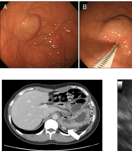
Fig. 1. Upper gastrointestinal endo- scopy findings. (A) A gastric SMT-like lesion was approximately 30 mm in diameter. (B) The mass was hard, and had a well-demarcated margin at the gastric fundus.
Fig. 2. Abdominal contrast-enhanced computed tomography revealed a well-marginated and enhanced ovoid mass close to the gastric fundus (white arrow).
Fig. 3. Endoscopic ultrasonography showed a hypoechoic mass in the fourth layer.
Fig. 4. Endoscopic ultrasonography-guided fine needle aspiration findings (white arrow: 25-G needle).
months prior she had undergone a simultaneous left hemi-colectomy and splenectomy for colorectal cancer and a wandering spleen, respectively. Her family history was
non-contributory, and she did not smoke or drink alcohol. On admission, the physical examination and laboratory results were all unremarkable. An upper gastrointestinal endoscopy identified a hard, elevated lesion approximately 30 mm in di- ameter with a well-demarcated margin at the gastric fundus, consistent with a gastric SMT (Fig. 1). An abdominal con- trast-enhanced CT revealed a well-marginated and en- hanced ovoid mass approximately 19 mm in diameter lo- cated closed to the gastric fundus (Fig. 2). An EUS was per- formed for further evaluation, and showed an approximately 27-mm hypoechoic mass in the fourth layer (Fig. 3). With find- ings of CT and EUS, we could not differentiate a gastric SMT and an accessory spleen. To obtain a definitive diagnosis, we performed EUS-guided FNA (Fig. 4). In the second EUS find- ing, the lesion was still found on the fourth layer of the stom- ach and there were no procedure-related complications.
Grossly, biopsied specimen was deep red-colored bloody, fri-
Ahn JY, et al. Accessory Spleen Mimicking a Gastric Submucosal Tumor 435
Vol. 59 No. 6, June 2012
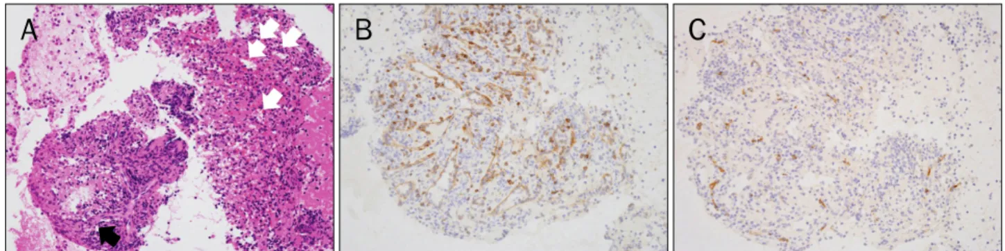
Fig. 5. Photomicrographs of biopsied submucosal tissue (×200). (A) Characteristic sinusoidal architectures (white arrows) in splenic red pulp were demonstrated. Transition of capillaries (black arrow) into sheathed capillaries was also present (H&E). (B) The endothelial cells of sinus were positive for anti-CD8 antibody. (C) But, the endothelial cells of sinus were negative for anti-CD34 antibody on immunohistochemical stains.
The cells showing positivity for anti-CD34 antibody were endothelial cells of perifollicular sinus.
able soft tissue. On microscopic evaluation, the tissue was mostly composed of monocytes and histiocytes. Many sinus- oidal spaces containing red blood cells were intervened be- tween those cells and the terminal end of the capillary branched from the arterioles sheathed by mononuclear phagocytic cells were also identified, which were frequently seen in red pulp or perifollicular zone of human spleen (Fig.
5A). On immunohistochemical stains, endothelial cells in si- nusoid architectures were stained with anti-CD8 antibody, which was characteristic immunophenotype of sinus endo- thelium in splenic red pulp (Fig. 5B). The sinus endothelial cells were not stained with anti-CD34 antibody (Fig. 5C). This pathology results indicated the mass contained splenic tis- sue, which confirmed it was an accessory spleen. After the procedure, the patient was carefully followed as an out- patient to monitor for any complications such as bleeding due to spontaneous rupture, despite this being a rare possibility.
The clinical course was uneventful, and there were no abnor- mal findings in physical and laboratory examinations during 6 months.
DISCUSSION
The present report describes the identification of an ac- cessory spleen adjoining the stomach fundus which ap- peared as a gastric SMT at endoscopy in a patient who had undergone a splenectomy 5 months prior. EUS-guided FNA was used to make a definitive diagnosis of an accessory spleen.
Accessory spleens can undergo compensatory hyper- trophy following a splenectomy, and sometimes reach 3-5 cm
in size.1 Accessory spleens have been reported to mimic gas- tric SMTs, enlarged lymph nodes, and tumors arising from ad- jacent organs such as the adrenal gland, pancreas, and kidney.5-9 Although an accessory spleen is an incidental find- ing with no clinical significance in most patients,2,10 they can sometimes become symptomatic due to torsion, sponta- neous rupture, hemorrhage and cystic formation.5-8 There- fore, detection and characterization can be clinically im- portant, especially in cases such as the present one where the lesion mimicked a gastric SMT.
CT and/or radionuclide imaging have been used to identify accessory spleens.1,9,10 CT is an imaging technique com- monly used to evaluate gastrointestinal tract diseases in- cluding SMT,11,12 and is useful in making a differential diag- nosis of an accessory spleen.2,10 Accessory spleens appear round or oval, and the attenuation is identical to that of nor- mal splenic parenchyma both before and after admin- istration of contrast medium in CT,2,10 as observed in the pres- ent patient. EUS is a better modality than CT for differ- entiating between an SMT and an extrinsic compression le- sion of stomach.13 However, when the findings of both diag- nostic methods are not consistent, pathology confirmation is required for a definite diagnosis.
Recently, EUS-guided FNA has been used for tissue sam- pling of intra- and extraluminal lesions of the gastrointestinal tract to assess SMT and splenic lesions, and it is recognized as a safe, convenient, and effective procedure.14-17 To date, there are no reports describing the use of EUS-guided FNA for diagnosing an accessory spleen mimicking a gastric SMT which is located on the fourth layer of the stomach in EUS findings.
436 안지용 등. 위점막하 종양 유사 부비장
The Korean Journal of Gastroenterology
This report describes the use of EUS-guided FNA for a defi- nite diagnosis of an accessory spleen. Even though we need more follow-up period consisted of careful outpatient mon- itoring, the post-procedure course was uneventful. So, this di- agnostic approach can be thought to be safe and effective when the diagnosis is unclear.
REFERENCES
1. Beahrs JR, Stephens DH. Enlarged accessory spleens: CT ap- pearance in postsplenectomy patients. AJR Am J Roentgenol 1980;135:483-486.
2. Halpert B, Gyorkey F. Lesions observed in accessory spleens of 311 patients. Am J Clin Pathol 1959;32:165-168.
3. Curtis GM, Movitz D. The surgical significance of the accessory spleen. Ann Surg 1946;123:276-298.
4. Verheyden CN, Beart RW Jr, Clifton MD, Phyliky RL. Accessory splenectomy in management of recurrent idiopathic thrombocy- topenic purpura. Mayo Clin Proc 1978;53:442-446.
5. Coote JM, Eyers PS, Walker A, Wells IP. Intra-abdominal bleeding caused by spontaneous rupture of an accessory spleen: the CT findings. Clin Radiol 1999;54:689-691.
6. Valls C, Monés L, Gumà A, López-Calonge E. Torsion of a wander- ing accessory spleen: CT findings. Abdom Imaging 1998;23:
194-195.
7. Seo T, Ito T, Watanabe Y, Umeda T. Torsion of an accessory spleen presenting as an acute abdomen with an inflammatory mass.
US, CT, and MRI findings. Pediatr Radiol 1994;24:532-534.
8. Brewster DC. Splenosis. Report of two cases and review of the literature. Am J Surg 1973;126:14-19.
9. Chin S, Isomoto H, Mizuta Y, Wen CY, Shikuwa S, Kohno S.
Enlarged accessory spleen presenting stomach submucosal tumor. World J Gastroenterol 2007;13:1752-1754.
10. Gayer G, Zissin R, Apter S, Atar E, Portnoy O, Itzchak Y. CT findings in congenital anomalies of the spleen. Br J Radiol 2001;
74:767-772.
11. Balthazar EJ. CT of the gastrointestinal tract: principles and interpretation. AJR Am J Roentgenol 1991;156:23-32.
12. Kim JY, Lee JM, Kim KW, et al. Ectopic pancreas: CT findings with emphasis on differentiation from small gastrointestinal stromal tumor and leiomyoma. Radiology 2009;252:92-100.
13. Motoo Y, Okai T, Ohta H, et al. Endoscopic ultrasonography in the diagnosis of extraluminal compressions mimicking gastric sub- mucosal tumors. Endoscopy 1994;26:239-242.
14. Yoshida S, Yamashita K, Yokozawa M, et al. Diagnostic findings of ultrasound-guided fine-needle aspiration cytology for gastro- intestinal stromal tumors: proposal of a combined cytology with newly defined features and histology diagnosis. Pathol Int 2009;59:712-719.
15. Chatzipantelis P, Salla C, Karoumpalis I, et al. Endoscopic ultra- sound-guided fine needle aspiration biopsy in the diagnosis of gastrointestinal stromal tumors of the stomach. A study of 17 cases. J Gastrointestin Liver Dis 2008;17:15-20.
16. Schreiner AM, Mansoor A, Faigel DO, Morgan TK. Intrapancre- atic accessory spleen: mimic of pancreatic endocrine tumor di- agnosed by endoscopic ultrasound-guided fine-needle aspira- tion biopsy. Diagn Cytopathol 2008;36:262-265.
17. Fritscher-Ravens A, Mylonaki M, Pantes A, Topalidis T, Thonke F, Swain P. Endoscopic ultrasound-guided biopsy for the diagnosis of focal lesions of the spleen. Am J Gastroenterol 2003;98:1022- 1027.