• 검색 결과가 없습니다.

Anomalous Origin of the Right Subclavian Artery in a Patient with D-transposition of the Great Arteries

N/A
N/A
Protected

Academic year: 2021

Share "Anomalous Origin of the Right Subclavian Artery in a Patient with D-transposition of the Great Arteries"

Copied!
3
0
0

로드 중.... (전체 텍스트 보기)

전체 글

(1)

ISSN: 2233-601X (Print) ISSN: 2093-6516 (Online)

− 403 −

Received: May 24, 2018, Revised: August 23, 2018, Accepted: August 27, 2018, Published online: December 5, 2018

Corresponding author: Jinyoung Song, Department of Pediatrics, Samsung Medical Center, Heart Vascular Stroke Institute, Grown-up Congenital Heart Clinic, Sungkyunkwan University School of Medicine, 81 Irwon-ro, Gangnam-gu, Seoul 06351, Korea

(Tel) 82-2-3410-3539 (Fax) 82-2-3410-0043 (E-mail) amyjys@naver.com

© The Korean Society for Thoracic and Cardiovascular Surgery. 2018. All right reserved.

This is an open access article distributed under the terms of the Creative Commons Attribution Non-Commercial License (http://creativecommons.org/

licenses/by-nc/4.0) which permits unrestricted non-commercial use, distribution, and reproduction in any medium, provided the original work is properly cited.

Anomalous Origin of the Right Subclavian Artery in a Patient with D-transposition of the Great Arteries

Hyojung Park, M.D. 1 , Jinyoung Song, M.D., Ph.D. 1 , June Huh, M.D., Ph.D. 1 , I-Seok Kang, M.D. 1 , Tae-Gook Jun, M.D., Ph.D. 2 , Ji-Hyuk Yang, M.D., Ph.D. 2

Departments of

1

Pediatrics and

2

Thoracic and Cardiovascular Surgery, Samsung Medical Center, Sungkyunkwan University School of Medicine

This case report concerns a young patient with an extremely rare combination of d-transposition of the great arteries (d-TGA) and anomalous origin of the right subclavian artery. In our patient, the right sub- clavian artery originated from the pulmonary artery, which is why he did not show reversed differential cyanosis. We conclude that the presence of an aortic arch anomaly should be considered in patients with d-TGA who do not present with reversed differential cyanosis. A further imaging work-up, including com- puted tomography or magnetic resonance imaging, might be helpful.

Key words: 1. Anomalous origin of right subclavian artery 2. Transposition of the great arteries

3. Reversed differential cyanosis

Case report

A 1-day-old boy was transferred to Samsung Medical Center for evaluation of desaturation. He was born by cesarean section due to fetal bradycardia at a local hospital and weighed 3.7 kg at birth. At the time of arrival, his blood pressure was 68/32 mm Hg, his body temperature was 36°C, his pulse rate was 140 beats per minute, and his respiration rate was 50 beats per minute. Oxygen saturation of the right arm was 90% in room air, and that of the right leg was 65%. There were no significant physical ex- amination findings apart from a grade 2 systolic murmur on the left sternal border. A chest X-ray showed neither cardiomegaly nor increased pulmo- nary vascularity of either lung. Transthoracic echo- cardiography revealed d-transposition of the great ar-

teries (d-TGA) and patent ductus arteriosus.

However, he did not show reversed differential cya- nosis during hospitalization, as is expected in pa- tients with d-TGA. On the third day after hospital- ization, balloon atrial septostomy was performed due to low oxygen saturation of 70% in the right upper limb and 40% in the lower limb. Subsequently, right upper limb saturation improved to 80%, while lower limb saturation was only 70%. After we conducted another echocardiographic evaluation due to the per- sistently unusual presentation of saturation, we sus- pected that an aberrant origin of the right subclavian artery (RSCA) from the descending aorta was the reason for the unusual saturation pattern. No further imaging work-up was carried out because that lesion did not need to be corrected clinically. At 12 days of age, an arterial switch operation for d-TGA was

Korean J Thorac Cardiovasc Surg 2018;51:403-405 □ CASE REPORT □

https://doi.org/10.5090/kjtcs.2018.51.6.403

(2)

Hyojung Park, et al

− 404 −

Fig. 1. Intraoperative image of the RSCA connected to the pulmo- nary artery. RSCA, right subclavian artery; MPA, main pulmonary artery; Ao, aorta; PDA, patent ductus arteriosus.

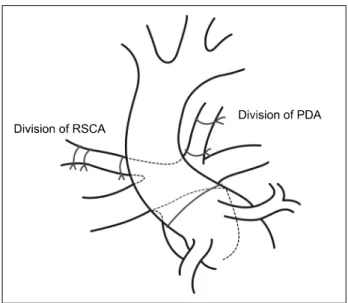
Fig. 2. Sketch of the operative procedure showing arterial switch and RSCA division. RSCA, right subclavian artery; PDA, patent ductus arteriosus.

planned. However, during the operation, it was found that the RSCA originated from the pulmonary artery, and RSCA division was therefore conducted at the same time (Figs. 1, 2). The surgeon performed dou- ble ligation and division of the RSCA using 5-0 polypropylene. During the operation, the car- diopulmonary bypass time and aortic cross-clamp time were 200 minutes and 143 minutes, respectively. The patient’s postoperative course was smooth and stable.

This case report received an exemption from local ethics committee and the informed consent was waived.

Discussion

Our report details an extremely rare combination of d-TGA and anomalous origin of the RSCA (specifically, the RSCA originating from the pulmo- nary artery). Our preoperative evaluation was in- sufficient because an aberrant origin of the RSCA from the descending aorta was suspected.

Generally, higher oxygen saturation in the leg can only occur if oxygen saturation in the pulmonary ar- tery is higher than that in the ascending aorta, which is indicative of d-TGA [1,2]. However, in our patient, oxygen saturation in the right upper limb was higher, even though he demonstrated d-TGA. This un- expected finding caused confusion in the diagnosis;

with this in mind, the presence of an anomalous RSCA should be considered prior to surgery. In pa- tients with d-TGA, the deoxygenated blood from the

right ventricle is pumped into the systemic circu- latory system through the aorta to supply the head, neck, and upper limbs, while oxygenated blood from the left ventricle is pumped into the pulmonary cir- culation via the pulmonary artery [3]. This leads to higher saturation in the lower limbs than in the arms and generally suggests reversed differential cyanosis. In our case, the pulmonary artery, which received oxygenated blood from the left atrium and left ventricle, was connected to the RSCA. Thus, satu- ration was higher in the right hand than in the foot.

We reviewed the literature on d-TGA available

through PubMed and found that associated anomalies

of the aortic arch are seen in approximately 15% of

patients with d-TGA, with coarctation of the aorta

and right aortic arch being the most common abnor-

malities [4]. However, to our knowledge, no previous

studies have described an anomalous origin of the

subclavian artery combined with d-TGA in South

Korea. Generally, ‘isolation’ should not be confined to

a subclavian artery that does not connect to the aor-

ta, but rather to the homolateral pulmonary artery

by the ductus arteriosus, whether closed or patent

[5,6]. In situations where pulmonary vascular resist-

ance is high, there may be antegrade flow from the

pulmonary artery to the RSCA, resulting in differ-

ential cyanosis with diminished oxygen saturation. It

must be noted that, in d-TGA, there is also reversed

(3)

Aberrant RSCA in d-TGA

− 405 − differential cyanosis, as the pulmonary artery re-

ceives well-oxygenated blood from the left ventricle [7].

Theoretically, it might be correct to describe the RSCA as originating from the right pulmonary artery by way of the ductus arteriosus. However, the sur- geon described the origin of the RSCA in our patient as the main pulmonary artery or between both pul- monary arteries, instead of the right pulmonary artery. Nonetheless, the position of an abnormal RSCA could be different in a heart with anomalous great arteries. Therefore, it was not easy to define the origin precisely in our patient, and we think that it is most accurate to say that it originated from the pulmonary artery.

The diagnosis of anomalous origin of the RSCA from the pulmonary artery can be predicted by the clinical presentation of a patient with d-TGA. The ability to make a definitive diagnosis based on echo- cardiography might be limited. Generally, an aberrant origin of the RSCA can be suspected when the RSCA is not traced from the innominate artery, but echo- cardiographic confirmation of its origin can be highly challenging. This was the reason why an aberrant RSCA originating from the descending aorta was sus- pected in our patient after repeated echocardio- graphic evaluations. Therefore, further imaging mo- dalities such as computed tomography, magnetic res- onance imaging, or angiography should be recom- mended when a patient shows an unexpected satu- ration pattern. Techniques for managing this con- dition include simple ligation of the subclavian artery and surgical reimplantation of the RSCA [8].

In conclusion, this case represents an instance of anomalous origin of the RSCA in a patient with d-TGA. An aortic arch anomaly should be considered in patients with d-TGA who do not present with re- versed differential cyanosis, and a further imaging

workup, including computed tomography and/or magnetic resonance imaging, should be performed prior to the arterial switch operation.

Conflict of interest

No potential conflict of interest relevant to this ar- ticle was reported.

References

1. Files MD, Arya B. Preoperative physiology, imaging, and management of transposition of the great arteries. Semin Cardiothorac Vasc Anesth 2015;19:210-22.

2. Martins P, Castela E. Transposition of the great arteries.

Orphanet J Rare Dis 2008;3:27.

3. Al-Mukhaini KS, Mohamed AM. Transposition of the great arteries and coarctation of the aorta in an infant present- ing with bronchiolitis: an incidental finding. Sultan Qaboos Univ Med J 2017;17:e348-51.

4. Cui W, Patel D, Husayni TS, Roberson DA. Double aortic arch and d-transposition of the great arteries. Echocardio- graphy 2008;25:91-5.

5. McElhinney DB, Silverman NH, Brook MM, Reddy VM, Hanley FL. Rare forms of isolation of the subclavian ar- tery: echocardiographic diagnosis and surgical considera- tions. Cardiol Young 1998;8:344-51.

6. Lee JS, Park JY, Ko SM, Seo DM. Isolation of the left sub- clavian artery with right aortic arch in association with bi- lateral ductus arteriosus and ventricular septal defect.

Korean J Thorac Cardiovasc Surg 2015;48:415-8.

7. Mosieri J, Chintala K, Delius RE, Walters HL 3rd, Hakimi M.

Abnormal origin of the right subclavian artery from the right pulmonary artery in a patient with D-transposition of the great vessels and left juxtaposition of the right at- rial appendage: an unusual anatomical variant. J Card Surg 2004;19:41-4.

8. Arunamata A, Perry SB, Kipps AK, Vasanawala SS, Axelrod

DM. Isolation of the right subclavian artery in a patient

with d-transposition of the great arteries. Ann Pediatr

Cardiol 2015;8:161-3.

수치

Fig. 1. Intraoperative image of the RSCA connected to the pulmo- pulmo-nary artery. RSCA, right subclavian artery; MPA, main pulmopulmo-nary  artery; Ao, aorta; PDA, patent ductus arteriosus.

참조

관련 문서

Kuwait will celebrate on Sunday the fourth anniversary of the UN honoring and proclamation of His Highness the Amir, Sheikh Sabah Al-Ahmad Al-Jaber Al-Sabah as

• 이명의 치료에 대한 매커니즘과 디지털 음향 기술에 대한 상업적으로의 급속한 발전으로 인해 치료 옵션은 증가했 지만, 선택 가이드 라인은 거의 없음.. •

 If the total charge is zero, the dipole moment is independent of the choice of origin. Let’s displace the origin by

A source of light waves moving to the right with velocity 0.7c. The frequency is higher on the right, and lower

One might try to solve the problem by copying the first d elements of x to a temporary array, moving the remaining n − d elements left d places, and then copying the first d

The proposal of the cell theory as the birth of contemporary cell biology Microscopic studies of plant tissues by Schleiden and of animal tissues by Microscopic studies of

Since every classical or virtual knot is equivalent to the unknot via a sequence of the extended Reidmeister moves together with the forbidden moves, illustrated in Section 2,

웹 표준을 지원하는 플랫폼에서 큰 수정없이 실행 가능함 패키징을 통해 다양한 기기를 위한 앱을 작성할 수 있음 네이티브 앱과