1)
Effect of Microcurrent Therapy on Interleukin-6 Expression in Adjuvant Induced
Rheumatoid Arthritis Rat Model
Hyun-min Lee, PT, PhD, Sang-yeol Lee, PT, PhD1, Jong-sung Chang, PT, PhD2 Myoung-hee Lee, PT, PhD3, Jong-ho Kang, PT, PhD4
Department of Physical Therapy, Honam University
1Department of Physical Therapy, Gimhae College
2Department of Physical Therapy, Yeungnam College of Science & Technology
3Department of Physical Therapy, Daegu Health College
4Department of Physical Therapy, Nambu University
미세전류치료가 아주반트 유도 류마티스관절염 유발 흰쥐의 인터루킨-6 발현에 미치는 영향
이현민⋅이상열1⋅장종성2⋅이명희3⋅강종호4
호남대학교 물리치료학과, 1김해대학 물리치료과, 2영남이공대학 물리치료과,
3대구보건대학 물리치료과, 4남부대학교 물리치료학과
<Abstract>
연구목적:미세전류 자극이 아주반트로 류마티스 관절염을 유발한 실험동물의 염증반응과 통증에 미치는 효과 를 알아보기 위하여 실험동물의 발허리발가락관절내 염증반응 정도를 나타내는 인터루킨-6(interleukin-6)의 발 현과 핫플레이트(hot plate)를 이용한 발도피지연시를 측정하여 미세전류의 효과에 대하여 알아보고자 하는데 목적이 있다.
연구방법:실험동물은 무작위로 대조군(n=18)과 미세전류를 적용한 실험군(n=18)으로 구분하였고, 각 군당 6마 리씩 1일군, 7일군, 14일군으로 배정하였다. 류마티스 관절염 유발후 1일, 7일, 14일에 모든 실험동물의 열통각 역치를 나타내는 발도피지연시와 발허리발가락관절내 인터루킨-6의 발현정도를 측정하였다. 각 집단 내의 기간 에 따른 발도피지연시와 인터루킨-6의 면역반응성은 일원배치 분산분석을 실시하였고, 사후분석으로는 Duncan 의 다중범위검정을 실시하였다. 실험군과 대조군을 비교하기 위하여 독립표본 t-test를 실시하였다.
연구결과:실험결과는 다음과 같다. 1) 아주반트 주사 1일후, 실험군과 대조군에서 인터루킨-6 면역반응성과
교신저자:강종호, E-mail: [email protected]
논문접수일:2010년 09월 06일 / 수정접수일:2010년 10월 28일 / 게재승인일:2010년 11월 11일
발도피지연시는 비슷한 양상을 보였다. 2) 인터루킨-6 면역반응성은 아주반트 주사 7일, 14일 후 대조군이 실 험군보다 유의하게 증가되었다(p<.05). 3) 발도피지연시는 아주반트 주사 7일, 14일 후 실험군이 대조군보다 유의하게 증가되었다(p<.05).
결론:이상의 결과로부터, 미세전류 자극이 아주반트로 유발된 류마티스관절염 모델에서 활액 조직내 염증반 응을 감소시키고 열통각역치는 증가되는 것을 알 수 있었다.
Key Words:류마티스관절염, 미세전류, 인터루킨-6
Ⅰ. Introduction
Rheumatoid arthritis (RA), which inflicts morbidity on approximately 1% of the population (Kannan et al., 2005), is a progressive, systemic autoimmune disease, causing synovial joint inflammation. This inflammation causes irreversible joint destruction and functional disability. Patients with RA experience pain, swelling, deformity, and loss of physical function right from the earliest stage of the disease(Vliet Vlieland, 2003). Therefore, RA patients have difficulty performing household chores, shopping, and enjoying social and recreational activities, and 31% of patients find it necessary to reduce their working hours or stop working altogether(Scott et al., 2005).
RA treatment usually consists of disease-modifying and symptomatic medication. Nonpharmacological treatments, including exercise therapy, physical modalities, joint protection, energy conservation strategies, aids, and orthoses are also used (Vliet Vlieland, 2003; Gossec et al., 2006). Physical therapy reduces RA patients’ pain, and improves range of motion and muscle strength. Physical therapy consists of electrical therapy (ultrasound, transcutaneous nerve stimulation: (TENS), low level laser therapy), thermotherapy (heat or ice pack, paraffin wax), manual therapy (massage, joint mobilization), and exercise.
Electrical stimulation is one of several treatments particularly recommended for RA patients. Electrical stimulation reduces pain or facilitates joint motion prior to exercise (Fransen, 2004). However, the
efficacy of this therapy is still controversial and has limited evidence. Recently, Microcurrent Neuromuscular Electrical Stimulation (MNES) has received attention as an alternate type of electrical stimulation. MNES, also known as Microcurrent Electrical Stimulation (MES), is defined as a new form of electromedical intervention using less than one milliampere of current delivered in biocompatable waveforms (Frick and McCauley, 2005). It has been used to enhance soft tissue healing, for pain control, and to treat fractures (Bonacci and Higibie, 1997; Frick and McCauley, 2005). MNES intensity is below the sensation threshold and also significantly lower than other electrical stimulation, such as TENS (Allen et al., 1999). For this reason, no adverse effects result from MNES therapy.
Most MNES studies have been carried out on damaged muscles, tendons, and ligaments (Zvaifler et al, 1997; Allen et al., 1999; Chapman-Jones, 2002; El-Husseini et al., 2007; Rexing et al., 2010).
To date, there are few studies regarding RA treatment with MNES. Thus, the objective of this paper is to examine the effect of MNES using a pro-inflammatory cytokine, such as interleukin-6 (IL-6), in an adjuvant induced RA rat model.
Analgesia was determined using hot plate assay after induction.
Ⅱ. Materials and Methods
1. Experimental animalsThe experimental animals used in this study included 36 Sprague-Dawley rats, regardless of gender, 8-10 weeks old, and reared under identical conditions, with an average weight 250-330g. The experimental animals were randomly divided into a control group (n=18) and an experimental group (n=18), each of which had three subgroups of 1-day, 7-day, and 14-day groups with six rats in each subgroup. The experimental group received microcurrent stimulus after rheumatoid arthritis induction and was bred in a standard cage (290mm
×430mm×180mm). The control group was bred in a standard cage after rheumatoid arthritis was induced by the same method as in the experimental group.
During the experimental period, water and feed were supplied ad libitum and the temperature and humidity of the experimental lab were maintained at 25±2℃ and 65±5%, respectively. The dark cycle was set to correnspond to the life cycle of the rats so that they could maintain their daily routine.
2. Experiment method
1) Establishment of rheumatoid arthritis model A rheumatoid arthritis model was built following the study by Simões et al. (2005). A 0.1㎖ adjuvant (Complete Freund Adjuvant, Sigma-Aldrich, U.S.A.) was hypodermically injected to the right hind plantar.
The subjects without redness or edema after adjuvant injection were excluded from the experiment.
2) Application of microcurrent treatment
The experimental group received microcurrent treatment one day after the rheumatoid arthritis induction. Using an Ag-AgCl electrode (Biopac, U.S.A.) with a diameter of 1 cm, the negative terminal was attached to the tarsal bone and the positive terminal was attached to the metatarsal bone in the animals in the experimental group under general anesthesia. A microcurrent (Multi Ⅲ,
Excel Tech Ltd., U.S.A.) was subsequently applied (20 minutes, 20 ㎂, 0.3 ㎐). The control group did not receive any treatment after the general anesthesia.
3. Measurement
1) Hot plate test
Paw withdrawal latency of the hind foot of the affected side was measured using a hot plate (ⅡTC Model 39, Life Science Instruments, U.S.A) both in the experimental group and the control group at 1 day, 7 days, and 14 days after rheumatoid arthritis induction. After placing a rat on an unheated hot plate for 30 minutes, the rat was placed on a hot plate heated to a constant temperature (51.2℃) where the paw withdrawal latency of the damaged side was measured. Detachment of paw from the plate for the rat`s movement was excluded from the measurement. Measurement was performed within 30 seconds to prevent tissue damage.
2) Immunohistochemistry
Rats were anesthetized with an intraperitoneal injection of pentobarbital sodium (50 mg/kg), and perfused via the left ventricle with about 20 ml of physiological saline to flush out the blood. This was immediately followed by 4% paraformaldehyde (PFA, Merck, Germany). The feet of the subjects were removed and decalcified with EDTA solution for 3 weeks. The extracted and decalcified tissues received the standard tissue section processing of dehydration, clearing, and paraffin-embedment. The paraffin-embedded tissues were cut into 10 μm wide transverse sections using a microcutter (Model SM2000R, Leica, Germany) and mounted on slides.
Immunohistochemistry was conducted in order to examine the immune response of IL-6.
A primary antibody of mouse anti-IL-6 IgG (Chemicon International, U.S.A) was deposited on
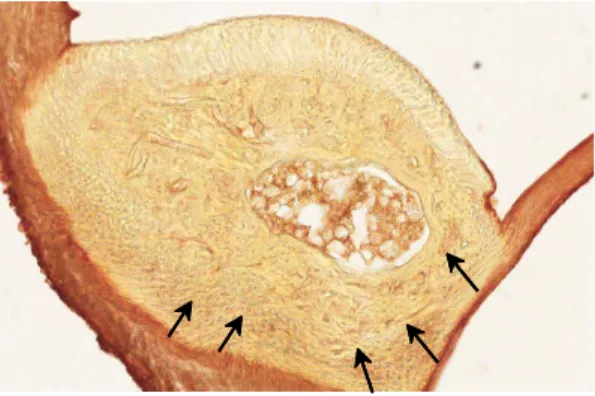
Fig 1. Expression of IL-6
Group 1 day 7 days 14 days
Control group 114.23±3.69a 159.63±4.22b 182.58±4.98c MNES group 117.28±4.15a 135.82±5.24b 155.58±4.28b Mean±SD. Values with different superscripts in the same column are significant (p<0.05) by Duncan`s multiple range test.
Table 1. IL-6 reactive cell ratio in each group
(unit : pixel) the tissue sections at a concentration of 1:1,000.
They were treated for 24 hours at room temperature.
After the primary antibiotic treatment, biotinylated anti-mouse IgG (Vector Lab, Inc., U.S.A.) was used for a 90-minute treatment at a concentration of 1:25, after which, the slides were treated with a teritiary antibody for one hour using Vectastatin Elite ABC Reagent (Vetor Lab. Inc., U.S.A.). During the antibody treatment procedures, the slides were washed three times for 10 minutes each using 10 mM PB. Tissue samples undergoing the complete antibiotic treatment received 0.3% DAB (3, 3‘-diaminobenzidine tetrahydrochloride) treatment to induce color reaction. The samples were dried after washing with distilled water three times for 10 minutes each time. The samples were subsequently encapsulated for observation after a regular dehydration process.
4. Treatment results and analysis
In order to examine the impact of microcurrent treatments on the IL-6 immune response of metatarsophalangeal tissue, the Aperio ImageScope v9.1.19.1571 (Aperio Technologies, U.S.A.) program was used by connecting ScanScope CS (Aperio Technologies, U.S.A.) to a personal computer to quantitatively analyze, by observation, the number of cells manifested in the immune response. SPSS 12.0 for Windows was used for the statistical analysis. One-way ANOVA was conducted to examine the paw withdrawal latency and immune reaction of IL-6, according to time, in each group.
Duncan’s multiple range test was performed for the
ex-post analysis. An independent sample t-test was used for a comparison between the experimental group and the control group (p<.05).
Ⅲ. Results
1. IL-6 immunohistochemistry
Immunoreactive cell bodies colored by the immunohistochemistric method with IL-6 as the antigen appeared as small brown-colored granular structures, which were observed most frequently surrounding the synovial membrane. Both the experimental and control groups exhibited the least cells 1 day after rheumatoid arthritis induction. The control group showed a significant increase at 7 days and 14 days, indicating a persistent increase in inflammatory response. The experimental group showed a significant increase at 1 day and 7 days, while the increase between 7 days and 14 days was not statistically significant.
Group 1 day 7 days 14 days Control group 4.48±.68a 4.93±1.02a 5.87±.79b
MNES group 4.58±.58a 6.18±1.24b 7.64±.1.93c Mean±SD. Values with different superscripts in the same column are significant (p<.05) by Duncan`s multiple range test.
Table 2. Paw withdraw latency in each group
(unit : second)
Expression of IL-6 in cells stained with IL-6 antibodies are shown in brown. Intense staining was observed in the lining layer of the synovium(X40).
2. Paw withdrawal latency
Paw withdrawal latency, which indicates pain threshold to thermal stimulus, showed an increasing trend as time went on both in the control group and in the experimental group. In the experimental group, there were significant differences between the groups at 1 day, 7 days, and 14 days, with an increasing pattern of pain threshold. However, the control group showed a significant increase only at 1 day and 7 days.
Ⅳ. Discussion
Rheumatoid arthritis is a chronic disease characterized by progressive destruction of joints and synovial proliferation. It is a well-known fact, supported by several previous studies (Choy and Panayi, 2001; Okamoto and Nishida, 2001), that cytokines plays a central role in maintaining the inflammatory reaction of the synovium in rheumatoid arthritis. In particular, IL-6 leads the inflammatory reaction by stimulating acute phase protein production along with proinflammatory cytokines, such as TNF- α and IL-1 (Jazayeri et al., 2010). Moreover, IL-6 indirectly participates in bone remodeling through stimulation of cell formation by increasing RANK-L released by and synovial cells in rheumatoid arthritis. There is a recent report that shows direct correlation between IL-6 activity in synovial fluid
and osteoclast severity among rheumatoid arthritis patients, according to the activity of the disease.
IL-6 measurement is being used in diagnosis or prognosis prediction (Milman et al., 2010). We attempted to evaluate the clinical validity of this practice by using the IL-6 manifestations in the joint tissue of rheumatoid arthritis induced rats.
The reported benefits of microcurrent treatment include creation of adenosine triphosphate in tissue and increased protein synthesis and membrane transport (Cheng et al., 1982). Microcurrent treatment repairs and recovers damaged tissue using the biocurrent that flows in cells of the body (Park, 2003).
It also restores homeostasis by enabling smooth flow in damaged sites, as it reduces resistance. As such, microcurrent is known to play the role of a catalyst that forms chemical and electronic responses in the recovery procedure (Aaron et al., 2004).
Hence, the application of microcurrent increases the intrinsic current flow in the damaged area, which enables easy flow of biocurrent to stimulate homeostasis of the recovery area by reducing the resistance of damaged tissue and restoring normal cell capacity (Frick and McCauley 2005). The benefits of microcurrent treatment can be summarized as tissue repair and pain relief. As for tissue repair, treatment effects are reported regarding inflammatory tissues in wounds (Oh et al., 2008), damaged muscles (Zuim et al., 2006) and tendons (Chan et al, 2007), and lateral epicondylitis (Ho et al, 2007). In an experiment with a sample of cervical spine trauma patients, McMakin et al. (2005) confirmed the anti-inflammatory effect of microcurrent application through a decrease in the transmission of the
inflammatory substances of IL-1, IL-6, and TNF- α in blood plasma, and confirmed the pain relief effect through a decrease in substance p and an increase in β-endorphin. Similar to previous studies, this study showed minimum immune response of the proinflammatory cytokine IL-6 one day after adjuvant induced rheumatoid arthritis after which the reaction gradually increased. Moreover, the experimental group receiving microcurrent treatment showed inflammatory reaction relief when compared to the control group. This is consistent with the study by Lee and Chae (2009), where the application of microcurrent to tissue decreased IL-1, which is a proinflammatory cytokine, along with the edema index.
In their clinical studies, Allen et al. (1999), Rockstroh et al. (2010) reported that microcurrent treatment is highly effective for pain relief in a variety of pain models. Kim et al. (2004) reported that microcurrent suppresses pain transmission by oppressing the manifestation of a pain substance, c-fos, in the dorsal horn in a pain model using rats.
Kim et al. (2007) confirmed the pain suppression function of electroacupuncture and microcurrent stimulus by proving an increase in the mechanical and thermal pain thresholds in a pain-induced rat model. The present study showed results that are consistent with previous studies through the increase in paw withdrawal latency, which indicates an increased pain threshold to thermal stimulus. Hence, it is speculated that microcurrent application contributed to the increase of the pain threshold by suppressing the inflammatory activation in peripheral neuropathy.
The results above indicate that microcurrent stimulus on rats with adjuvant induced rheumatoid arthritis is effective in preventing inflammatory reaction and relieving pain. Even though rapid progress was seen in the experimental animals within a few weeks, it is difficult to generalize the
positive results for humans considering the differences in rat and human pathology. Moreover, we were unable to verify the functional effect of microcurrent treatment using behavioral tests as we focused on inflammatory reaction and pain threshold.
More research is needed to clarify optimal treatment regimes and the effectiveness of microcurrent therapy compared to conventional physical modalities.
Ⅴ. Conclusion
The results of this study imply that microcurrent treatment can contribute to the prevention of inflammatory reaction and pain relief in rheumatoid arthritis. Moreover, this therapy can delay the limiting of range of motion or deformation due to the progression of disease. We believe that more studies are required suggesting microcurrent stimulation as a preservative treatment method for rheumatoid arthritis.
References
Aaron RK, Boyan BD, Ciombor DM et al.
Stimulation of growth factor synthesis by electric and electromagnetic fields. Clin Orthop Relat Res. 2004;419:30-7.
Allen JD, Mattacola CG, Perrin DH. Effect of Microcurrent Stimulation on Delayed-Onset Muscle Soreness : A Double-Blind Comparison. J Athl Train. 1999;34(4):334-7.
Bonacci JA. & Higbie EJ. Effects of Microcurrent Treatment on Perceived Pain and Muscle Strength Following Eccentric Exercise. J Athl Train. 1997;
32(2):119-23.
Chan HK, Fung DT, Ng GY. Effects of low-voltage microamperage stimulation on tendon healing in rats. J Orthop Sports Phys Ther, 2007;37(7):399- 403.
Chapman-Jones D, Hill D. Novel Microcurrent
Treatment is More Effective than Conventional Therapy for Chronic Achilles Tendinopathy : Randomised comparative trial. Physiotherapy.
2002;88(8):471-80.
Cheng N, Van Hoof H, Bockx E et al. The effects of electric currents on ATP generation, protein synthesis, and membrane transport of rat skin.
Clin Orthop Relat Res. 1982;171:264-72.
Choy EH. & Panayi GS. Cytokine pathways and joint inflammation in rheumatoid arthritis. N Engl J Med. 2001;344(12):907-16.
El-Husseini T, El-Kawy S, Shalaby H et al.
Microcurrent skin patches for postoperative pain control in total knee arthroplasty : a pilot study.
Int Orthop. 2007;31(2):.229-33.
Fransen M. When is physio therapy appropriate?
Best Pract Res Clin Rheumatol. 2004;18(4):477-89.
Frick, A. & McCauley D. Microcurrent electrical therapy. Journal of Equine Veterinary Science.
2005;25(10):418-22.
Gossec L, Pavy S, Constantin A et al. Nonphar- macological treatments in early rheumatoid arthritis : clinical practice guidelines based on published evidence and expert opinion. Joint Bone Spine.
2006;73(4):396-402.
Ho LOL, Kwong WL, Cheing GLY. Effectiveness of Microcurrent Therapy in the Management of Lateral Epicondylitis : A Pilot Study. HongKong Physiotherapy Journal. 2007;25:14-20.
Jazayeri JA, Carroll GJ, Vernallis AB. Interleukin-6 subfamily cytokines and rheumatoid arthritis : role of antagonists. Int Immunopharmacol. 2010;
10(1):1-8.
Kannan K, Ortmann RA, Kimpel D. Animal models of rheumatoid arthritis and the irrelevance to human disease. Pathophysiology. 2005;12(3):167-81.
Kim GY, Kim YE, Kim SE et al. The Effect of Microcurrent Stimulation on Immediately Early Gene in Pain Induced Model. J Kor Soc Phys Ther. 2004;16(3):449-58.
Kim YP, Lee JW, Seo SK et al. Anti-hyperalgesic Effects of Electroacupuncture Combination of Microcurrent Stimulation in Rat with Induced Inflammation. J Kor Soc Phys Ther. 2007;19(1):
67-78.
Lee HM, Chae YW. Influence of Microcurrent Therapy in Interleukin-1 Expression in Rhueumatoid Arthritis Rats. J Kor Soc Phys Ther. 2009;21(20):
103-8.
McMakin CR, Gregory WM, Phillips TM. Cytokine changes with microcurrent treatment of fibromyalgia associated with cervical spine trauma. Journal of Bodywork and Movement Therapies. 2005;9(3):
169-176.
Milman N, Karsh J, Booth RA. Correlation of a multi-cytokine panel with clinical disease activity in patients with rheumatoid arthritis. Clin Bio chem. 2010:1-6.
Oh HJ, Kim JW, Kim MS et al. Microcurrent stimulation, Woundhealing, Intensity. J Kor Soc Phys Ther. 2008;20(1):1-7.
Okamoto Y, Nishida M. Cytokine balance in the pathogenesis of rheumatoid arthritis. Yakugaku- Zasshi. 2001;121(2):131-8.
Park RJ. Electrotherapy. Seoul. Hyunmoon. 2003:
424-35.
Rexing J, Dunning D, Siegel AM et al. Effects of cold compression, bandaging, and microcurrent electrical therapy after cranial cruciate ligament repairindogs. Vet Surg. 2010;39(1):54-8.
Rockstroh G, Schleicher W, Krummenauer F.
Effectiveness of microcurrent therapy as a constituent of post-hospital rehabilitative treatment in patients after total knee alloarthroplasty - a randomized clinical trial. Rehabilitation(Stuttg).
2010;49(3):173-9.
Scott DL, Smith C, Kingsley G. What are the consequences of early rheumatoid arthritis for the individual? Best Pract Res Clin Rheumatol.
2005;19(1):117-36.
Simões SI, Delgado TC, Lopes RM et al. Develop- ments in the rat adjuvant arthritis model and its use in therapeutic evaluation of novel non- invasive treatment by SOD in Transfersomes. J Control Release. 2005;103(2):419-34.
Vliet Vlieland TP. Rehabilitation of people with rheumatoid arthritis. Best Pract Res Clin Rheumatol.
2003;17(5):847-61.
Zuim PR, Garcia AR, Turcio KH et al. Evaluation
of microcurrent electrical nerve stimulation(MENS) effectiveness on muscle pain in temporomandibular disorders patients. J Appl Oral Sci. 2006;14(1):
61-6.
Zvaifler NJ, Tsai V, Alsalameh S et al. Pannocytes : distinctive cells found In rheumatoid arthritis articular cartilage erosions. Am J Pathol. 1997;150 (3):1125-38.