• 검색 결과가 없습니다.

요로결석의 성분분석: 최근 5년간 단일기관 분석 결과(2009년- 2013년)

N/A
N/A
Protected

Academic year: 2021

Share "요로결석의 성분분석: 최근 5년간 단일기관 분석 결과(2009년- 2013년)"

Copied!
6
0
0

로드 중.... (전체 텍스트 보기)

전체 글

(1)

Original Article Korean J Urogenit Tract Infect Inflamm 2014;9(1):44-49

요로결석의 성분분석: 최근 5년간 단일기관 분석 결과(2009년- 2013년)

강필문, 서원익, 강동일

인제대학교 의과대학 비뇨기과학교실

Analysis of Urinary Stone Composition: A Retrospective Single Center Study during the Last Five Years (2009-2013)

Pil Moon Kang, Won Ik Seo, Dong Il Kang

Department of Urology, Inje University College of Medicine, Busan, Korea

Purpose: Analysis of urinary stone composition is essential for treatment and prevention of urolithiasis. We evaluated the effect of age, gender, and stone location on urinary stone components during the last five years.

Materials and Methods: We conducted a retrospective analysis of the medical records of 375 patients ranging in age from 15 years old to 93 years old (241 males and 134 females) who underwent conventional or endoscopic surgery, shock wave lithotripsy, or passed their stones spontaneously between 2009 and 2013. Stone analysis was performed using a chemical analysis method to determine the molecular composition of each stone.

Results: Of the 375 patients with urolithiasis, 152 patients (40.5%) had predomi- nantly calcium oxalate, 85 patients (21.5%) had uric acid, 17 patients (4.5%) had calcium phosphate, 17 patients (4.5%) had magnesium ammonium phosphate, 16 patients (4.3%) had calcium carbonate, and 86 patients (22.9%) had mixed stone type. There was a decreasing prevalence of calcium oxalate stone with age group (42.5% in young adult (20-39 years, n=40), 45.0% in adult (40-60 years, n=151), and 32.7% in elderly (≥60 years, n=180) as opposed to increasing prevalence in uric acid stone (7.5% in young adult, 15.2% in adult and 32.2% in elderly). Uric acid stones were more common in males and in lower urinary tract stones.

Conclusions: In this study, the incidence of calcium oxalate decreased with age and that of uric acid increased with age and in male. In comparison of the results of previous studies for several decades, the incidence of uric acid increased in the last five years.

Keywords: Urinary calculi; Analysis; Uric acid

Copyright 2014, Korean Association of Urogenital Tract Infection and Inflammation. All rights reserved.

This is an open access article distributed under the terms of the Creative Commons Attribution Non-Commercial License (http://creativecommons.org/licenses/by-nc/3.0) which permits unrestricted non-commercial use, distribution, and reproduction in any medium, provided the original work is properly cited.

Received: 14 February, 2014 Revised: 24 March, 2014 Accepted: 27 March, 2014

Correspondence to: Dong Il Kang

Department of Urology, Inje University Busan Paik Hospital, 75, Bokji-ro, Busanjin-gu, Busan 614-735, Korea

Tel: +82-51-890-6439, Fax: +82-51-892-9887

E-mail: urokang@lycos.co.kr

(2)

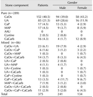
Table 1. Components of 375 urinary calculi and distribution according to the gender

Stone component Patients Gender

Male Female

Pure (n=289) CaOx UA CaP MAP AAU Cystine CaCarb Mixed (n=86) CaOx+UA CaOx+CaP CaOx+MAP CaOx+CaCarb UA+CaP UA+MAP UA+Cystine UA+CaCarb CaP+Cystine CaP+CaCarb MAP+CaCarb CaOx+UA+CaCarb CaOx+CaP+CaCarb Total

152 (40.5) 85 (21.5) 17 (4.5) 17 (4.5) 0 2 (0.5) 16 (4.3) 23 (6.1) 6 (1.6) 1 (0.3) 14 (3.7) 2 (0.5) 4 (1.1) 1 (0.3) 1 (0.3) 1 (0.3) 13 (3.5) 7 (1.9) 2 (0.5) 11 (2.9) 375

94 (39.0) 69 (28.6) 13 (5.3) 9 (3.7) 0 2 (0.8) 4 (1.7)   19 (7.9) 3 (1.2) 0 8 (3.3) 2 (0.8) 4 (1.7) 0 0 0 4 (11.7) 3 (1.2) 2 (0.8) 5 (2.0) 241

58 (43.2) 16 (11.9) 4 (2.9) 8 (5.9) 0 0 12 (8.9)   4 (2.9) 3 (2.2) 1 (0.7) 6 (4.5) 0 0 1 (0.7) 1 (0.7) 1 (0.7) 9 (6.7) 4 (3.0) 0 6 (4.5) 134 Values are presented as number (%).

CaOx: calcium oxalate, UA: uric acid, CaP: calcium phosphate, MAP:

magnesium ammonium phosphate, AAU: ammonium acid, CaCarb:

calcium carbonate.

서 론

요로결석은 비뇨기과 영역에서 흔한 질환으로 유병률은 지역 및 국가에 따라 5-15%, 발병률은 100-300명/100,000명/

년으로 각각 보고하고 있으며 한국에서의 유병률은 3.5% (남 성 6.0%, 여성 1.8%)로 보고하고 있다.

1,2

요석의 원인과 발생기전에 대해서는 아직까지도 명확하게 밝혀지지 않았으나 성별, 연령, 기후, 사회-경제적 상태, 식이 등 다양한 원인에 의하여 발생하는 것으로 알려져 있으며, 사회-경제적 상태와 식이 등 생활방식의 변화는 요석의 유병률 과 결석의 성분의 변화에도 영향을 미치게 된다. 체외충격파쇄 석술과 내비뇨기과학과 같은 치료의 발달에도 불구하고 여전 히 높은 유병률과 재발률은 생활방식, 특히 서구화된 식생활로 인한 고지혈증, 고혈압, 동물성 단백질의 과다섭취와 관련 있을 것으로 추측된다.

3,4

치료에 있어 결석의 성분을 파악하는 것이 앞으로의 치료방 침의 결정과 재발 방지를 위한 계획을 세우는 데 도움을 얻을 수 있을 것으로 생각되며, 이에 저자들은 최근 5년간 요로결석 의 성분분석을 통하여 요로결석의 원인과 발생기전에 대한 이해를 넓히고자 하였다.

대상 및 방법

2009년 1월부터 2013년 8월까지 본원에서 보존적 치료, 체외충격파쇄석술 및 수술 등을 통하여 배출된 결석중 결석의 성분 분석이 가능했던 결석 375예를 대상으로 결석의 성분과 임상적 특징을 분석하였다.

결석의 성분은 서울의과학연구소(Seoul Medical Science Institute)에 의뢰하여 화학적 분석법을 이용하여 분석하였으 며, 구성성분의 수에 따라 한가지 성분만으로 이루어진 결석을 단일석, 두 가지 이상의 성분이 혼합된 경우를 혼합석으로 정의하여 분석하였다.

결석의 성분에 따른 임상적 특징으로 나이별, 성별, 장기별 로 나누어 분석하였으며 통계적 분석은 SAS ver. 9.3 (SAS Institute, Cary, NC, USA)을 사용하여 Fisher’s exact test를 이용하였으며, p값이 0.05 미만인 경우 유의한 것으로 간주하 였다.

결 과

1. 결석의 역학

요로결석을 가지고 성분분석이 가능했던 375예의 환자들 중 남자는 241예, 여자는 134예로서 남녀비는 1.8:1이었다.

환자들의 연령은 15세부터 93세로서 평균 57.6±14.4세였다.

결석의 위치는 신장 115예(30.6%), 요관 184예(49.0%), 방광

70예(18.7%)와 요도 6예(1.7%)였다.

2. 결석 성분 분석

전체 375예의 결석 중 단일성분으로 이루어진 결석이 77%

(289예)였고 혼합성분의 결석이 33% (86예)였다. 전체적으로 는 수산칼슘 단일결석이 가장 많아서 152예(40.5%)였으며, 요산 85예(21.5%), 수산칼슘과 요산이 혼합된 결석이 23예 (6.1%)였다(Table 1).

단일성분결석 289예 중에는 수산칼슘으로만 이루어진 결석 이 152예(52.6%)로 가장 많았으며 요산 85예(29.4%), 인산칼 슘 17예(5.9%), 인산마그네슘암모늄 17예(5.9%), 탄산칼슘 16예(5.5%)의 순서였다.

혼합성분의 결석 86예 중에서는 수산칼슘과 요산의 혼합결 석이 23예(26.7%)로 가장 많았으며 수산칼슘과 탄산칼슘 14예 (16.2%), 인산칼슘과 탄산칼슘 13예(15.1%) 등의 순서로 관찰 되었다.

3. 성별에 따른 성분 분포

성별에 따른 결석 성분의 분포는 남성에서 요산이 차지하는 비율이 69예(28.6%)로 16예(11.9%)인 여성보다 높았으며(p

<0.001), 혼합석에서도 수산칼슘과 요산이 혼합석이 차지하

는 비율이 각각 19예(7.9%), 4예(3.0%)로 상대적으로 높은

(3)

Table 2. Distribution of urinary stone according to the age

Stone component Age

Child Young Adult Elderly Pure

CaOx UA CaP MAP AAU Cystine CaCarb Mixed CaOx+UA CaOx+CaP CaOx+MAP CaOx+CaCarb UA+CaP UA+MAP UA+Cystine UA+CaCarb CaP+Cystine CaP+CaCarb MAP+CaCarb CaOx+UA+CaCarb CaOx+CaP+CaCarb Total

3 (75.0) 1 (25.0)

- - - - - - - - - - - - - - - - - 4

17 (42.5) 3 (7.5) 6 (15) 2 (5) 2 (5) - 2 (5) 1 (2.5)

- - 1 (2.5) 1 (2.5) 1 (2.5) - 1 (2.5) 1 (2.5) 1 (2.5)

- - - 40

68 (45.0) 23 (15.2) 6 (3.9) 4 (2.6)

- - 8 (5.2) 9 (5.9) 8 (5.2) 4 (2.6) 7 (4.6)

- 2 (1.3)

- - 5 (3.3) -

-  - - 151

59 (32.7) 58 (32.2) 3 (1.6) 11 (6.1)

- - 9 (5.0) 13 (7.2) 3 (1.7)

- 6 (3.3) 1 (0.6) 3 (1.7) 1 - 8 - 2 - -  180 Values are presented as number (%).

Child (0-19 years), young (20-39 years), adult (40-59 years), elderly (over 60 years).

CaOx: calcium oxalate, UA: uric acid, CaP: calcium phosphate, MAP:

magnesium ammonium phosphate, AAU: ammonium acid, CaCarb:

calcium carbonate.

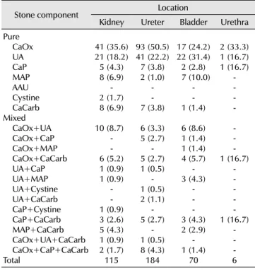
Table 3. Distribution of urinary stone according to the location

Stone component Location

Kidney Ureter Bladder Urethra Pure

CaOx UA CaP MAP AAU Cystine CaCarb Mixed CaOx+UA CaOx+CaP CaOx+MAP CaOx+CaCarb UA+CaP UA+MAP UA+Cystine UA+CaCarb CaP+Cystine CaP+CaCarb MAP+CaCarb CaOx+UA+CaCarb CaOx+CaP+CaCarb Total

41 (35.6) 21 (18.2) 5 (4.3) 8 (6.9) 2 (1.7) - 8 (6.9)   10 (8.7)

- - 6 (5.2) 1 (0.9) 1 (0.9)

- - 1 (0.9) 3 (2.6) 5 (4.3) 1 (0.9) 2 (1.7) 115

93 (50.5) 41 (22.2) 7 (3.8) 2 (1.0)

- - 7 (3.8)

  6 (3.3) 5 (2.7)

- 5 (2.7) 1 (0.5) 1 (0.5) - 2 (1.1) 5 (2.7) - 1 (0.5) - 8 (4.3) 184

17 (24.2) 22 (31.4) 2 (2.8) 7 (10.0)

- - 1 (1.4)

  6 (8.6) 1 (1.4) 1 (1.4) 4 (5.7)

- 3 (4.3)

- - 3 (4.3) - 2 (2.9) 1 (1.4) - 70

2 (33.3) 1 (16.7) 1 (16.7) -  - - - - - - 1 (16.7)

- - - - 1 (16.7) -

-  - - 6 Values are presented as number (%).

CaOx: calcium oxalate, UA: uric acid, CaP: calcium phosphate, MAP:

magnesium ammonium phosphate, AAU: ammonium acid, CaCarb:

calcium carbonate.

비율을 보였다(Table 1).

4. 나이에 따른 성분 분포

나이에 따른 결석 성분의 분포는 Tunisian classification

5

에 따라 소아층군(0-19세), 청년층군 (20-39세), 중년층군(40-59 세), 노년층군(60세 이상)으로 4개의 군으로 나누어 비교하였 다. 각 군에서 수산칼슘 성분이 가장 많았고, 연령이 증가함에 따라 요산이 차지하는 비율이 높았으며(p<0.001), 수산칼슘 이 차지하는 비율은 낮았다(p=0.047) (Table 2).

5. 결석의 위치에 따른 성분 분포

결석의 위치에 따른 결석 성분의 분포는 차이가 없었다.

다만 요관 결석은 수산칼슘이 93예(50.5%)로 전체보다 많은 비율을 차지하였으며(p<0.001), 방광에 분포하는 결석은 수 산칼슘이 차지하는 비율 17예(24.2%)보다 요산이 차지하는 비율 22예(31.4%)가 높게 나타났다(Table 3).

고 찰

요로결석은 진단 및 치료의 발달에도 불구하고 비뇨기과 영역에서 여전히 높은 유병률과 재발률을 보이는 질환이다.

요로결석의 재발률은 연간 약 10%로 알려져 있으며, 요석 환자의 절반은 10년 내에 재발하며 20-30년 내에 요석 환자의 80%에서 한 번 이상 재발하는 것으로 보고되고 있다.

6

이러한 이유에서 요석 형성을 예방하는 것은 매우 중요하며, 정확한 요로 결석의 성분을 아는 것은 요로 결석의 형성 기전에 대한 이해를 통해 과학적인 근거를 바탕으로 한 최선의 치료선택에 도움을 준다.

7

요로결석 성분분석법에는 chemical analysis, polarization microscopy, Infrared spectroscopy, X-ray diffraction, thermo-gravimetry, scanning electron microscopy 등 여러 가지 방법이 사용되고 있으며 각각의 분석법은 각각의 장단점 을 가지고 있다. 본 연구에서는 화학적 분석법을 이용하였으 며, 이 방법은 결석 성분에 대한 정량적 분석이나 형태적 분석은 어렵지만 타 검사에 비해 경제적이며 비교적 간단히 시행할 수 있는 장점이 있는 검사법이다.

8

결석의 성분에서 수산칼슘은 요로결석의 가장 흔한 성분으

로 요로결석의 75-90%를 차지하고 있으며, 다음으로 많은

성분으로는 인산칼슘 및 요산으로 알려져 있다.

9-11

국내에서는

수산칼슘의 단일석은 7.6%부터 74.0%까지 다양하게 보고된

바 있으며 본 연구에서는 40.5%로 가장 높은 빈도로 관찰되었

으며 수산칼슘의 혼합석을 포함하였을 때 수산칼슘을 근간으

로 한 결석은 55.7%였다.

12,13

(4)

칼슘석 환자에서 가장 많이 발견되는 대사 이상은 고칼슘뇨 증이며, 이 외에도 고요산뇨증, 고수산뇨증, 저구연산뇨증 등이 위험인자로 알려져 있다. 고칼슘뇨증은 대부분 특발성이 며 흡수성 고칼슘뇨증(absorptive hypercalciuria)과 신성고칼 슘뇨증(renal hypercalciuria), 골흡수의 증가 등이 있으며 단 독 혹은 복합적으로 작용하여 나타난다. 고뇨산뇨증은 요산석 뿐만 아니라 칼슘수산석의 결정화를 촉진하는데 요산은 이질 핵형성(heterogenous nucleation)으로 두 성분의 격자 구조가 매우 유사하기 때문으로 알려져 있다. 고수산뇨증의 원인은 수산 생성의 증가, 장에서 수산 흡수 증가 그리고 대사성 고수산뇨증 등 세 가지 형태로 구분지을 수 있다. 수산은 칼슘보다 상대적으로 적은 배설량으로 쉽게 요중 용해도의 변화를 유도하여 칼슘보다 수산이 수산칼슘뇨의 과농축에 더 큰 영향을 미치며 과칼슘뇨보다 과수산뇨가 더 중요한 결석 형성 인자로 알려져 있다. 구연산은 칼슘과 복합체를 형성하여 결정체 형성을 억제하는 인자로 신세뇨관증, 만성설 사, thiazide 이뇨제 투여로 인한 대사성 산증이나 동물성 단백의 과량섭취, 고염분식, 요로감염, 장흡수 감소 등으로 인한 저구연산뇨증 역시 위험인자가 될 수 있다.

14-17

요산의 단일석은 전체 결석 성분 중에서 21.5%로 확인되었 다. 연령이 증가할수록, 여성보다는 남성에서 요산석이 차지 하는 비율이 증가하였으며 방광 결석인 경우 수산칼슘보다 발생빈도가 높았다. 요산은 퓨린의 최종 대사 산물로 비가역적 이며 사람에게는 요산분해효소가 없어 모두 소변으로 배설된 다. 요산석 형성에 가장 중요한 인자는 과요산뇨증, 요량감소, 산성뇨로 알려져 있다. 요산은 약산성 물질로 소변의 pH가 6 이하에서는 대부분에서 과포화 상태로 존재하며 pH 6을 기준으로 약 5배의 용해도 차이를 보인다. 산성뇨가 나타나는 원인은 신세뇨관의 주요한 완충제인 암모니아의 생성이 감소 된 것과 연관이 있다. 퓨린의 과잉섭취 및 요산배설에 대한 근위신세뇨관의 장애 등이 과요산뇨증과 관련이 있으며, 만성 설사나 장기간의 탈수로 인한 요량감소가 상대적인 요산의 포화도를 증가시키고 산성뇨를 조장할 수 있다.

18

통풍 및 당뇨 환자, 비만 환자, 대사성 증후군 환자 등에서 산성뇨와 요산석이 흔한데, 이는 신장에서의 암모니아 배설을 감소시키 는 인슐린 저항성에 기인한다.

19,20

또한 동물성 단백질이 요산 석의 생성을 증가시키는데 이는 동물성 단백질이 요산의 전구 물질을 다량 포함하며, 황을 포함한 단백질과 인지질이 용해되 지 않는 요산의 중요한 공급원이 되고, 요중 요산의 배설을 촉진시키기 때문이다.

본 연구 결과에서는 연령이 증가할수록 요산석이 차지하는 비율이 증가하였는데, 요석의 종류와 발생 연령과의 관련성에 대하여 Negri 등

21

은 순수 요산석군이 순수 수산칼슘석군에 비해 연령이 높았다고 보고하였으며, Zechner 등

22

은 요산석 과 연령의 증가와의 관련성에 대해 강조하였는데, 이들은 특발

성 요산석 환자 264명에 대하여 역학적 조사 및 대사검사를 시행하였을 때 순수 요산석 환자군이 다른 유형의 요석 환자군 에 비하여 연령이 높았다고 보고하였다. 연령에 따른 요산석이 차지하는 비율이 높은 이유는 아직 정확히 밝혀진 것이 없으나 연령이 증가할수록 식습관과 같은 생활 환경인자에 더 많은 노출이 되며, 대사질환에 이환되는 비율이 높아지는 것이 원인 이 될 것으로 생각한다. 남성에 있어 요산석 비중이 높은 이유로는 여성보다 남성에게 고요산뇨증 유병률이 높은 것과 관련이 있을 것으로 생각되며, 이와 같은 성별에 따른 고요산뇨 증 유병률의 차이는 요산의 배설을 증가시키는 에스트로겐의 영향으로 설명한다.

23

과거에는 방광 결석이 방광출구폐색으 로 인한 요의 정체가 원인으로 생각하였으나 최근 연구에 의하면 요 정체가 있는 환자에서 방광 결석은 단지 3-8%으로 발생하며, 방광 결석이 생긴 환자들의 여러 인자들을 분석했을 때 고요산뇨증과 산성뇨와 같은 대사이상이 더 밀접한 관계가 있다는 보고가 방광 결석에 요산석 빈도가 높은 이유를 뒷받침 해준다.

24

과칼슘뇨증과 알카리뇨를 특징으로 하는 인산칼슘석과 요 소분해균의 감염과 관련이 있다고 알려진 인산마그네슘 암모 늄석은 단일석으로 각각 17예(4.5%)였으며 혼합석을 포함해 서는 37예(9.4%), 29예(7.3%)였다.

상염색체열성으로 전달되는 유전질환인 시스틴뇨증으로 발생하는 시스틴석은 아미노산의 신세뇨관과 장에서의 흡수 이상으로 인해 과량으로 요중에 배설되므로 발생하는데, 발생 빈도는 매우 드물어 전체 요석의 1% 미만을 차지하는 것으로 알려져 있다. 본 연구에서도 단일석으로는 0.5%, 혼합석을 포함해서는 1%를 차지하였다.

설사제의 남용, 반복되는 요로감염, 요산석의 재발, 염증성 장질환과 관련된 요산암모늄산은 본 연구에서 나타나지 않았 다.

Byeon 등

12

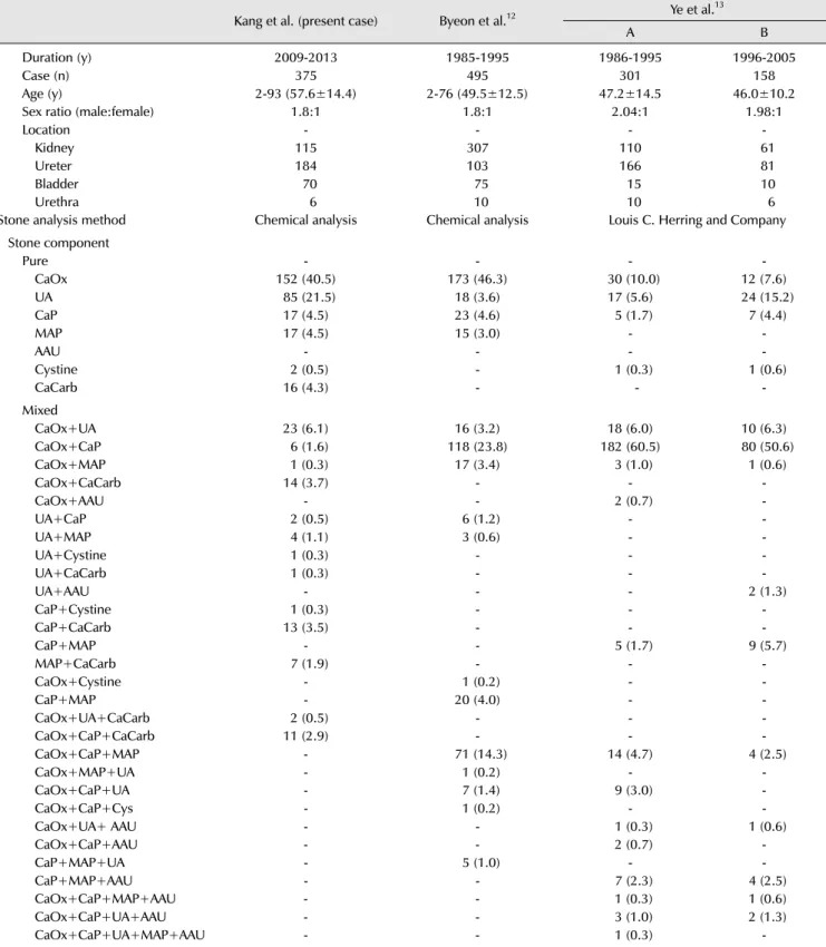
이 보고한 1985년에서 1995년까지 화학적 분석 법을 이용한 요로결석의 성분분석 495예와 비교할 때 본 연구 에서는 수산칼슘 단일석은 40.5%로 과거의 46.3%에 비해 낮은 비율을 보였으며, 요산석은 기존의 3.6%에 비교하면 21.5%로 높은 비율을 보였다. 그 외 인산칼슘이나 인산마그네 슘암모늄 등은 차이가 없었다. 이 결과는 Ye 등

13

이 1986년에 서 1995년까지(A군), 1996년에서 2005년까지(B군)으로 10년 단위로 나누어 분석한 연구와 같이 수산칼슘은 A군에서 73.1%, B군에서 56.3%로 감소하며, 요산석은 A군에서 12.3%, B군에서 22.2%로 유의한 증가를 보이는 것과 유사한 추이를 보였다(Table 4).

이런 변화는 앞서 설명한 요로 결석의 발생 기전에 있어

식생활의 변화와 더불어 동물성 단백질 및 퓨린 함유음식

섭취 증가와 관련 있을 것으로 생각하며 향후 체질량지수에

따른 요산석의 발생 비율과 비만과 요 중 pH의 상관관계에

(5)

Table 4. Compairing data - current and previous periods

    Kang et al. (present case) Byeon et al.

12

Ye et al.

13

A B

Duration (y) 2009-2013 1985-1995 1986-1995 1996-2005

Case (n) 375 495 301 158

Age (y) 2-93 (57.6±14.4) 2-76 (49.5±12.5) 47.2±14.5 46.0±10.2

Sex ratio (male:female) 1.8:1 1.8:1 2.04:1 1.98:1

Location - - - -

Kidney 115 307 110 61

Ureter 184 103 166 81

Bladder 70 75 15 10

Urethra 6 10 10 6

Stone analysis method   Chemical analysis Chemical analysis Louis C. Herring and Company

Stone component      

Pure - - - -

CaOx 152 (40.5) 173 (46.3) 30 (10.0) 12 (7.6)

UA 85 (21.5) 18 (3.6) 17 (5.6) 24 (15.2)

CaP 17 (4.5) 23 (4.6) 5 (1.7) 7 (4.4)

MAP 17 (4.5) 15 (3.0) - -

AAU - - - -

Cystine 2 (0.5) - 1 (0.3) 1 (0.6)

CaCarb 16 (4.3) -   - - 

Mixed

CaOx+UA 23 (6.1) 16 (3.2) 18 (6.0) 10 (6.3)

CaOx+CaP 6 (1.6) 118 (23.8) 182 (60.5) 80 (50.6)

CaOx+MAP 1 (0.3) 17 (3.4) 3 (1.0) 1 (0.6)

CaOx+CaCarb 14 (3.7) - - -

CaOx+AAU - - 2 (0.7) -

UA+CaP 2 (0.5) 6 (1.2) - -

UA+MAP 4 (1.1) 3 (0.6) - -

UA+Cystine 1 (0.3) - - -

UA+CaCarb 1 (0.3) - - -

UA+AAU - - - 2 (1.3)

CaP+Cystine 1 (0.3) - - -

CaP+CaCarb 13 (3.5) - - -

CaP+MAP - - 5 (1.7) 9 (5.7)

MAP+CaCarb 7 (1.9) - - -

CaOx+Cystine - 1 (0.2) - -

CaP+MAP - 20 (4.0) - -

CaOx+UA+CaCarb 2 (0.5) - - -

CaOx+CaP+CaCarb 11 (2.9) - - -

CaOx+CaP+MAP - 71 (14.3) 14 (4.7) 4 (2.5)

CaOx+MAP+UA - 1 (0.2) - -

CaOx+CaP+UA - 7 (1.4) 9 (3.0) -

CaOx+CaP+Cys - 1 (0.2) - -

CaOx+UA+ AAU - - 1 (0.3) 1 (0.6)

CaOx+CaP+AAU - - 2 (0.7) -

CaP+MAP+UA - 5 (1.0) - -

CaP+MAP+AAU - - 7 (2.3) 4 (2.5)

CaOx+CaP+MAP+AAU - - 1 (0.3) 1 (0.6)

CaOx+CaP+UA+AAU - - 3 (1.0) 2 (1.3)

  CaOx+CaP+UA+MAP+AAU - - 1 (0.3) -

Values are presented as number (%), range (mean±standard deviation) or mean±standard deviation only.

CaOx: calcium oxalate, UA: uric acid, CaP: calcium phosphate, MAP: magnesium ammonium phosphate, AAU: ammonium acid, CaCarb: calcium

carbonate.

(6)

대한 추가적인 연구가 이루어진다면 과거에 비해 요산석의 생성 비율이 증가하는 현상에 대한 이해에 도움이 될 것으로 생각된다.

결 론

최근 5년간 요로결석 성분을 분석한 결과 단일 성분의 결석 이 77.0%였고 수산칼슘 단일석이 40.5%로 가장 흔한 결석의 성분임을 알 수 있었으며, 과거 요로결석 성분분석 결과를 비교했을 때 요산석이 차지하는 비율이 증가하였고 연령이 증가함에 따라 남성일 경우 요산석이 차지하는 비율이 높아짐 을 알 수 있었다. 이와 같은 요산석의 증가는 동물성 단백질 및 퓨린 함유음식 섭취 증가와 같은 식생활 변화와 밀접한 관련성이 있을 것으로 생각되며, 요로 결석의 치료 및 재발 방지에 있어 적절한 식이 조절의 역할이 더욱 중요할 것으로 생각한다.

CONFLICT OF INTEREST

No potential conflict of interest relevant to this article was reported.

ACKNOWLEDGMENTS

The statistical analysis was supported by the Clinical Trial Center, Inje University Busan Paik Hospital.

REFERENCES

1. Saita A, Bonaccorsi A, Motta M. Stone composition: where do we stand? Urol Int 2007;79(Suppl 1):16-9.

2. Kim Hh, Jo MK, Kwak C, Park SK, Yoo KY, Kang D, et al.

Prevalence and epidemiologic characteristics of urolithiasis in Seoul, Korea. Urology 2002;59:517-21.

3. Johnson CM, Wilson DM, O'Fallon WM, Malek RS, Kurland LT.

Renal stone epidemiology: a 25-year study in Rochester, Minnesota. Kidney Int 1979;16:624-31.

4. Smith LH. The medical aspects of urolithiasis: an overview. J Urol 1989;141:707-10.

5. Alaya A, Nouri A, Belgith M, Saad H, Jouini R, Najjar MF. Chan- ges in urinary stone composition in the Tunisian population: a retrospective study of 1,301 cases. Ann Lab Med 2012;32:

177-83.

6. Goldfarb DS, Coe FL. Prevention of recurrent nephrolithiasis.

Am Fam Physician 1999;60:2269-76.

7. Kourambas J, Aslan P, Teh CL, Mathias BJ, Preminger GM. Role

of stone analysis in metabolic evaluation and medical treatment of nephrolithiasis. J Endourol 2001;15:181-6.

8. Basiri A, Taheri M, Taheri F. What is the state of the stone analysis techniques in urolithiasis? Urol J 2012;9:445-54.

9. Asper R. Epidemiology and socioeconomic aspects of uroli- thiasis. Urol Res 1984;12:1-5.

10. Hesse A, Siener R. Current aspects of epidemiology and nutrition in urinary stone disease. World J Urol 1997;15:

165-71.

11. Daudon M, Donsimoni R, Hennequin C, Fellahi S, Le Moel G, Paris M, et al. Sex- and age-related composition of 10 617 calculi analyzed by infrared spectroscopy. Urol Res 1995;23:

319-26.

12. Byeon SS, Kim HH, Kim SW. Analysis of the urinary stone components using chemical analysis method. Korean J Urol 1996;37:179-86.

13. Ye SJ, Yoo ES, Park YK. Analysis of urinary stone components during the last two decades. Korean J Urol 2007;48:1285-88.

14. Balaji KC, Menon M. Mechanism of stone formation. Urol Clin North Am 1997;24:1-11.

15. Menon M, Mahle CJ. Oxalate metabolism and renal calculi. J Urol 1982;127:148-51.

16. Finlayson B. Symposium on renal lithiasis. Renal lithiasis in review. Urol Clin North Am 1974;1:181-212.

17. Siener R, Glatz S, Nicolay C, Hesse A. The role of overweight and obesity in calcium oxalate stone formation. Obes Res 2004;12:106-13.

18. Coe FL, Evan A, Worcester E. Kidney stone disease. J Clin Invest 2005;115:2598-608.

19. Abate N, Chandalia M, Cabo-Chan AV Jr, Moe OW, Sakhaee K.

The metabolic syndrome and uric acid nephrolithiasis: novel features of renal manifestation of insulin resistance. Kidney Int 2004;65:386-92.

20. Maalouf NM, Sakhaee K, Parks JH, Coe FL, Adams-Huet B, Pak CY. Association of urinary pH with body weight in nephro- lithiasis. Kidney Int 2004;65:1422-5.

21. Negri AL, Spivacow R, Del Valle E, Pinduli I, Marino A, Fradinger E, et al. Clinical and biochemical profile of patients with "pure"

uric acid nephrolithiasis compared with "pure" calcium oxalate stone formers. Urol Res 2007;35:247-51.

22. Zechner O, Pfluger H, Scheiber V. Idiopathic uric acid lithiasis:

epidemiologic and metabolic aspects. J Urol 1982;128:1219- 23.

23. Poletto J, Harima HA, Ferreira SR, Gimeno SG. Hyperuricemia and associated factors: a cross-sectional study of Japanese- Brazilians. Cad Saude Publica 2011;27:369-78.

24. Childs MA, Mynderse LA, Rangel LJ, Wilson TM, Lingeman JE,

Krambeck AE. Pathogenesis of bladder calculi in the presence

of urinary stasis. J Urol 2013;189:1347-51.

수치

Table 1. Components of 375 urinary calculi and distribution according  to the gender
Table 2. Distribution of urinary stone according to the age
Table 4. Compairing data - current and previous periods

참조

관련 문서

Season, sex, age, and education as modifiers of the effects of outdoor air pollution on daily mortality in Shanghai, China: The Public Health and Air Pollution in Asia

In the late nineteenth century, although the proportion of ether was increased and that of chloroform decreased due to the raised awareness of the high

The foreign policy triumphs with China and the Soviet Union and the administration’s announcement that peace “is at hand” in Vietnam helped reelect Nixon as

Comparison of clinical risk factors and distribution of age, sex of patients aged ≧80 years and <80 years... Comparison of the stroke subtype and thrombolysis of

Drug-intoxicated patients were predominantly male, with the highest incidence of intoxication among patients above the seventh decade of age(21.5%) and the

- Renewable E, waste recycling, technology / industrial clusters, infrastructure integrated design Underground transportation

First, after the Group Art Therapy, the scale of hopelessness depression in the test group decreased significantly in comparison with that before the group

With these results, we can suggest the hypothesis that increased cardiac output in the patients who administered ketamine increased muscle blood flow, and this is the