mia and hyperphosphatemia [1]. The identification of the PTH re- ceptor and its signal transduction pathway facilitated better un- derstanding of PHP pathophysiology. PHP types I and II are dis- tinguished by cyclic AMP (cAMP) levels in response to exogenous PTH [2]. The main PHP subtypes, Ia and Ib (PHP-Ia and PHP-Ib, respectively), are caused by genetic and/or epigenetic alterations within or upstream of the GNAS locus. Most patients with PHP-Ia exhibit Albright’s hereditary osteodystrophy (AHO) and multi-hor- mone resistance, while 2/3 of patients with PHP-Ib exhibit only PTH resistance [1]. PHP exemplifies a quite unusual form of hor- mone resistance as the molecular cause is not a deficiency of the hormone receptor itself, but instead is a partial deficiency of the α -subunit of the stimulatory G protein (Gs α ), encoded by the GNAS imprinted locus (MIM#139320), a key regulator of the hormone- activated cAMP signaling pathway. The GNAS complex locus has multiple promoters and differentially methylated regions (DMRs) and produces several parent-of-origin products [3]. Using parent- specific methylation patterns in the DMRs, the GNAS complex produces stimulatory G protein, neuroendocrine secretory pro-
INTRODUCTION
Pseudohypoparathyroidism (PHP) is characterized by periph- eral parathyroid hormone (PTH) resistance along with hypocalce-
GNAS 메틸화 이상으로 인한 거짓부갑상선기능저하증 Ib 1예
A Case of Pseudohypoparathyroidism Type Ib Caused by Aberrant Methylation in the GNAS Complex Locus
조성진1·한은희1·장우리1·채효진1·김용구1·이건동1·조원경2·서병규2·김명신1
Sung Jin Jo, M.D.1, Eunhee Han, M.D.1, Woori Jang, M.D.1, Hyojin Chae, M.D.1, Yonggoo Kim, M.D.1, Gun Dong Lee, M.T.1, Won Kyoung Cho, M.D.2, Byung-Kyu Suh, M.D.2, Myungshin Kim, M.D.1
가톨릭대학교 의과대학 진단검사의학과1, 소아청소년과2
Departments of Laboratory Medicine1 and Pediatrics2, College of Medicine, The Catholic University of Korea, Seoul, Korea https://doi.org/10.3343/lmo.2017.7.2.83
Corresponding author: Myungshin Kim
Department of Laboratory Medicine, College of Medicine, The Catholic University of Korea, Seoul St. Mary’s Hospital, 222 Banpo-daero, Seocho- gu, Seoul 06591, Korea
Tel: +82-2-2258-1645, Fax: +82-2-2258-1719, E-mail: [email protected] Co-corresponding author: Byung-Kyu Suh
Department of Pediatrics, College of Medicine, The Catholic University of Korea, Seoul St. Mary’s Hospital, 222 Banpo-daero, Seocho-gu, Seoul 06591, Korea
Tel: +82-2-2258-6185, Fax: +82-2-537-4544, E-mail: [email protected] Received: June 13, 2016
Revision received: August 17, 2016 Accepted: August 18, 2016
This article is available from http://www.labmedonline.org 2017, Laboratory Medicine Online
This is an Open Access article distributed under the terms of the Creative Commons Attribution Non-Commercial License (http://creativecommons.org/licenses/by-nc/4.0/) which permits unrestricted non-commercial use, distribution, and reproduction in any medium, provided the original work is properly cited.
Pseudohypoparathyroidism (PHP) is a rare disorder caused by genetic and epigenetic aberrations in the GNAS complex locus resulting in impaired expression of stimulatory G protein (Gsα). PHP type Ib (PHP-Ib) is characterized by hypocalcemia and hyperphosphatemia due to renal resistance to the parathyroid hormone, and is distinguished from PHP-Ia by the absence of osteodystrophic features. An 11-yr-old boy presented with poor oral intake and cramping lower limb pain after physical activity. Laboratory studies revealed hypocalcemia, hyperphosphatemia, and increased para- thyroid hormone levels. The GNAS complex locus was evaluated using the methylation-specific multiplex ligation-dependent probe amplification (MS-MLPA) assay. Gain of methylation in the NESP55 domain and loss of methylation in the antisense (AS) transcript, XL, and A/B domains in the maternal allele were observed. Consequently, we present a case of PHP-Ib diagnosed using MS-MLPA.
Key Words: Pseudohypoparathyroidism, Methylation, GNAS complex locus, MLPA
tein 55 (NESP55), the extra-large variant of Gs α (XL α s), A/B tran- script, and the GNAS antisense (AS) transcript (Fig. 1) [3]. PHP-Ia is mainly caused by inactivation of maternally inherited mutations affecting Gs α coding exons. Loss of imprinting in the exon A/B DMR due to microdeletions in STX16 is the most frequent mecha- nism of familial PHP-Ib [4-8], while the most sporadic cases of PHP-Ib are caused by imprinting abnormalities of DMRs in the
GNAS complex [4, 9, 10]; however, no clinical difference was ob-served between the familial and sporadic forms of PHP-Ib [11].
Endocrine levels and other blood biochemical parameters vary according to the stage of life and disease severity [1]. Although pa- tients with PHP-Ia typically express AHO features, physical fea- tures, as well as biochemical and molecular findings in some cases are too ambiguous to distinguish it from PHP-Ib. The long-term effects of elevated PTH levels on the bones are also unclear [1] be- cause various skeletal phenotypes have been observed [12-15].
Therefore, identification of the molecular cause of PHP aids in confirmation of the diagnosis and the understanding of clinical characteristics. In this report, we present a patient with PHP-Ib caused by impaired imprinting in the GNAS complex.
CASE
1. Patient
An 11-yr-old boy presented with poor oral intake and cramping pain in both the lower limbs after physical activity. The parents ex- pressed concern that the patient had a slight physique compared to other children in the same age group. However, the patient show ed a normal growth rate according to the Korean growth chart (height
=
142.6 cm, 25-50th percentile, SDS -0.49; body wei ght
=34.7 kg, 10-25th percentile, SDS -0.78). The patient had no family history of any relevant diseases. The patient’s blood biochemical investiga- tions revealed hypocalcemia (6.6 mg/dL, reference range 8.0-10.0 mg/dL) and hyperphosphatemia (7.6 mg/dL, reference range 2.6- 4.5 mg/dL) with an elevated PTH level (127 pg/mL, reference range 13-54 pg/mL). Thyroid stimulating hormone (TSH) level was in-
creased (8.42 mIU/L, reference range 0.17-4.05 mIU/L), but free T4 (1.14 ng/dL, reference range 0.85-1.86 ng/dL) and T3 (1.20 ng/mL, reference range 0.78-1.82 ng/mL) levels were within the normal range. Serum cortisol (5.41 μg/dL, reference range 1.81-12.67 μg/
dL), 17 α -OH progesterone (4 ng/dL, reference range
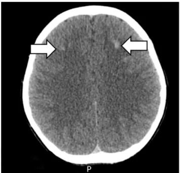
≤20 ng/dL), and adrenocorticotropic hormone (ACTH, 33.89 pg/mL, reference range 6.00-56.70 pg/mL) levels were also normal. To rule out PHP, further evaluations were performed. Ultrasonography and a scan (Tc-99m) of the thyroid and parathyroid glands were performed, but no abnormal findings were found. There was no evidence of AHO features in either the hand X-rays or physical examination of the patient. However, computed tomography (CT) scan of the brain revealed multiple high-density nodular lesions in the bilat- eral basal ganglia and subcortical white matter of the frontal lobe (Fig. 2). The cAMP level in the 24-hr urine sample was slight ly de- creased (1.60 μmol/day, reference range 1.8-6.3 μmol/day).
Fig. 1. Multiple methylated regions and GNAS gene of the GNAS complex locus. The general organization and imprinting patterns of GNAS alleles are shown. NESP55 is imprinted in the paternal allele and AS, XL and A/B are imprinted in the maternal allele (grey colored box ).
12 254
255
Fig. 2. The brain CT scan. Multiple high density nodular lesions in the bilateral basal ganglia
256
and subcortical white matter of the frontal lobe.
257
STX16 NESP55 AS XL A/B GNAS
P M
Fig. 2. The brain CT scan. Multiple high density nodular lesions in the bilateral basal ganglia and subcortical white matter of the frontal lobe.
2. Genetic Evaluation
First, we performed direct sequencing of GNAS and STX16.
Amplification of exons was performed using primers designed to anneal sequences in the pre- and post-exon introns. PCR prod- ucts were bidirectionally sequenced with the Big-Dye Terminator v3.1 Cycle Sequencing Kit (Applied Biosystems, Foster City, CA, USA) using the ABI PRISM 3130xl Genetic Analyzer (Applied Bio- systems). The Sequencher program (GeneCodes Corp., Ann Ar- bor, MI, USA) was used to align the derived and reference sequen- ces (NM_000516.4). In the direct sequencing of GNAS and STX16, no pathogenic mutations were detected.
A multiplex ligation-dependent probe amplification (MLPA) as- say was performed to identify the presence of deletions and du- plications, including DMRs, in the GNAS complex and STX16. Me- thylation-specific-MLPA (MS-MLPA) was performed to identify any abnormal methylation of the sequence and copy-number changes within the GNAS complex and STX16. MLPA and MS-MLPA were performed using the SALSA MS-MLPA probemix ME031-B1 GNAS kit (MRC-Holland, Amsterdam, The Netherlands) according to the manufacturer’s protocol. The ME031-B1 probemix contains 25 pro- bes specific for the GNAS locus and six probes specific for STX16 with amplicon sizes between 125 and 500 nucleotides. DNA was denatured at 98˚C for 5 min and hybridized with the probe set over- night at 60˚C. The ligation reaction using ligase was performed for 30 min at 48˚C, followed by 5 min at 98˚C for heat inactivation of the enzyme. PCR was performed with the specific SALSA PCR pri- mers for 35 cycles (95˚C for 30 sec; 60˚C for 30 sec; 72˚C for 1 min).
MLPA fragment analysis data was generated using the Applied
Biosystems 3130xl Genetic Analyzer and analyzed using Gene- Marker software (SoftGenetics, State College, PA, USA). A sample from a healthy male was used as the normal control in both MLPA and MS-MLPA assays.
The results of the MLPA analysis revealed a normal copy num- ber for both the GNAS complex and STX16. For the MS-MLPA as- say, the restriction enzyme HhaI was used to assess methylation status within a given sample. Un-methylated DNAs were digested by HhaI, and the digested probes, not amplified by PCR, did not generate a signal. In the MS-MLPA, the control sample showed a 50% reduction of each DMR (NESP55, NESPAS, XL, A/B domains), which is consistent with the previously reported normal methyla- tion pattern (Fig. 3). In the patient’s sample, the NESP55 peak was 100% preserved, but peaks of NESPAS, XL, and A/B domains show ed a 100% reduction, compared to those in the non-HhaI treated sam- ple. This suggests gain of methylation in the NESP55 domain and loss of methylation in the NESPAS, XL, and A/B domains of the maternal allele.
DISCUSSION
PHP presents different disease features according to its subtypes owing to genetic and epigenetic aberrations in the GNAS complex [1, 3]. The GNAS complex, a highly imprinted region, provides a better understanding of the pathogenesis of this disease. PHP-Ia and PHP-Ib are caused by distinct mechanisms, but some clini- cally overlapping cases have been reported [8]. In cases with an uncertain diagnosis, careful clinical examination, including as-
Fig. 3. MS-MLPA analysis of GNAS and STX16. (A) MS-MLPA peak ratio of patient treated after HhaI enzyme. Peaks for NESP55 (black colored box ■) and AS, XL, A/B (grey colored box ) are expressed. (B) Relative copy numbers of DMRs (NESP55, AS, XL, and A/B) after HhaI restriction enzyme treat- ed to the each allele compared to healthy control. NESP55 conserved about a pair of alleles, suggesting gain of methylation in the paternal allele.AS, XL and AS showed a 100% reduction of relative peaks, suggesting that loss of methylations in the maternal allele.
A B
2.5 2.0 1.5 1.0 0.5 0
Peak ratio
Size (bp)
100 200 300 400 500
1.0
0.5
0
Relative amplification products
NESP55 AS XL A/B
Normal man Patient
sessment of AHO-specific manifestations and further laboratory and radiological investigations, are needed. Blood biochemical parameters and urinary calcium excretion should be monitored in patients with PHP-I. In case of children, height, growth velocity, and pubertal development should be closely observed. High birth weight and/or early-onset obesity and macrocephaly are features of paternal uniparental disomy (UPD) of chromosome 20q. The most sporadic cases of PHP-Ib demonstrate broad GNAS methyla- tion defects, and no specific gene mutation has been identified as a cause of the methylation defects. Some cases of PHP-Ib have been identified to have paternal UPD as the underlying cause (10-25%
according to various reports) [16, 18, 19].
In Korea, several cases of PHP-Ib have been previously reported [20]. Cho et al. described six patients with PHP-Ib exhibiting vari- ous symptoms at diagnosis, such as seizures or carpal spasm. Two of the patients with PHP-Ib also showed intracranial calcifications that were revealed by a brain CT scan. Molecular studies also re- vealed methylation defects of the GNAS DMRs and STX16 dele- tions in the patients with PHP-Ib. Cho et al. suggested a diagnos- tic progression of direct sequencing of the GNAS gene followed by MS-MLPA of both GNAS DMRs and STX16 for the diagnosis of patients without AHO features. Microsatellite analyses were rec- ommended to exclude paternal disomy if parental DNA was avail- able. Garin et al. recommended single CpG bisulphite-based meth- ods to confirm the results in patients with partial methylation de- fects [19]. A pyrosequencing method with bisulphite treatment is useful for the detection of more subtle methylation defects. How- ever, MS-MLPA has the advantage of providing quantification of methylation along with detection of gene deletions and duplica- tions. MS-MLPA analysis consists of two parts—determining copy numbers by comparing different undigested samples, and deter- mining methylation patterns by comparing each undigested sam- ple to its digested counterpart. The second part is unique to MS- MLPA probe mixes and serves to semi-quantify the percentage of methylation within the given sample. In our case, we performed simultaneous direct sequencing of GNAS and STX16 because the patient had no familial history and had ambiguous clinical features, such as high-density lesions revealed by a brain CT scan. After di- rect sequencing, MS-MLPA was performed, but without parental DNA owing to its unavailability. In previously reported Korean cases of PHP-Ib and in our case, the most frequent aberrant meth- ylation pattern was gain of methylation in the NESP55 domain and
loss of methylation in the NESPAS, XL, and A/B domains. The cases of PHP are not sufficient to prepare an appropriate diagnostic work- flow; it only allows us to ascertain the origin of the disease and provides the evidence for further genetic counseling. Each study presents a different work-flow in use for PHP diagnosis, but all studies emphasize the use of MS-MLPA. In our case, the patients showed elevated TSH levels and normal free T4 and T3 levels. TSH resistance is frequently noted in patients with PHP-Ia, while less commonly in patients with PHP-Ib. Compared to previously re- ported Korean cases of PHP-Ib [20], our case showed significantly low PTH levels (127 mmol/L vs. 306.3
±119.1 mmol/L, P
=0.014) and relatively high TSH levels (8.42 mIU/L vs. 5.75
±5.71 mIU/L,
P=0.3). Intracranial calcification can be observed in both sub- types. Overlapping laboratory and physical findings in PHP sub- types have made differential diagnosis challenging. Nonetheless, molecular analyses are reliable methods for PHP diagnosis. In con- clusion, in addition to the GNAS and STX16 analyses using direct sequencing, quantitative analysis of gene copy number and de- tection of methylation defects are required for the diagnosis of PHP. Thus, MS-MLPA is a suitable method for identifying and un- derstanding the disease.
AUTHORS’ DISCLOSURE OF POTENTIAL CONFLICTS OF INTEREST
No potential conflicts of interest relevant to this article were re- ported.
ACKNOWLEDGMENTS
We would like to thank all patients who participated in this study, the referring clinicians, and The Catholic Genetic Labora- tory Center for assisting us with this study. This study was sup- ported by Research Fund of Seoul St.Mary’s Hospital, The Catho- lic University of Korea.
REFERENCES
1. Mantovani G. Clinical review: Pseudohypoparathyroidism: diagnosis and treatment. J Clin Endocrinol Metab 2011;96:3020-30.
2. Drezner M, Neelon FA, Lebovitz HE. Pseudohypoparathyroidism type II: a possible defect in the reception of the cyclic AMP signal. N Engl J
Med 1973;289:1056-60.
3. Bastepe M. The GNAS Locus: Quintessential complex gene encoding Gsalpha, XLalphas, and other imprinted transcripts. Curr Genomics 2007;8:398-414.
4. Yuno A, Usui T, Yambe Y, Higashi K, Uqi S, Shinoda J, et al. Genetic and epigenetic states of the GNAS complex in pseudohypoparathy- roidism type Ib using methylation-specific multiplex ligation-depen- dent probe amplification assay. Eur J Endocrinol 2013;168:169-75.
5. Bastepe M, Fröhlich LF, Hendy GN, Indridason OS, Josse RG, Koshi- yama H, et al. Autosomal dominant pseudohypoparathyroidism type Ib is associated with a heterozygous microdeletion that likely disrupts a putative imprinting control element of GNAS. J Clin Invest 2003;112:
1255-63.
6. Linglart A, Bastepe M, Jüppner H. Similar clinical and laboratory find- ings in patients with symptomatic autosomal dominant and sporadic pseudohypoparathyroidism type Ib despite different epigenetic changes at the GNAS locus. Clin Endocrinol 2007;67:822-31.
7. Liu J, Nealon JG, Weinstein LS. Distinct patterns of abnormal GNAS imprinting in familial and sporadic pseudohypoparathyroidism type IB. Hum Mol Genet 2005;14:95-102.
8. Mantovani G, Bondioni S, Linglart A, Maghnie M, Cisternino M, Cor- betta S, et al. Genetic analysis and evaluation of resistance to thyrotro- pin and growth hormone-releasing hormone in pseudohypoparathy- roidism type Ib. J Clin Endocrinol Metab 2007;92:3738-42.
9. Bastepe M, Pincus JE, Sugimoto T, Tojo K, Kanatani M, Azuma Y, et al.
Positional dissociation between the genetic mutation responsible for pseudohypoparathyroidism type Ib and the associated methylation defect at exon A/B: evidence for a long-range regulatory element within the imprinted GNAS1 locus. Hum Mol Genet 2001;10:1231-41.
10. Liu J, Litman D, Rosenberg MJ, Yu S, Biesecker LG, Weinstein LS. A GNAS1 imprinting defect in pseudohypoparathyroidism type IB. J Clin Invest 2000;106:1167-74.
11. Linglart A, Bastepe M, Jüppner H. Similar clinical and laboratory find- ings in patients with symptomatic autosomal dominant and sporadic
pseudohypoparathyroidism type Ib despite different epigenetic changes at the GNAS locus. Clin Endocrinol 2007;67:822-31.
12. Kidd GS, Schaaf M, Adler RA, Lassman MN, Wray HL. Skeletal respon- siveness in pseudohypoparathyroidism: a spectrum of clinical disease.
Am J Med 1980;68:772-81.
13. Murray TM, Rao LG, Wong MM, Waddell JP, McBroom R, Tam CS, et al. Pseudohypoparathyroidism with osteitis fibrosa cystica: direct dem- onstration of skeletal responsiveness to parathyroid hormone in cells cultured from bone. J Bone Miner Res 1993;8:83-91.
14. Balkissoon AR, Hayes CW. Case 14: intramedullary osteosclerosis. Ra- diology 1999;212:708-10.
15. Sbrocchi AM, Rauch F, Lawson ML, Hadjiyannakis S, Lawrence S, Bastepe M, et al. Osteosclerosis in two brothers with autosomal dominant pseu- dohypoparathyroidism type 1b: bone histomorphometric analysis. Eur J Endocrinol 2011;164:295-301.
16. Bastepe M, Altug-Teber O, Agarwal C, Oberfield SE, Bonin M, Jüppner H. Paternal uniparental isodisomy of the entire chromosome 20 as a molecular cause of pseudohypoparathyroidism type Ib (PHP-Ib). Bone 2011;48:659-62.
17. Bastepe M, Lane AH, Jüppner H. Paternal uniparental isodisomy of chromosome 20q--and the resulting changes in GNAS1 methylation- -as a plausible cause of pseudohypoparathyroidism. Am J Hum Genet 2001;68:1283-9
18. Dixit A, Chandler KE, Lever M, Poole RL, Bullman H, Mughal MZ, et al. Pseudohypoparathyroidism type 1b due to paternal uniparental di- somy of chromosome 20q. J Clin Endocrinol Metab 2013;98:E103-8.
19. Garin I, Mantovani G, Aguirre U, Barlier A, Brix B, Elli FM, et al. Euro- pean guidance for the molecular diagnosis of pseudohypoparathyroid- ism not caused by point genetic variants at GNAS: an EQA study. Eur J Hum Genet 2015;23:438-44.
20. Cho SY, Yoon YA, Ki CS, Huh HJ, Yoo HW, Lee BH, et al. Clinical char- acterization and molecular classification of 12 Korean patients with pseudohypoparathyroidism and pseudopseudohypoparathyroidism.
Exp Clin Endocrinol Diabete 2013;121:539-45.