KJTCVS
The Korean Journal of Thoracic and Cardiovascular SurgeryClinical Research Role and Prognosis of Extracorporeal Life Support in Patients Who Develop Cardiac Arrest during or after Office-Based
Cosmetic Surgery
Seong Soon Kwon, M.D.
1, Byoung-Won Park, M.D., Ph.D.
1, Min-Ho Lee, M.D.
1, Duk Won Bang, M.D., Ph.D.
1, Min-Su Hyon, M.D., Ph.D.
1, Won-Ho Chang, M.D., Ph.D.
2, Hong Chul Oh, M.D.
2, Young Woo Park, M.D., Ph.D.
21
Division of Cardiology, Department of Internal Medicine and
2Department of Chest Surgery, Soonchunhyang University Seoul Hospital, Seoul, Korea
ARTICLE INFO
Received September 27, 2019 Revised April 25, 2020 Accepted May 4, 2020 Corresponding author Duk Won Bang Tel 82-2-709-9216 Fax 82-2-709-9554 E-mail [email protected] ORCID
https://orcid.org/0000-0002-6691-7546
Background: Cardiac arrest during or after office-based cosmetic surgery is rare, and little is known about its prognosis. We assessed the clinical outcomes of patients who de- veloped cardiac arrest during or after cosmetic surgery at office-based clinics.
Methods: Between May 2009 and May 2016, 32 patients who developed cardiac arrest during or after treatment at cosmetic surgery clinics were consecutively enrolled. We com- pared clinical outcomes, including complications, between survivors (n=19) and non-sur- vivors (n=13) and attempted to determine the prognostic factors of mortality.
Results: All 32 of the patients were female, with a mean age of 30.40±11.87 years. Of the 32 patients, 13 (41%) died. Extracorporeal life support (ECLS) was applied in a greater per- centage of non-survivors than survivors (92.3% vs. 47.4%, respectively; p=0.009). The mean duration of in-hospital cardiopulmonary resuscitation (CPR) was longer for the non-sur- vivors than the survivors (31.55±33 minutes vs. 7.59±9.07 minutes, respectively; p=0.01).
The mean Acute Physiology and Chronic Health Evaluation score was also higher among non-survivors than survivors (23.85±6.68 vs. 16.79±7.44, respectively; p=0.01). No predictor of death was identified in the patients for whom ECLS was applied. Of the 19 survivors, 10 (52.6%) had hypoxic brain damage, and 1 (5.3%) had permanent lower leg ischemia.
Logistic regression analyses revealed that the estimated glomerular filtration rate was a predictor of mortality.
Conclusion: Patients who developed cardiac arrest during or after cosmetic surgery at office-based clinics experienced poor prognoses, even though ECLS was applied in most cases. The survivors suffered serious complications. Careful monitoring of subjects and ac- tive CPR (when necessary) in cosmetic surgery clinics may be essential.
Keywords: Plastic surgery, Cardiac arrest, Extracorporeal circulation
Copyright
© The Korean Society for Thoracic and Cardiovascular Surgery. 2020. All right reserved.
This is an Open Access article distributed under the terms of the Creative Commons Attribution Non-Commercial License (http://creativecommons.org/licenses/
Introduction
Extracorporeal life support (ECLS) is a modification of the cardiopulmonary bypass circuit that is commonly used in various critical cardiac situations [1], including post-car- diotomy, post-heart transplantation, after the development of severe cardiac failure attributable to decompensated car- diomyopathy, in patients with myocarditis, in patients with acute coronary syndrome accompanied by cardiogenic shock, and in patients exhibiting cardiac depression attrib- utable to drug overdose or sepsis. A form of ECLS termed
extracorporeal cardiopulmonary resuscitation (CPR) is also used in cases of cardiac arrest [2]. ECLS is becoming an increasingly popular treatment for adults with cardiac and/or respiratory failure, as its outcomes have improved over time. The survival rates of adults with cardiac and re- spiratory failure are 39% and 55%, respectively [3]. Howev- er, the rate of survival after out-of-hospital cardiac arrest (OHCA) remains poor [4]. The prognostic factors for poor outcomes (including mortality) are advanced age, stroke, renal failure, acidosis, and hypoglycemia [5].
Cardiac arrest during non-cardiac surgery is rare. In a
https://doi.org/10.5090/kjtcs.19.077 pISSN: 2233-601X eISSN: 2093-6516
Korean J Thorac Cardiovasc Surg. 2020;53(5):277-284
KJTCVS https://doi.org/10.5090/kjtcs.19.077
recent study, the incidence was 7.22 cases per 10,000, and the 30-day mortality rate was 63% [6]. The risk of cardiac arrest during or after surgery increases with advanced age, impaired functional status, the presence of comorbidities, the level of surgical risk, and the need for blood transfu- sion [6]. A quality assurance program administered by the American Association for Accreditation of Ambulatory Surgery Facilities in the accredited facilities of that organi- zation included more than 1 million outpatient procedures performed between 2001 and 2006 and reported a mortali- ty rate of 0.002% [7]. However, little is known regarding the risk of complications or overall prognosis of patients who develop cardiac arrest during or after office-based cosmetic surgery.
We retrospectively reviewed our 8-year data on patients who developed cardiac arrest during or after office-based cosmetic surgery with the objectives of identifying the mortality and complication rates after the use of ECLS and defining factors that predict survival.
Methods
Between May 2009 and May 2016, we enrolled and retro- spectively reviewed 32 consecutive patients who developed cardiac arrest during or after cosmetic surgery at primary clinics. A total of 21 patients underwent ECLS to treat re-
fractory cardiac arrest and/or shock. Our hospital is locat- ed near more than 200 office-based cosmetic surgery clin- ics that are within 15–20 minutes’ drive by ambulance (Fig.
1) [8]. The baseline clinical characteristics, types of cos- metic surgery, and types of anesthesia employed were re- viewed, in addition to the duration of CPR, the time inter- val between cardiac arrest and arrival at our hospital, the Acute Physiology and Chronic Health Evaluation (APACHE) score, and propofol usage. However, we lacked information regarding both the doses of anesthetic agents administered and the surgical duration.
The 32 patients were divided into those who survived and were discharged (survivors, n=19) and those who died in the hospital (non-survivors, n=13). Two extracorporeal systems were employed. The first featured a centrifugal pump; a polypropylene, hollow-fiber membrane oxygen- ator; and a heparin-coated circuit (Capiox EBS circuit; Ter- umo Inc., Tokyo, Japan). The second included a Rotaflow centrifugal pump and a Quadrox PLS oxygenator (Maquet, Hirrlingen, Germany). Arterial cannulae (14F–22F) were percutaneously inserted into the femoral artery using the Seldinger technique. Heparin (5,000 U) was administered 5 minutes prior to femoral arterial cannulation. Once the cannula was in place, the ECLS system was activated. Per- cutaneous femoral ECLS was instituted in the emergency room while CPR was being administered or after the re-
Plastic surgery clinic LQ LQ<1 1 1.99 2 2.99 3 3.99 Our hospital
Fig. 1. Regional distribution of plas-
tic surgery clinic and location of
Soonchunhyang University Seoul
Hospital. LQ, location quotient (a
simple tool used to determine the
spatial distribution of a particular
industry, cluster, occupation, or de-
mo graphic group).
Seong Soon Kwon, et al. Prognosis of Young Patients with Cardiac Arrest KJTCVS
turn of spontaneous circulation. If not contraindicated, low-molecular-weight or unfractionated heparin was given to reduce the risk of thromboembolic complications during ECLS. The activated partial thromboplastin time was monitored at least every 6–12 hours, and the intravenous heparin infusion rate was 12–15 U/kg/hr in all cases. No patient underwent distal perfusion to prevent lower leg ischemia. At each weaning attempt, transthoracic echocar- diography was used to monitor heart function. During weaning, the flow was gradually reduced to 1 L/min/m
2. Inotropic agents were used to facilitate weaning in most cases. Patients who maintained adequate ventricular func- tion and exhibited stable vital signs were decannulated [9].
The results for continuous variables are reported as means±standard deviations, whereas the results for cate- gorical variables are presented as frequencies and percent- ages. Comparisons between continuous variables were made using the Student t-test, while comparisons between categorical variables were evaluated using the Fisher exact test or the Pearson chi-square test, as appropriate. Survival analysis was performed using Kaplan-Meier analysis. Vari-
ables with p<0.2 were entered into the multivariable logis- tic regression analysis to calculate the odds ratios (ORs) and 95% confidence intervals (CIs). A p-value <0.05 was considered to indicate a statistically significant difference.
All statistical analyses were performed using IBM SPSS ver.
21.0 (IBM Corp., Armonk, NY, USA). This study was per- formed in accordance with the ethical guidelines of the 1975 Declaration of Helsinki and was approved by the In- stitutional Review Board of Soonchunhyang University Seoul Hospital (SCHUH 2019-01-009). Due to the retro- spective nature of this study, informed consent was not re- quired.
Results
Patient characteristics
The baseline characteristics and clinical features of all 32 patients are summarized in Table 1. All were female, with a mean age of 30.40±11.87 years. The types of plastic surgery performed were liposuction (n=12), rhinoplasty (n=5), Table 1. Baseline characteristics of all patients and comparison between survivors and non-survivors
Characteristic All (n=32) Survival
p-value Yes (n=19) No (n=13)
Clinical
Age (yr) 30.40±11.87 29.58±7.71 33.54±14.99 0.334
Extracorporeal life support applied 21 (65.6) 9 (47.4) 12 (92.3) 0.009
Out-of-hospital arrest 27 (84.4) 14 (73.7) 13 (100.0) 0.064
Pulmonary edema 16 (50) 7 (36.8) 9 (69.2) 0.072
Cardiopulmonary resuscitation duration at hospital (min) 17±24.81 7.59±9.07 31.55±33.87 0.010 Acute Physiology and Chronic Health Evaluation 19.66±7.86 16.79±7.44 23.85±6.68 0.010
Left ventricular ejection fraction (%) 40.59±19.24 45.0 ±18.91 33.36±18.32 0.115
Survival time (day) 18.2±33.7
Time interval between cardiac arrest and arrival at the hospital (min) 49±75 64±93 27±20 0.039
Anesthesia 0.266
Local 11 (35) 8 (42) 3 (23)
General 21 (65) 11 (58) 10 (77)
Propofol 20 (63) 11 (58) 9 (69) 0.515
Laboratory (initial)
pH 7.12±0.23 7.21±0.19 6.99±0.22 0.006
Partial pressure of oxygen (mm Hg) 109.41±119.32 143.37±143.56 62.39±47.62 0.061
Partial pressure of carbon dioxide (mm Hg) 78.13±110.33 87.71±142.99 64.86±33.69 0.578
Bicarbonate (mmol/L) 24.6±15.64 16.18±7.78 14.89±6.02 0.291
Total carbon dioxide (mmol/L) 15.06±7.01 16.26±4.50 13.31±4.80 0.086
Serum creatinine (mg/dL) 1.6±0.87 0.79±0.13 0.97±0.26 0.015
Estimated glomerular filtration rate (mL/min) 92.89±20.38 99.98±17.04 82.53±21.0 0.015
Hemoglobin (g/dL) 13.27±1.86 13.44±1.57 12.27±1.20 0.031
Hematocrit (%) 39.65±4.88 41.10±4.88 37.52±4.18 0.040
Creatine kinase-MB (ng/mL) 3.78±3.86 4.35±4.86 2.93±1.36 0.316
Troponin T (ng/mL) 0.39±0.08 0.11±0.12 0.04±0.06 0.086
Values are presented as mean±standard deviation for continuous variables and number (%) for categorical variables.
KJTCVS https://doi.org/10.5090/kjtcs.19.077
breast augmentation (n=5), maxillofacial surgery (n=5), scar revision (n=3), facelift (n=1), and hair transplantation (n=1). Of the 32 patients, 13 died (41%). ECLS was used in a total of 21 patients (65.6%). The mean age of the survi- vors was 29.58±7.71 years, while that of the non-survivors was 33.54±14.99 years. ECLS was applied in a greater per- centage of non-survivors than survivors (92.3% versus 47.4%, respectively; p=0.009). No predictor of death was identified in the patients for whom ECLS was applied. The characteristics of patients who received ECLS are shown in Table 2. Neither the OHCA rate nor the extent of pulmo- nary edema evident on chest X-ray differed significantly between the survivors and the non-survivors. The mean in-hospital CPR duration was longer in the non-survivors than in the survivors (31.55±33.87 minutes in non-survi- vors versus 7.59±9.07 minutes in survivors, p=0.01). The APACHE score was also higher in non-survivors than in survivors (23.85±6.68 versus 16.79±7.44, respectively;
p=0.01). Finally, the time taken to arrive at the hospital was similarly longer in non-survivors than in survivors (64±93 minutes versus 27±20 minutes, respectively; p=0.039). The left ventricular ejection fraction did not differ significantly between the 2 groups. A total of 11 (35%) and 21 (65%) pa- tients underwent local and general anesthesia, respectively.
Propofol was administered to 20 patients (63%). Neither the anesthesia type nor the frequency of propofol usage differed significantly between groups. Blood pH, serum creatinine and hemoglobin levels, and hematocrit were lower in the non-survivors than in the survivors. The other laboratory findings did not differ significantly between groups.
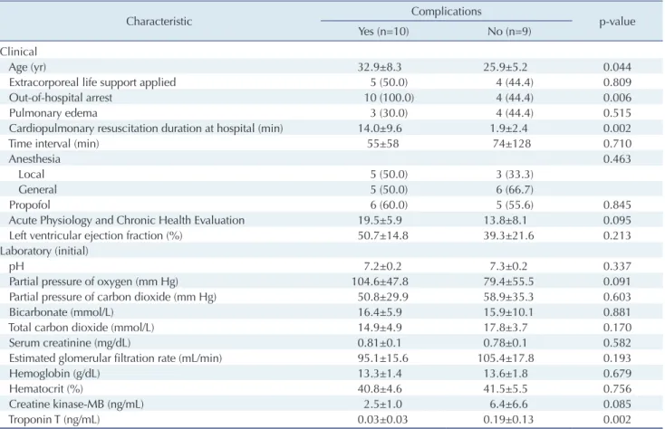
Complications in survivors
The characteristics according to the presence or absence of complications among survivors are shown in Table 3.
Survivors with complications tended to be older (mean age, 32.9±8.3 years in patients with complications versus 25.9±5.2 years in patients with no complications; p=0.044), to have had OHCA (100% in patients with complications versus 44.4% in patients with no complications, p=0.006), and had a longer mean CPR duration (14.0±9.6 minutes in patients with complications versus 1.9±2.4 minutes in pa- tients with no complications, p=0.002). The frequency of ECLS application, the APACHE score, and the type of an- esthesia employed did not differ significantly between groups. With the exception of troponin T levels, the labo- ratory data also lacked significant differences between groups.
Analysis of mortality
The Kaplan-Meier curves showed that mortality was sig- nificantly higher in patients for whom ECLS was applied (n=21) than in those for whom ECLS was not applied (n=11) (log-rank p=0.026) (Fig. 2).
Univariable logistic regression analysis showed that ECLS application (OR, 13.333; 95% CI, 1.434–123.989;
p=0.023), CPR duration (OR, 1.091; 95% CI, 0.998–1.120;
p=0.025), pH (OR, 0.006; 95% CI, 0.000–0.377; p=0.016), APACHE score (OR, 1.164; 95% CI, 1.022–1.326; p=0.022), estimated glomerular filtration rate (eGFR; OR, 0.949; 95%
CI, 0.907–0.994; p=0.027), and glucose levels (OR, 1.005;
95% CI, 1.000–1.011; p=0.045) were associated with mor- tality. In the multivariable logistic analysis, which included variables with p-values <0.2, eGFR was the only significant predictor of mortality (OR, 0.925; 95% CI, 0.864–0.990;
p=0.025) (Table 4).
Discussion
We found that patients entering cardiac arrest during or Table 2. Characteristics of patients who received ECLS
Characteristic All (n=21) Survival
p-value
Yes (n=9) No (n=12)
Age (yr) 31.14±12.31 21.56±8.09 32.33±14.98 0.622
Initial rhythm
Asystole 13 (60.90) 7 (77.78) 6 (50.00) -
Pulseless electrical activity 2 (9.52) 0 2 (16.67) -
Shockable rhythm 6 (28.57) 2 (22.22) 4 (33.33) -
Bystander initiated cardiopulmonary resuscitation 21 (100.00) 9 (100.00) 12 (100.00) - Time from cardiac arrest to ECLS initiation
a)97.12±142.29 35.14±7.01 140.50±175.73 0.091 Values are presented as mean±standard deviation or number (%).
ECLS, extracorporeal life support.
a)
Time of cardiac arrest was defined as the time of arrival to the emergency room of Soonchunhyang University Seoul Hospital.
Seong Soon Kwon, et al. Prognosis of Young Patients with Cardiac Arrest KJTCVS
soon after cosmetic surgery exhibited poor prognoses (overall mortality rate, 41%), even though ECLS was ap- plied in most cases. Of the survivors, 69% had serious complications, including hypoxic brain damage and lower
leg ischemia. This study revealed that eGFR was a signifi- cant clinical predictor of mortality, and CPR duration was also an important factor associated with mortality.
Although ECLS is often used to treat OHCA, the out- comes are poor [4]. Recent studies have found a survival rate of >30% among patients with OHCA who received ECLS [10,11]. The prognosis is affected primarily by the underlying cause of OHCA. A recent study suggested that ECLS was a useful rescue strategy for select patients with refractory OHCA, but a multidisciplinary team approach was required [12]. In the present study, despite prolonged CPR (>30 minutes), ECLS was performed in almost all of the patients because they were young and had no underly- ing disease.
Although cardiac arrest during office-based cosmetic surgery is uncommon, additional studies are needed due to the high level of associated mortality [13]. A retrospective analysis of a resuscitation registry containing data on 2,524 cardiopulmonary arrest patients treated in compliance with current guidelines revealed that the prognosis was poor, the survival-to-discharge rate was low (31.7%), and Table 3. Comparison between patients who did and did not experience complications
Characteristic Complications
p-value
Yes (n=10) No (n=9)
Clinical
Age (yr) 32.9±8.3 25.9±5.2 0.044
Extracorporeal life support applied 5 (50.0) 4 (44.4) 0.809
Out-of-hospital arrest 10 (100.0) 4 (44.4) 0.006
Pulmonary edema 3 (30.0) 4 (44.4) 0.515
Cardiopulmonary resuscitation duration at hospital (min) 14.0±9.6 1.9±2.4 0.002
Time interval (min) 55±58 74±128 0.710
Anesthesia 0.463
Local 5 (50.0) 3 (33.3)
General 5 (50.0) 6 (66.7)
Propofol 6 (60.0) 5 (55.6) 0.845
Acute Physiology and Chronic Health Evaluation 19.5±5.9 13.8±8.1 0.095
Left ventricular ejection fraction (%) 50.7±14.8 39.3±21.6 0.213
Laboratory (initial)
pH 7.2±0.2 7.3±0.2 0.337
Partial pressure of oxygen (mm Hg) 104.6±47.8 79.4±55.5 0.091
Partial pressure of carbon dioxide (mm Hg) 50.8±29.9 58.9±35.3 0.603
Bicarbonate (mmol/L) 16.4±5.9 15.9±10.1 0.881
Total carbon dioxide (mmol/L) 14.9±4.9 17.8±3.7 0.170
Serum creatinine (mg/dL) 0.81±0.1 0.78±0.1 0.582
Estimated glomerular filtration rate (mL/min) 95.1±15.6 105.4±17.8 0.193
Hemoglobin (g/dL) 13.3±1.4 13.6±1.8 0.679
Hematocrit (%) 40.8±4.6 41.5±5.5 0.756
Creatine kinase-MB (ng/mL) 2.5±1.0 6.4±6.6 0.085
Troponin T (ng/mL) 0.03±0.03 0.19±0.13 0.002
Values are presented as mean±standard deviation for continuous variables and number (%) for categorical variables.
Fig. 2. Kaplan-Meier estimates for overall survival stratified ac- cording to whether ECLS was applied. Crosses indicate censored events. ECLS, extracorporeal life support.
1.0
0.8
0.6
0.4
0.2
0.0
1,200
Survival rate
Time (day)
ECLS applied ECLS not applied
0 200 400 600 800 1,000
Log-rank p=0.026