PREVENTION RESEARCH □ ORIG INAL ARTICLE □
39
INTRODUCTION
Arsenical compounds are distributed as a natural toxicant with no color, no taste, and no smell. Arsenic trioxide, As
2O
3has been reported to induce almost complete remission from
acute promyelocytic leukemia (APL).
1∼3)Cytopathological stu- dies also showed that As
2O
3induces apoptosis in APL cells.
Recent reports have shown that As
2O
3down-regulates bcl-2 gene expression and induces the expression of apoptosis-related proteins, caspases as well as degradation of promyelocytic leukemia (PML) and promyelocytic leukemia gene/retinoic acid
Apoptosis-induced Cell Growth Inhibitory Effects of a Novel Compound, As 4 O 6 in a Cervical
Cancer Cell Line, SiHa in Vitro
Hong-Seok Chang1, Su-Mi Bae2, Sun-Young Kwak2, Ae Ju Lee2, Aery Lee2
Yong Seok Lee3, Il-Ju Bae4, Jin-Young Yoo5, Young-Joo Lee6, Chong-Kook Kim7 and Woong-Shick Ahn3
1Department of Therapeutic Radiology, 2Catholic Research Institutes of Medical Science,
3Department of Obstetrics and Gynecology, The Catholic University of Korea College of Medicine, Seoul 137-040, 4Laboratory of Chonjisan Institute, Seoul 150-727,
5Department of Pathology, The Catholic University of Korea College of Medicine, Seoul 137-040,
6Department of Bioscience and Biotechnology, Institute of Biotechnology, College of Life Science, Sejong University, Seoul 143-747, 7College of Pharmacy, Seoul National University, Seoul 151-742, Korea
As
2O
3has been reported to be effective for treating acute leukemia and induce apoptosis in many tumor cells. In this study, we evaluated the ability of a novel arsenical compound, As
4O
6, along with As
2O
3to induce cell growth inhibition as well as apoptosis in human cervical cancer cells, SiHa cells
in vitro. To examine the levels of apoptosis, SiHa cells were given two sensitive doses, 0.5 and 1μMof arsenical compounds, and then, DNA fragmentation assay and FACS analysis were conducted. In addition, Western blotting assay was done for the identification of target molecules for apoptosis. Both As
2O
3and As
4O
6caused dose-dependent inhibition of SiHa cell proliferation. In particular, As
4O
6was more effective for suppressing SiHa cell growth, as compared to As
2O
3. In parallel with inhibition of cell proliferation, As
4O
6caused an increase of the sub-G1 cell population significantly more than As
2O
3, as determined by propidium iodide DNA staining. This was confirmed by DNA fragmentation assay and annexin V staining. Western blotting analysis also showed that the proliferating cell nuclear antigen (PCNA) expression was suppressed by As
4O
6significantly more than As
2O
3, and that Bcl-
XLwith se- quence homology to Bcl-2 was significantly suppressed by As
4O
6. However, apoptosis-related proteins, p21 and Bax, were expressed by As
4O
6significantly more than As
2O
3. Taken together, these findings suggest that a novel arsenic compound, As
4O
6possesses more potent anti-proliferative effects on human cervical cancer cells with induction of apoptosis at least through activation in p21 and Bax proteins
in vitro. (Cancer Prev Res 11, 39-45, 2006)ꠏꠏꠏꠏꠏꠏꠏꠏꠏꠏꠏꠏꠏꠏꠏꠏꠏꠏꠏꠏꠏꠏꠏꠏꠏꠏꠏꠏꠏꠏꠏꠏꠏꠏꠏꠏꠏꠏꠏꠏꠏꠏꠏꠏꠏꠏꠏꠏꠏꠏꠏꠏꠏꠏꠏꠏꠏꠏꠏꠏꠏꠏꠏꠏꠏꠏꠏꠏꠏꠏꠏꠏꠏꠏꠏꠏꠏꠏꠏꠏꠏꠏꠏꠏꠏꠏꠏꠏꠏꠏꠏꠏꠏꠏꠏꠏꠏꠏꠏꠏꠏꠏ Key Words: Cervical cancer, Arsenic trioxide, Arsenic hexoxide, Apoptosis
책임저자:안웅식 ꂕ 137-040, 서울시 서초구 반포동 505 가톨릭대학교 의과대학 산부인과학교실 Tel: 02-590-2786, Fax: 02-599-4120 E-mail: [email protected]
접수일:2005년 12월 20일, 게재승인일:2006년 1월 18일
Correspondence to:Woong-Shick Ahn
Department of Obstetrics and Gynecology, The Catholic University of Korea College of Medicine, 505, Banpo-dong, Seocho-gu, Seoul 137-040, Korea Tel: +82-2-590-2786, Fax: +82-2-599-4120
E-mail: [email protected]
receptor (PML/RAR) α proteins in APL cells.
2,4)Similarily, arsenic trioxide suppresses the growth of tumor cells by cell cycle arrest, induction of cyclin-dependent kinase (CDK) inhi- bitors and apoptosis in a myeloma cell line, MC/CAR.
5)Arsenic trioxide also causes cell death through apoptosis in a human leukemia cell line, NB4,
6)a human papillomavirus (HPV) 16 infected cervical carcinoma cells,
7)and a human pancreatic can- cer cells.
8,9)These collective studies suggest that arsenic trioxide has an anti-tumor activity in a variety of tumor cells.
Cervical cancer is caused mostly by infection with a high risk group of HPV.
10∼12)After high risk HPV infection, two viral oncogenic proteins, E6 and E7 play a critical role in inducing cervical cancers by interacting with p53 and retinoblastoma protein (pRb) for inactivation of these cellular regulatory pro- teins, respectively.
13∼15)Presently, surgical, radiation, chemo- therapies have been approached with limited success. However, an early detection of cervical cancer using the Pap smear has contributed to cut down the incidence of cervical cancers.
However, relapsing cervical cancers have been problematic, adding importance to developing anti-cervical cancer drugs.
Here we evaluated the ability of a new arsenical compound, As
4O
6along with As
2O
3to suppress cell growth in human cervical carcinoma cells, SiHa cells. We observe that As
4O
6is more effective for inhibiting the SiHa cell growth, as compared to As
2O
3. Furthermore, As
4O
6caused an increase of the sub-G1
cell population, DNA ladder formation and annexin V staining significantly more than As
2O
3. Furthermore, Western blotting analysis showed that expression of a cell proliferation marker and Bcl-
XLis suppressed by As
4O
6significantly more than As
2O
3. However, expression of apoptosis-related proteins was increased by As
4O
6significantly more than As
2O
3. Thus, these data suggest that a novel arsenic compound, As
4O
6possesses more potent anti-proliferative effects on human cervical cancer cells with induction of apoptosis in vitro, as compared to As
2O
3.
MATERIALS AND METHODS
1. Cell cultureHPV 16 immortalized human cervical carcinoma cell lines, SiHa cells containing wild type P53 were incubated DMEM supplemented with 5% fetal bovine serum, 0.37% sodium bicarbonate, 30 mM HEPES, and 100μg/ml streptomycin/
penicillin (cDMEM) at 37
oC in the CO
2incubator.
2. Chemical reagents
As
2O
3was purchased from Sigma Chemical Co. As
4O
616∼19)was provided from Chonjisan., Co., Seoul, Korea. The chemical structures of both As
2O
3and As
4O
6are inserted in Fig. 1.
These chemicals were diluted in phosphate-buffered saline (PBS) to a final concentration of 10
-3M and kept at 4
oC.
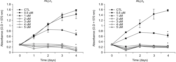
Fig. 1. Growth inhibition patterns of As2O3 and As4O6 in SiHa cells in vitro. Cells were treated with the indicated amount of arsenic compounds, As2O3 and As4O6 for 4 day incubation periods. Cell growth suppression was measured as described in “Methods and Materials.” OD was measured at 570 nm. The assay was performed in triplicate and average OD values and SD were recorded.
This was repeated two more times with similar results.
*Statistically significant at p<0.05 using the paired Student's t test compared to no drug treatment (control, CTL). The chemical structures of As4O6 and As4O6 were inserted in each panel. The shaded bigger circles and empty smaller circles represent As and O, respectively.
ꠏꠏꠏꠏꠏꠏꠏꠏꠏꠏꠏꠏꠏꠏꠏꠏꠏꠏꠏꠏꠏꠏꠏꠏꠏꠏꠏꠏꠏꠏꠏꠏꠏꠏꠏꠏꠏꠏꠏꠏꠏꠏꠏꠏꠏꠏꠏꠏꠏꠏꠏꠏꠏꠏꠏꠏꠏꠏꠏꠏꠏꠏꠏꠏꠏꠏꠏꠏꠏꠏꠏꠏꠏꠏꠏꠏꠏꠏꠏꠏꠏꠏꠏꠏꠏꠏꠏꠏꠏꠏꠏꠏꠏꠏꠏꠏꠏꠏꠏꠏꠏꠏꠏꠏꠏꠏꠏꠏꠏꠏꠏꠏꠏꠏꠏ
3-(4,5-dimethylthiazol-2-yl)-2,5-diphenyltetrazolium bromide (MTT) was purchased from Sigma and dissolved in PBS at a final concentration of 5 mg/ml.
3. Cell growth inhibition assay
For cell growth inhibition assay, MTT assay was performed.
First, SiHa cells (1×10
3cells/well) were divided into 96 well plate in 100μl of cDMEM. After 24 h incubation, cells were treated with different amount of arsenic compounds. After 4 days incubation, each well of plates were added with 20μl of MTT solution for 4 h at 37
oC. The cell media were replaced with 100μl of dimethyl sulfoxide (Sigma) per well. The plate was shaken for 10 sec, and then optical density (OD) was measured at a wavelength of 570 nm using enzyme-linked immunosorbent assay-Reader (Spectromax 250, Molecular De- vices). The growth inhibition rate (%) was calculated as follows:
OD of non-treatment -OD of drug treatment/OD of non- treatment ×100.
4. DNA fragmentation assay
SiHa cells were divided into 5×10
5cells per 100 mm dish plate. After 24 h incubation, cells were added with different amounts of arsenic compounds for 48 h at 37
oC. The cells were then centrifuged and subsequent cell pellets were added with lysis buffer [0.8% SDS, 0.1 M NaCl, 0.1 M EDTA, 50 mM Tris-HCl (pH8.0)], followed by addition of 20μg/ml proteinase K (Sigma). This was incubated for 4 h at 56
oC. DNA was ex- tracted by phenol/chloroform treatment. Five μg of extracted DNA was analyzed on a 2% agarose gel containing ethidium bromide (0.1μg/ml). DNA ladder formation was visualized under UV light.
5. FACS analysis
Cells were washed twice with PBS and then resuspended in 1×binding buffer (10 mM Hepes/NaOH, pH 7.4, 140 mM NaCl, 2.5 mM CaCl
2). One×10
5cells per tube were added with 5μl of Annexin V-FITC and 10μl of propidium iodide (BD, San Jose, CA), followed by incubation at 22
oC for 15 min.
Each tube was added with 100μl of 1×binding buffer and then cells were analyzed by flow cytometer (BD). For DNA contents, ethanol-fixed cells were incubated with RNase A (10 mg/ml) and propidium iodide (400μg/ml) and shaken for 1 h at 37
oC in the dark. The samples were read using flow
cytometer (BD). Cell debris and fixation artifacts were gated out and G0/G1, S and G2/M populations were quantified using the CellQuest program.
6. Western blotting analysis
SiHa Cells were treated with 0.5 and 1μM of As
2O
3and As
4O
6for 48 h. The cell lysates (approximately 30μg of protein) was separated in 12% polyacrylamide SDS-gels and transferred to the nitrocellulose membrane (Schleicher &
Schuell, Dassel, Germany). This was then immersed in blocking buffer (5% skim milk and 0.1% Tween 20 in PBS, pH 7.4) for 1 h at room temperature and incubated with primary antibodies (SantaCruz Biotechnology, Inc., California, USA), PCNA (1:200), CDK4 (1:200), p21 (1:200), Bax (1:
200), Bcl-
XL/Bcl-
XS(1:500) and actin (1:5,000) in blocking buffer overnight at 4
oC . After the incubation, the membrane was probed with horseradish peroxidase-labeled anti-mouse IgG antibody (1:5,000) in PBS (containing of 0.05% Tween 20 and 5% skim milk powder) for 30 min at room temperature.
The proteins in the membrane were detected by enhanced chemiluminescence detection system (Amersham, Buckingham- shire, UK) and bands were visualized by autoradiography using X-ray film (Amersham).
RESULTS
1. As4O6 inhibited SiHa cell growth to a more significant level than As2O3
To investigate whether a new arsenical compound, As
4O
6could suppress the growth of human cervical cancer cells, SiHa
cells were treated with an increasing amount (0.5∼5μM) of
As
4O
6for 48 h. As a positive control, cells were also treated
with As
2O
3. As shown in Fig. 1, the growth of SiHa cells was
unaffected by treating cells with 0.5μM of As
2O
3. However,
1μM of As
2O
3showed approximately 50% cell growth
inhibition while As
2O
3ranging from 2 to 5μM displayed
complete cancer cell growth inhibition. In the case of As
4O
6,
0.5μM of As
4O
6displayed approximately 50% cancer cell
growth inhibition. In contrast, As
4O
6ranging from 1 to 5μM
showed complete cancer cell growth inhibition. This illustrates
that As
4O
6is more effective than As
2O
3for inhibiting human
cervical cancer cell growth in vitro.
2. As4O6 induced apoptosis more significant than As2O3 in SiHa cells
We were next interested in examining the levels of apoptosis achieved by addition of two most sensitive doses, 0.5 and 1μM
of arsenical compounds (As
2O
3and As
4O
6). As shown in Fig.
2A, As
4O
6induced a DNA ladder formation at 0.5μM, which is indicative of apoptosis. However, little DNA ladders were formed by addition of 0.5μM As
2O
3. This suggests that As
4O
6is more effective for induction of apoptosis, as compared to
Fig. 2. Induction of DNA ladder formation (A) and sub-G1 cell population (B) in SiHa cells by As2O3 and As4O6. Cells were treated with 0.5 and 1μM of As2O3 and As4O6 for 48 h. (A) DNA was analyzed on a 2% agarose gel and photographed under UV light (Con: control, SM: size marker). (B) Cells were stained with propidium iodide and analyzed using flow cytometer for detection of sub-G1 population.B
0 0.5 1
SubG1 cells (%) -
Concent ation (uM)r 0
10 20 30 40 50 60 70
As O4 6 As O2 3
A
As O As O As O As O2 3 4 6 2 3 4 6 0.5 Mµ 1 Mµ
Fig. 3. Induction of early and late apoptotic cells in SiHa cells by As2O3and As4O6. Cells were treated with 0.5 and 1μM of As2O3
and As4O6 for 48 h. Cells were stained with both annexin V-FITC and propidium iodide and then analyzed for different apoptotic cell populations using flow cytometer.
PI
Annexin V 100 101 102 103 104
100 101 102 103 104
FL3H-
100 101 102 103 104 100
101 102 103 104
FL3H-
Control
Control
17.2 3.7
1.4
17.2 3.7
1.4
100 101 102 103 104 100
101 102 103 104
FL3H-
100 101 102 103 104 100
101 102 103 104
FL3H-
As O 0.5 uM2 3'
As O 0.5 uM4 6'
6.1 3.7
1.9
9.0 8.0
2.5
100 101 102 103 104 100
101 102 103 104
FL3H-
100 101 102 103 104 100
101 102 103 104
FL3H-
As O 1 uM2 3'
As O4 6' 1 uM
4.2 4.8
4.4
6.8 11.5
8.1
FL1-H
FL1-H FL1-H
FL1-H
FL1-H FL1-H
ꠏꠏꠏꠏꠏꠏꠏꠏꠏꠏꠏꠏꠏꠏꠏꠏꠏꠏꠏꠏꠏꠏꠏꠏꠏꠏꠏꠏꠏꠏꠏꠏꠏꠏꠏꠏꠏꠏꠏꠏꠏꠏꠏꠏꠏꠏꠏꠏꠏꠏꠏꠏꠏꠏꠏꠏꠏꠏꠏꠏꠏꠏꠏꠏꠏꠏꠏꠏꠏꠏꠏꠏꠏꠏꠏꠏꠏꠏꠏꠏꠏꠏꠏꠏꠏꠏꠏꠏꠏꠏꠏꠏꠏꠏꠏꠏꠏꠏꠏꠏꠏꠏꠏꠏꠏꠏꠏꠏꠏꠏꠏꠏꠏꠏꠏ
As
2O
3. This apoptosis pattern was confirmed by flow cytometry (Fig. 2B). In particular, As
4O
6displayed 25% sub-G1 cell populations at 0.5μM. However, little sub-G1 cell populations were observed by 0.5μM of As
2O
3.Similarly, 1μM of As
2O
3and As
4O
6showed 30 and 50% sub-G1 cell populations, respectively. Therefore, this data confirms that As
4O
6is a more potent arsenic compound to induce apoptosis in SiHa cells.
3. As4O6 induced more early and late apopto- tic cell populations in SiHa cells, as compa- red to As2O3
We next counted different apoptotic cell populations induced by these two drugs. As shown in Fig. 3, propidium iodide (+)/Annexin V (+) double positive cell populations (late apoptotic group) were 3.7, 3.7 and 4.8% at 0, 0.5 and 1μM of As
2O
3, respectively. Propidium iodide (-)/Annexin V (+) cell populations (early apoptotic group) were 1.4, 1.9 and 4.4%
at 0, 0.5 and 1μM of As
2O
3, respectively. However, pro- pidium iodide (+)/Annexin V (+) double positive cell popu- lations were 3.7, 8.0 and 11.5% at 0, 0.5 and 1μM of As
4O
6, respectively. In similar, propidium iodide (-)/Annexin V (+) cell populations were 1.4, 2.5 and 8.1% at 0, 0.5 and 1μM of As
4O
6, respectively. This shows that As
4O
6induced more increased early and late apoptotic cells, as compared to As
2O
3.
4. Comparison of expression of cell growth- and apoptosis-related proteins by As2O3
and As4O6 in SiHa cells
To compare anti-growth effects induced by As
2O
3and As
4O
6at the levels of cell proliferation- and apoptosis-related proteins, cells were treated with two arsenical compounds at 0.5 and 1μM. As shown in Fig. 4, expression of the cell proliferation marker, PCNA, and the anti-apoptotic protein, Bcl-
XL, was decreased by these arsenic compounds, whereas expression of apoptosis- related proteins, Bax and p21 was instead increased.
In particular, As
4O
6inhibited PCNA and Bcl-
XLexpression significantly more than As
2O
3at 0.5 M and 1μM, respectively.
Similarly, Bax and p21 expression was increased by As
4O
6significantly more than As
2O
3. However, expression of CDK4, Bcl-
XSand actin was unaffected by these two arsenic com- pounds. This illustrates that As
4O
6can induce apoptosis through activation of apoptosis-related proteins, Bax and p21 to a more significant level, as compared to As
2O
3.
DISCUSSION
Anti-tumor functions of an arsenic compound, As
2O
3have been reported in leukemia in vivo and in vitro.
1∼4)We also observed that As
2O
3induced apoptosis and cell growth inhibition in SiHa cell lines. This supports previous findings that arsenic trioxide induces anti-tumor effects through induc- tion of tumor cell apoptosis.
1∼5,8,9)In the case of promyeloleu- kemic cells, As
2O
3down-regulates the expression of bcl-2 and PML/RARα/PML proteins which are correlated with apopto- sis.
2,4)As
2O
3also induces apoptosis in human pancreatic cancer cells through changes in cell cycle and caspase activation.
8,9)Dai et al. also reported that glutathione redox system is associated with apoptosis by As
2O
320). In one literature, As
2O
3can inhibit cell growth and induce apoptosis in different cancers including leukemia, myeloma, lymphoma, and other solid
Fig. 4. Western blots of cell proliferation marker and apopto- sis-related proteins in SiHa cells by As2O3 and As4O6. Cells were treated with 0.5 and 1μM of As2O3 and As4O6 for 48 h.The cells were harvested and cell lysates were run on 12%
SDS-PAGE. Subsequent protein bands were transblotted into nitrocellulose membrane for immunoblot assay.
PCNA
CDK4
p21
Bax
BclXS
Actin 0 As O2 3 As O4 6 As O2 3 As O4 6
0.5 uM 1 uM
tumors
21), suggesting that apoptosis is a likely mechanism of arsenic trioxide to suppress tumor cell growth in vivo and in
vitro.In our observation, As
4O
6was more effective for suppressing SiHa cells proliferation in vitro, as compared to As
2O
3. In particular, As
4O
6showed a significant anti-growth activity (ap- proximately 50% cell growth inhibition) at a dose of 0.5
μM, at which As
2O
3displayed little anti-growth effects. Instead, 1 μM of As
2O
3showed approximately 50% growth inhibition.
This is consistent with our observation that at a dose of 0.5 μM both DNA fragments and sub-G1 cell population were generated by As
4O
6significantly more than As
2O
3. This is in line with more induction of early and late apoptotic cells by As
4O
6we observed. These data support the notion that As
4O
6could be a more potent drug against cervical cancer cell growth through apoptosis. The dose effects of arsenic trioxide on growth inhibition are consistent with many previous reports.
5,7)We also observed that the expression of cell proliferation marker, PCNA,
22)and the anti-apoptotic protein, Bcl-
XL23)was decre- ased by these arsenic compounds. In particular, As
4O
6inhibited PCNA and Bcl-
XLexpression significantly more than As
2O
3. This is in line with our observation that As
4O
6inhibited cancer cell growth significantly more than As
2O
3. Furthermore, the expression of apoptosis-related proteins, Bax and p21
22,24)was increased by arsenic compounds. In particular, Bax and p21 expression was increased by As
4O
6significantly more than As
2O
3. This correlates well with our observation that As
4O
6induced apoptosis in SiHa cells significantly more than As
2O
3. This is also compatible with the previous finding that As
2O
3increased the expression of p21 and Bax proteins in human pancreatic cancer cells.
9)However, expression of CDK4, Bcl-
XSand actin was unaffected by these two arsenic compounds.
Thus, these data suggest that As
4O
6mediates apoptosis at least through activation of Bax and p21 pathways in human cervical cancer cells, SiHa cells.
In conclusion, the data presented here suggest that As
4O
6is more effective for suppressing SiHa cell growth, as compared to As
2O
3. In parallel with inhibition of cell proliferation, As
4O
6caused an increase of the sub-G1 cell population, DNA ladder formation and annexin V staining significantly more than As
2O
3. Furthermore, Western blotting analysis showed that expression of apoptosis-related proteins, p21 and Bax was in- creased by As
4O
6significantly more than As
2O
3.Taken toge- ther, these findings suggest that a novel arsenic compound,
As
4O
6possesses more potent anti-growth effects on human cervical cancer cells with induction of apoptosis in vitro, which might provide a new drug choice for treating HPV-associated cervical cancer.
ACKNOWLEDGEMENT
This work was supported by Korea Research Foundation Grant (KRF-2000-015-FP0047).
REFERENCES
1) Shen ZX, Chen GQ, Ni JH, Li XS, Xiong SM, Qiu QY, Zhu J, Tang W, Sun GL, Yang KQ, Chen Y, Zhou L, Fang ZW, Wang YT, Ma J, Zhang P, Zhang TD, Chen SJ, Chen Z, Wang ZY. Use of arsenic trioxide (As2O3) in the treatment of acute promyelocytic leukemia (APL): II. Clinical efficacy and pharmacokinetics in relapsed patients. Blood 89, 3354- 3360, 1997.
2) Soignet SL, Maslak P, Wang ZG, Jhanwar S, Calleja E, Da- rdashti L, Corso D, Deblasio A, Gabrilove J, Scheinberg DA, Pandolfi PP, Warrell RP Jr. Complete remission after trea- tment of acute promyelocytic leukemia with arsenic trioxide.
N Engl J Med 339, 1341-1348, 1998.
3) Zhang P, Wang SY, Hu LH, Shi FD, Qiu FD, Hong RJ, Han XY, Yang HF, Song YZ, Liu YP, Zhou J, Jin ZJ.
Arsenic trioxide treated 72 cases of acute promyelocytic leukemia. Chinese J Hematol 17, 58-70, 1996.
4) Chen GQ, Zhu J, Shi XG, Ni JH, Zhong HJ, Si GY, Jin XL, Tang W, Li XS, Xong SM, Shen ZX, Sun GL, Ma J, Zhang TD, Gazin C, Naoe T, Chen SJ, Wang ZY, Chen Z.
In vitro studies on cellular and molecular mechanisms of arse- nic trioxide (As2O3) in the treatment of acute promyelocytic leukemia: As2O3 induces NB4 cell apoptosis with downregula- tion of Bcl-2 expression and modulation of PML-PARalpha/
PML proteins. Blood 88, 1052-1061, 1996.
5) Park WH, Seol JG, Kim ES, Hyun JM, Jung CW, Lee CC, Kim BK, Lee YY. Arsenic trioxide-mediated growth inhibition in MC/CAR myeloma cells via cell cycle arrest in association with induction of cyclin-dependent kinase inhibitor, p21, and apoptosis. Cancer Res 60, 3065-3071, 2000.
6) Gurr JR, Bau DT, Liu F, Lynn S, Jan KY. Dithiothreitol enhances arsenic trioxide-induced apoptosis in NB4 cells. Mol Pharmacol 56, 102-109, 1999.
7) Zheng J, Deng YP, Lin C, Fu M, Xiao PG, Wu M. Arsenic trioxide induces apoptosis of HPV16 DNA-immortalized human cervical epithelial cells and selectively inhibits viral gene expression. Int J Cancer 82, 286-292, 1999.
8) Li X, Ding X, Adrian TE. Arsenic trioxide inhibits pro- liferation and induces apoptosis in pancreatic cancer cells.
Anticancer Res 22, 2205-2213, 2002.
ꠏꠏꠏꠏꠏꠏꠏꠏꠏꠏꠏꠏꠏꠏꠏꠏꠏꠏꠏꠏꠏꠏꠏꠏꠏꠏꠏꠏꠏꠏꠏꠏꠏꠏꠏꠏꠏꠏꠏꠏꠏꠏꠏꠏꠏꠏꠏꠏꠏꠏꠏꠏꠏꠏꠏꠏꠏꠏꠏꠏꠏꠏꠏꠏꠏꠏꠏꠏꠏꠏꠏꠏꠏꠏꠏꠏꠏꠏꠏꠏꠏꠏꠏꠏꠏꠏꠏꠏꠏꠏꠏꠏꠏꠏꠏꠏꠏꠏꠏꠏꠏꠏꠏꠏꠏꠏꠏꠏꠏꠏꠏꠏꠏꠏꠏ
9) Li X, Ding X, Adrian TE. Arsenic trioxide induces apoptosis in pancreatic cancer cells via changes in cell cycle, caspase activation and GADD expression. Pancreas 27, 174-179, 2003.
10) Cullen AP, Reid R, Campion M, Lorincz AT. Analysis of the physical state of different human papillomavirus DNAs in intraepithelial and invasive cervical neoplasia. J Virol 65, 606-612, 1991.
11) Lorincz AT, Temple GF, Kurman RJ, Jenson AB, Lancaster WD. Oncogenic association of specific papillomavirus types with cervical neoplasia. J Natl Cancer Inst 79, 671-677, 1987.
12) zur Hausen H. Papillomaviruses in anogenital cancer as a model to understanding the role of viruses in human cancers.
Cancer Res 49, 4677-4681, 1989.
13) Scheffner M, Werness BA, Heibregtse JM, Levine AJ, Howley PM. The E6 oncoprotein encoded by human papillomavirus types 16 and 18 promotes the degradation of p53. Cell 63, 1129-1136, 1990.
14) Scheffner M, Munger K, Bryne JC, Howley PM. The state of the p53 and retinoblastoma genes in human cervical carci- noma cell lines. Proc Natl Acad Sci USA 88, 5523-5527, 1991.
15) Werness BA, Levine AJ, Howley PM. Association of HPV type 16 and 18 E6 protein with p53. Science 248, 76-79, 1990.
16) Park IC, Park MJ, Woo SH, Lee HC, An S, Gwak HS, Lee SH, Hong SI, Bae IJ, Seo KM, Rhee CH. Tetraarsenic oxide induces apoptosis in U937 leukemic cells through a reactive oxygen species-dependent pathway. Int J Oncol 23, 943-948, 2003.
17) Park MJ, Park IC, Bae IJ, Seo KM, Lee SH, Hong SI, Eun CK, Zhang W, Rhee CH. Tetraarsenic oxide, a novel orally
administrable angiogenesis inhibitor. Int J Oncol 22, 1271- 1276, 2003.
18) Ahn WS, Bae SM, Lee KH, Kim YW, Lee JM, Namkoong SE, Lee IP, Kim CK, Seo JS, Sin JI. Comparison of effects of As2O3 and As4O6 on cell growth inhibition and gene expression profiles by cDNA microarray analysis in SiHa cells.
Oncol Rep 12, 573-580, 2004.
19) Yoo MH, Kim JT, Rhee CH, Park MJ, Bae IJ, Yi NY, Jeong MB, Jeong SM, Nam TC, Seo KM. Reverse effects of tetra- arsenic oxide on the angiogenesis induced by nerve growth factor in the rat cornea. J Vet Med Sci 66, 1091-1095, 2004.
20) Dai J, Weinberg RS, Waxman S, Jing Y. Malignant cells can be sensitized to undergo growth inhibition and apoptosis by arsenic trioxide through modulation of the glutathione redox system. Blood 93, 268-277, 1999.
21) Murgo AJ. Clinical trials of arsenic trioxide in hematologic and solid tumors: overview of the national cancer institute cooperative research and development studies. Oncologist 6, 22-28, 2001.
22) Mitchell KO, Ricci MS, Miyashita T, Dicker DT, Jin Z, Reed JC, El-Deiry WS. Bax is a transcriptional target and mediator of c-myc-induced apoptosis. Cancer Res 60, 6318-6325, 2000.
23) Takehara T, Takahashi H. Suppression of Bcl-XL deamidation in human hepatocellular carcinomas. Cancer Res 63, 3054- 3057, 2003.
24) St. John LS, Sauter ER, Herlyn M, Litwin S, Adler-Storthz K. Endogenous p53 gene status predicts the response of human squamous cell carcinoma to wild-type p53. Cancer Gene Ther 7, 749-756, 2000.