INTRODUCTION
X-linked hyper-IgM syndrome (XHIM) is a primary immunodeficiency syndrome, characterized by recurrent infections, hypogammaglobulinemia, and normal or elevated serum levels of IgM (1, 2). Most patients with the syndrome suffer from infections by opportunistic pathogens, such as Cryptosporidium and Pneumocystis carinii (2, 3). XHIM results from mutations in the gene encoding for CD40 ligand (CD40L) (4, 5).
The CD40L is a membrane-bound protein found on acti- vated CD4+T lymphocytes (2), and plays critical roles in the generation of memory B cells and the formation of germinal centers (6). The CD40L defect in XHIM prevents B cells from undergoing isotype switching and explains the presence of IgM in the absence of other immunoglobulin classes (1). To date, about one hundred mutations in the CD40L gene have been described worldwide (7-9). However, little is known about Korean patients with XHIM in respect to the clinical
and immunologic characteristics, or the genetic polymor- phism. Here, we present the first mutation identified in a Korean patient with XHIM along with the clinical and immunologic manifestations.
CASE REPORT
A 3-yr-old boy visited Chonbuk University Hospital in 1991 due to recurrent upper respiratory infections, diarrhea, and fever over the previous 2 yr. The patient had a history of strong positive tuberculin skin test and was treated with isoniazid for 12 months. On physical examination, gross hepatosplenomegaly was evident and a complete blood count showed lymphocytosis, eosinophilia, neutropenia, and mild anemia. Peripheral blood mononuclear cells (PBMC) were obtained from the patient, and phenotypic expression of immune cells was evaluated by using fluorescein isothio- cyanate (FITC)-conjugated monoclonal antibodies: CD3
Eun-Kyeong Jo, Hyung-Seok Kim*, Min Young Lee�, Motohiro Iseki�, Jae-Ho Lee�, Chang-Hwa Song, Jeong-Kyu Park, Tai Ju Hwang‖, Hoon Kook‖
Department of Microbiology and Pediatrics�, College of Medicine, Chungnam National University, Taejon;
Department of Pathology*, Seonam University, Namwon;
Department of Pathology�and Pediatrics‖, Chonnam National University Medical School, Kwangju, Korea;
Department of Parasitology�, Faculty of Medicine, Kanazawa University, Kanazawa, Japan
Address for correspondence Eun-Kyeong Jo, M.D.
Department of Microbiology, College of Medicine, Chungnam National University, 6 Munhwa-dong, Jung-ku, Taejon 301-131, Korea
Tel : +82-42-580-8243, Fax : +82-42-585-3686 E-mail: [email protected]
*This study was financially supported by the Korea Science and Engineering Foundation in the 1998 program year (986-0700-005-2).
116
X-linked Hyper-IgM Syndrome Associated with Cryptosporidium
parvum and Cryptococcus neoformans Infections: the First Case with Molecular Diagnosis in Korea
X-linked hyper-IgM syndrome (XHIM) is a rare primary immunodeficiency disor- der, caused by mutations of the gene encoding CD40 ligand (CD40L; CD154).
We report the clinical manifestations and mutational analysis of the CD40L gene observed in a male patient from a XHIM family. Having hypogammaglobuline- mia and elevated IgM, the 3-yr-old boy exhibited the characteristic clinical fea- tures of XHIM. The patient suffered from frequent respiratory infections, and chronic enteritis caused by Cryptosporidium parvum. In addition, a lymph node biopsy and a culture from this sample revealed C. neoformans infection. Activat- ed lymphocytes from the patient failed to express CD40L on their surface as assessed by flow cytometry and a missence mutation (W140R) was found at the XHIM hotspot in his CD40L cDNA to confirm the diagnosis. Genetic analysis of the mother and sister showed a heterozygote pattern, indicating carrier sta- tus. To our knowledge, this is the first report on the molecular diagnosis of an XHIM patient in Korea.
Key Words : CD40 Ligand; Mutation; Immunoglobulin M; Flow Cytometry; Cryptosporidium parvum;
Cryptococcus neoformans
Received : 2 February 2001 Accepted : 13 April 2001
(85.9%); CD4 (60.0%); CD8 (16. 1%); CD19 (13.6%); CD 16/56 (4.5%). Immunologic examinations revealed clinical XHIM, with a decreased serum IgG (291.0 mg/dL; reference, 700-1,600 mg/dL) and increased IgM (1,220.0 mg/dL; ref- erence, 40-230 mg/dL). The serum IgA (117.0 mg/dL) and IgE (29.8 IU/mL) were within normal ranges. The patient had chronic neutropenia (lowest count, 220/mL). The clinical diagnosis was compatible with hyper-IgM syndrome and intravenous immunoglobulin (IVIG) therapy was started.
Subsequently, he had several episodes of bronchitis and chronic diarrhea. His fecal sample was processed using a quantitative centrifugation concentration flotation technique to identify Cryptosporidium parvum, which was enumerated using bright field and phase contrast microscopy. The C.
C
A B
Fig. 2.Cryptosporidium parvum oocysts from a fecal specimen (arrows). A: Oocysts seen using a quantitative centrifugation con- centration flotation technique. B: Oocysts in an acid-fast stain of a fecal smear. C: Oocysts in a fluorescent antibody-stained fecal smear (×500).
Fig. 1.Pedigree of the patient described in this study. Squares represent males and circles denote females. The shaded symbol represents patient with XHIM. Dot in the circle indicates that the individual is a carrier.
parvum oocysts were seen in sugar flotations as translucent, spherical bodies containing one to four dark granules (Fig.
2A). Each oocyst was visible with acid-fast stain (Fig. 2B) and fluorescent stain (Fig. 2C).
Nine years after the initial diagnosis, he visited the Depart- ment of Pediatrics to have a palpable lateral neck mass eval- uated. Chest and abdominal CT scans revealed widespread lymphadenopathy and hepatosplenomegaly. Cytology of a fine needle aspiration biopsy of a cervical lymph node revealed some granulomatous inflammation due to fungal infection with both May-Grunbald-Giemsa and Pap stains. A sequen- tial excision biopsy of the cervical lymph node was performed and samples were cultured. In the lower power view, we
observed the absence of germinal centers in the lymph node biopsy sample, a striking feature of the XHIM patient, sug- gesting the failure of generation of memory B lymphocytes (data not shown). Hematoxylin and eosin stain revealed numerous scattered multinucleated giant cells and granulo- matous inflammation throughout the cortex and medulla of the lymph node (Fig. 3A). In the high power view, the giant cells contained anucleated, silver-colored, round to oval foreign material surrounded by clear halos in the cytoplasm. The largest measured about 15 m in diameter (Fig. 3B). These morphologic features suggested cryptococcal infection and a culture from the lymph node biopsy sample showed C. neo- formans. The bone marrow examination revealed marked
Fig. 4.CD40L expression by activated PBMC from the father (normal), mother (carrier), and the patient. After stimulation with ionomycin and PMA for 8 hours, CD40L expression was evaluated by FACS analysis after immunostaining cells using MoAb 5c8.
CD40L
Father
61.2% 25.1% 0.1%
Mother
CD3
Patient
A B
Fig. 3. A photograph of a cervical lymph node showing granulomatous inflammation involving epithelioid histiocytes, lymphocytes, eosinophils, and neutrophils (A, H&E stain, ×200). Cryptococcus-like organisms were found in the cytoplasms of multinucleated giant cells (B, H&E stain, ×400).
eosinophilia with a left shift. No immature or abnormal cells were noted.
To evaluate CD40L expression, PBMC were prepared by Ficoll-Hypaque gradient centrifugation, and cultured with phorbol 12-myristate 13-acetate (10 ng/mL) (Sigma Chem- ical Co., St Louis, MO) and ionomycin (400 ng/mL) (Cal- biochem-Novabiochem Corp, La Jolla, CA) for 8 hr. In an immunophenotypic analysis of CD40L expression, activated T cells from the patient did not react to monoclonal antibody 5c8 (mouse IgG2a, Biogen, Cambridge, MA) (Fig. 4). In addition, only a small percentage (25.1%) of the patient’s mother’s PBMC expressed functional CD40L, whereas his father showed normal expression of CD40L (61.2%) (Fig. 4).
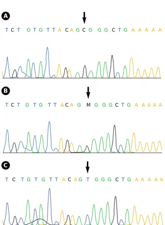
For mutation analysis, total RNA and genomic DNA were extracted from PBMC by conventional methods, and subjected to reverse transcriptase-polymerase chain reaction (RT-PCR), as described previously (11). We found a T-to-C transversion at nt 439, the first nucleotide of codon 140, by direct sequencing of the cDNA-PCR products (Fig. 5). This single base substitution resulted in a missense mutation from tryptophan to arginine (W140R). His mother and sister were heterozygotes in the mutation analysis, suggesting that they were obligate carriers (Fig. 5).
DISCUSSION
We performed a molecular characterization of a case of XHIM, and presented the clinical findings and family histo- ry. The patient showed typical serum immunoglobulin pro- files of XHIM, an elevated serum IgM with low IgG level.
IgA and IgE were in normal ranges. Although mutations in the CD40L gene explain the inability of XHIM patients to switch from IgM to IgG production both in vitro and in vivo, variability in the serum IgM profile in XHIM patients is often seen (10).
Our patient had chronic Cryptosporidium parvum enteritis, diagnosed by a quantitative centrifugation concentration flotation technique. Chronic diarrhea and severe hepatobiliary disease are common and often associated with a poor progno- sis in XHIM (2). In addition, Cryptosporidium infection is frequent and is associated with sclerosing cholangitis in some XHIM patients (2), as seen in patients with acquired immuno- deficiency syndrome (11). It is noteworthy that XHIM patients show inadequate T cell function and are often sus- ceptible to opportunistic infections typical of T cell deficien- cy such Pneumocystis carinii pneumonia and Cryptosporidium diarrhea (2). A recent study demonstrated that activated T cells from patients with XHIM produced markedly reduced levels of IFN- , and failed to induce antigen-presenting cells to synthesize IL-12 and TNF- , suggesting a basis for the increased susceptibility of XHIM patients to certain oppor- tunistic infections (3).
Cryptosporidial infection causing chronic diarrhea and possibly cholangiopathy or liver cirrhosis (12), malignancies (1, 12), and autoimmune disorders (1) are also known to occur in XHIM patients. To our knowledge, this is the first report on the isolation of C. parvum in a XHIM patient in Korea.
The patient in this case suffered from C. parvum enteritis in spite of the IVIG treatment. In agreement with our case, oth- ers have reported that some XHIM patients frequently suffer from infection with opportunistic pathogens, such as Cryp- tosporidium and P. carinii, despite IVIG treatment (2, 13).
In a previous study, we identified Cryptococcus in fine nee- dle aspiration smears of the cervical lymph node using May- Grunbald-Giemsa stain (14). The characteristic features of cryptococci were identified using several staining methods, including Gomori methenamine silver stain and periodic acid Schiff stain (14). In this case, we confirmed the cryptococ- cal infection with cultures, which are important for the char- acterization and diagnosis of cryptococcosis (15). The cervi- cal lymphadenopathy partially regressed three weeks after the initiation of antifungal medication. The patient also had chronic neutropenia, which might have predisposed the patient to cryptococcal infection, as has been reported in other chil- dren with XHIM (2, 16).
In the mutation analysis, we found a missense mutation at codon 140. Other patients with a mutation at codon 140 have been reported (4, 10, 17), confirming that codon 140
Fig. 5.Sequence analysis showing a missense mutation in the CD40L gene. A, Genomic sequence encompassing a point muta- tion (T to C) in exon 5 of the CD40L gene from the patient; B, mother (carrier); C, father (normal control).
T C T GT G T TA C AGC G G G C T G A A A A A
T C T G T G T T AC A G M G G G C TG A A A A A
T C T G T G T T AC AG T G G G C T G A A A A A A
B
C
represents a hotspot for XHIM mutations, although the mutations in XHIM are highly heterogenous. To date, about one hundred mutations in the CD40L gene have been described (7-9). The majority of mutations are missense mutations (7, 9), reading-frame terminations (7), or splicing defects that give rise to aberrant transcripts (5, 7, 9). In a recent report (9), activated PBMC from some patients had functional CD40L. These patients appeared to undergo milder clinical courses. Thus, nonfunctional CD40L is not an absolute diagnostic hallmark of XHIM, but molecular analysis of the CD40L gene may be required for the defini- tive diagnosis of XHIM (9). Using direct sequencing, we were able to confirm that the patient’s mother and sister had both normal and abnormal bands. Identification of muta- tions in the CD40L gene has allowed unambiguous assign- ment of carrier status and offers the possibility of prenatal diagnosis in this family.
In this paper, we described the clinical characteristics and molecular identification of an XHIM patient in Korea. It emphasizes the need for genetic evaluation of functional CD40L expression in male patients with a low serum IgG and normal or high IgM levels and with a normal propor- tion of circulating B cells.
REFERENCES
1. Notarangelo LD, Duse M, Ugazio AG. Immunodeficiency with hyper-IgM (HIM). Immunodefic Rev 1992; 3: 101-21.
2. Levy J, Espanol-Boren T, Thomas C, Fischer A, Tovo P, Bordigoni P, Resnick I, Fasth A, Baer M, Gomez L, Sanders EA, Tabone MD, Plantaz D, Etzioni A, Monafo V, Abinun M, Hammarstrom L, Abrabamsen T, Jones A, Finn A, Klemola T, DeVries E, Sanal O, Peitsch MC, Notarangelo LD. Clinical spectrum of X-linked hyper- IgM syndrome. J Pediatr 1997; 131: 47-54.
3. Jain A, Atkinson TP, Lipsky PE, Slater JE, Nelson DL, Strober W.
Defects of T-cell effector function and post-thymic maturation in X- linked hyper-IgM syndrome. J Clin Invest 1999; 103: 1151-8.
4. Korthauer U, Graf D, Mages HW, Briere F, Padayachee M, Malcolm S, Ugazio AG, Notarangelo LD, Levinsky RJ, Kroczek RA. Defec- tive expression of T-cell CD40 ligand causes X-linked immunodefi- ciency with hyper-IgM. Nature 1993; 361: 539-41.
5. DiSanto JP, Bonnefoy JY, Gauchat JF, Fischer A, de Saint Basile G. CD40 ligand mutations in X-linked immunodeficiency with hyper-IgM. Nature 1993; 361: 541-3.
6. Facchetti F, Appiani C, Salvi L, Levy J, Notarangelo LD. Immuno- histologic analysis of ineffective CD40-CD40 ligand interaction in lymphoid tissues from patients with X-linked immunodeficiency with hyper-IgM. Abortive germinal center cell reaction and severe deple- tion of follicular dendritic cells. J Immunol 1995; 154: 6624-33.
7. Notarangelo LD, Peitsch MC. CD40lbase: a database of CD40L gene mutations causing X-linked hyper-IgM syndrome. Immunol Today 1996; 17: 511-6.
8. Nonoyama S, Shimadzu M, Toru H, Seyama K, Nunoi H, Neubauer M, Yata J, Och HD. Mutations of the CD40 ligand gene in 13 Japanese patients with X-linked hyper-IgM syndrome. Hum Genet 1997; 99: 624-7.
9. Seyama K, Nonoyama S, Gangsaas I, Hollenbaugh D, Pabst HF, Aruffo A, Ochs HD. Mutations of the CD40 ligand gene and its effect on CD40 ligand expression in patients with X-linked hyper IgM syndrome. Blood 1998; 92: 2421-34.
10. Kroczek RA, Graf D, Brugnoni D, Giliani S, Korthuer U, Ugazio A, Senger G, Mages HW, Villa A, Notarangelo LD. Defective expression of CD40 ligand on T cells causes“X-linked immunodefi- ciency with hyper-IgM (HIGM1)”. Immunol Rev 1994; 138: 39-59.
11. Pol S, Romana CA, Richard S, Amouyal P, Deportes-Livage I, Carnot F, Pays JF, Berthelot P. Microsporidia infection in patients with the human immunodeficiency virus and unexplained cholangi- tis. N Engl J Med 1993; 328: 95-9.
12. Hayward AR, Levy J, Facchetti F, Notarangelo L, Ochs HD, Etzioni A, Bonnefoy JY, Cosyns M, Weinberg A. Cholangiopathy and tumors of the pancreas, liver, and biliary tree in boys with X-linked immunodeficiency with hyper-IgM. J Immunol 1997; 158: 977-83.
13. Cunningham CK, Bonville CA, Ochs HD, Seyama K, John PA, Rot- bart HA, Weiner LB. Enteroviral meningoencephalitis as a compli- cation of X-linked hyper IgM syndrome. J Pediatr 1999; 134: 584-8.
14. Lee MY, Chung JH, Shin JH, Hwang TJ, Kim KS, Lee JH, Nam JH, Lee MC, Park CS, Juhng SW, Choi C. Lymphonodular crypto- coccosis diagnosed by fine needle aspiration cytology in hyper-IgM syndrome: a case report. Acta Cytol 2001; 45: 241-4.
15. Das R, Dey P, Chakrabarti A, Ray P. Fine-needle aspiration biopsy in fungal infections. Diagn Cytopathol 1997; 16: 31-4.
16. Iseki M, Anzo M, Yamashita N, Matsuo N. Hyper-IgM immunode- ficiency with disseminated cryptococcosis. Acta Paediatr 1994; 83:
780-2.
17. Macchi P, Villa A, Strina D, Sacco MG, Morali F, Brugnoni D, Giliani S, Mantuano E, Fasth A, Andersson B, et al. Characterization of nine novel mutations in the CD40 ligand gene in patients with X- linked hyper IgM syndrome of various ancestry. Am J Hum Genet 1995; 56: 898-906.