www.ophthalmology.org 1032
대한안과학회지 2010년 제 51 권 제 7 호 J Korean Ophthalmol Soc 2010;51(7):1032-1035 pISSN: 0378-6471
eISSN: 2092-9374
DOI : 10.3341/jkos.2010.51.7.1032
= 증례보고 =
선천 상사근 결손 환아의 하사근 비측 전치술 치료 1예
박진환⋅차덕선⋅조윤애⋅서영우 고려대학교 의과대학 안과학교실
목적: 하사근 기능 항진을 동반한 단안 상사근 결손에 대해 하사근 비측 전치술을 시행하여 좋은 결과를 보였기에 보고하고자 한다.
증례요약: 우측으로의 머리 기울임과 좌안 상사시를 주소로 본원 내원한 2세 남아로 안과 검사상 제일안위에서 18 프리즘디옵터(PD)의 좌안 상사시, 우측 머리기울임시 정위, 좌측 머리기울임시 35PD의 좌안 상사시, 동향운동 검사상 좌안 상사근 기능 저하와 하사근 기능항진을 보였으며, 안와 전산화단층촬영(orbit computed topography (CT)) 상 좌측 상사근 결손 나타나 좌측 하사근 비측 전치술 을 시행하였다. 수술 3주 후 원거리, 근거리 교대프리즘 검사상 정위, Bielschowsky 머리기울임검사 음성, 정상 동향운동 소견 보였으 며, 우측으로의 머리기울임 및 복시는 관찰되지 않았다.
결론: 좌안에 단독으로 발생한 선천성 상사근 결손에 대해 좌안 비측 전치술을 통해 좌안 상사시, 우측 머리기울임을 치료하였기에 이를 보고하고자 하는 바이다.
<대한안과학회지 2010;51(7):1032-1035>
■ 접 수 일: 2009년 12월 14일 ■ 심사통과일: 2010년 5월 18일
■ 책 임 저 자: 서 영 우
서울 성북구 안암동 5가 126-1 고려대학교 안암병원 안과학교실 Tel: 02-920-5521, Fax: 02-924-6820 E-mail: [email protected]
* 본 논문의 요지는 2009년 대한안과학회 제102회 학술대회에서 포스터로 발표되었음.
상사근결손은임상적으로드물고선천적으로단독혹은 두개골안면골기형(craniofacial dystoses),1,2 다운증후군,3 무뇌아4등에서동반되어발생할수있다. Wallace and von Noorden5은 33예의 상사근 결손을 보고하였고, 국내에서 는2004년Cho and Kim6이1예를보고하였다. 이들은상 사근마비로진단된환자중수평사시, 약시, 제일안위에서 큰수직편위, 안구일치운동의확산(spread of comitance), 타안의 상사근 가성기능항진 등이 있으면 상사근 결손을 의심해야한다고 하였다.
상사근결손의경우일반적으로알려진하사근약화술 로는내전시 과상전, V-형사시를 교정하기 힘들고, 수 직근, 수평근의전치술을시행할경우안구회선정도를 악화시킬 수 있어 치료가 어렵다고 알려져 있다.8 이에 저자들은상사근결손에의한수직편위와하사근기능항 진을 관찰하고하사근 비측 전치술을통해 호전된 증례 를 경험하였고 이에 대한국내 보고가 없기에 보고하고 자 한다.
증례보고
2세남아가 생후 3개월부터 발생한 우측 머리기울임과 좌안의상사시를주소로내원하였다. 동반된안질환이나전 신질환은관찰되지않았고, 과거력상특이병력도없었다.
수술전양안나안시력모두0.8이었고우측머리기울임 (Fig. 1)은30°였다. 양안운동검사상좌안의하사근기능 항진이+3.5, 상사근기능저하가-3.5였고, 우안에는경도 의 하사근 기능항진, 상사근 기능저하가 의심되었다(Fig.
2). 교대프리즘검사상원거리18 프리즘디옵터(PD) 좌 안상사시, 근거리18PD 좌안상사시소견을보였으며, 머 리기울임검사(Fig. 3)상우측정위, 좌측35PD의좌안상 사시소견을보였다. 안저소견은정상이었으나좌안의시 신경과황반부의+3 외회선이있었다. 좌안상사근마비를 의심하였으나 동측 하사근의 과도한 기능항진 등 상사근 결손을배제할수없어안와전산화단층촬영을시행하였으 며, 좌안의상사근을 발견할수 없었다(Fig. 4).
수술시전신마취하시행한견인검사상상사근을인지할 수 없었다. 하사근을 하직근 부착부위의 내측으로부터 2 mm 비측, 2 mm 뒤쪽지점으로옮기는 전치술(Fig. 5)을 시행하였다.
수술후 3주째에교대가림검사상원거리, 근거리 모두 정위, 머리기울임검사(Fig. 3) 양측 정위소견 보였으며, 양안운동검사에서도하사근기능항진 없이정상적인안 구운동을보여주었고(Fig. 2), 우측으로의머리기울임도 나타나지않았다(Fig. 1). 안저소견상에서도좌안의시신
www.ophthalmology.org 1033 - 박진환 외 : 상사근 결손 환아의 하사근 비측전치술 -
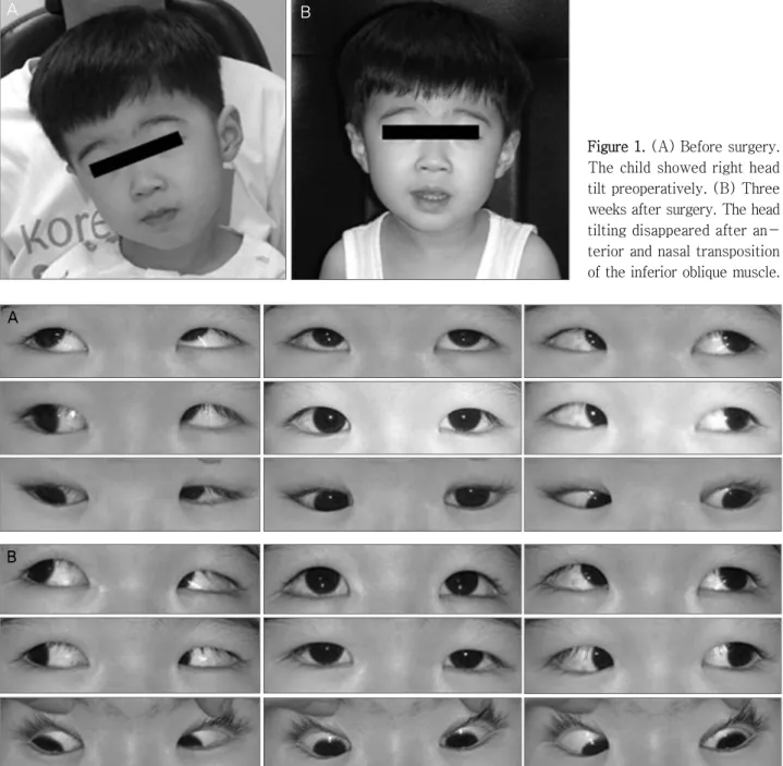
A B
Figure 1.(A) Before surgery.
The child showed right head tilt preoperatively. (B) Three weeks after surgery. The head tilting disappeared after an- terior and nasal transposition of the inferior oblique muscle.
A
B
Figure 2. (A) Before surgery. The patient shows severe superior oblique underaction and inferior oblique overaction of the left eye. (B) Three weeks after surgery. The dysfunction of both oblique muscles in the left eye improved significantly.
경, 황반부의외회선이+0.5로호전되었다. 동향운동검사 상 좌안의상전제한은 나타나지않았다.
고 찰
외안근의선천적결손은드물지만각외안근결손이모 두보고된바있다.6상사근의결손시에는결손된눈의상 사시, 내사시, 외회선이나타나고흔히임상적으로상사근마 비와유사한증상을보이게된다. Wallace and von Noorden5
은 소아에서심한수평사시, 제일안위에서심한수직편위, 동측하사근의기능항진, 타안상사근의가성기능항진, 오 래된 경우 비일치성 안근운동의 점진적인 일치화(spread of comitance), 약시등이있을때상사근결손을의심해야 한다고하였다.
Helveston et al9은수평사시, 약시, 안검하수증, 얼굴비 대칭등이있을때상사근결손을의심하고이를상사근마 비플러스(congenital superior oblique palsy plus)라하였 다. Mumma10는기능이항진된하사근약화술후에도상사
www.ophthalmology.org 1034
- 대한안과학회지 2010년 제 51 권 제 7 호 -
A B
Figure 3.(A) Preoperative forced head tilt test. The patient showshypertropia on left side head tilt. (B) Postoperative forced head tilt test. There is no vertical deviation on head tilt test.
Figure 5. The inferior oblique muscle is transposed anterior-nasally to the nasal border of the insertion of the inferior rectus muscle. A black arrow indicates the medial border of the inferior rectus muscle insertion, and a yellow arrowindicates the new insertion site of the inferior oblique muscle.
Figure 4. The superior oblique muscle of the left eye is not found on CT scan.
근기능저하와심한 머리기울임이계속될때상사근결손 을 의심하라고하였다. 본 연구의 증례에서는수평사시는 보이지않았으나심한수직편위와머리기울임, 상사근의심 한기능저하, 동측하사근의심한기능항진을보이고있어 상사근결손을의심해 술전안와전산화단층촬영을시행 하였고, 상사근 결손을확인할 수있었다.
상사근결손에서상사시의정도가심하고동측상직근의 운동이제한을받음에 따라경축이발생하여동측의하사 근약화술만으로는부족하고상직근약화술도시행하여야한 다는보고들이있다.5,6또한기존의하사근전치술은하사근 을내직근부착부의이측부근에부착시킴으로써, 하사근의 약화와 더불어하전 효과혹은 항-상전의효과를보이나 하사근이외회선근으로작용한다는점에는변함이없어상 사근결손 등심한상사근마비의 경우그효과가 부족할 수 있다. 저자들이 시행한하사근 비측 전치술은 2003년 Stager et al7이소개한것으로하사근을하직근부착부의 비측2 mm, 뒤쪽2 mm 지점에부착시키는방법으로그사 용 결과에대한보고가매우적다.7,8 하사근의비측전치술
의경우하사근의새로운부착부위가 하직근의내측이되 므로, 수술후하사근은전치술의효과인하사근약화와하 전및항상전효과를보일 뿐아니라신경섬유혈관다발이 기능적 기시부로 작용하는 내회선근으로서 역할도 할 수 있어, 상사근결손등에의한심한상사근마비에효과적일 것으로보인다.7,8
Helveston et al9에의하면완전한상사근건의결손이외 에얇아진건이뒤쪽테논에부착되거나, 상직근비측연에 약하게 부착되어 있는 경우도 있어 안와 전산화단층촬영 상상사근이보이지않는경우감별이필요할수있다. 그 러나 하사근비측전치술은상사근의해부학적결손이외에 매우심한상사근마비시에도좋은수술결과를보인다고 보고된바,7수술중탐색(exploration)을통한상사근결 손의확인에따라수술방법을바꿀필요가없다고판단하여, 본연구의증례에서는수술중탐색은 시행하지않았다.
본연구의증례에서는심한제일안위의상사시, 하사근 기능항진, 외회선등의소견을보인2세의환아로하사근비 측전치술을시행하여좋은결과를얻을수있었다. 하사근
www.ophthalmology.org 1035
=ABSTRACT=
A Case of Congenital Absence of the Superior Oblique Muscle Treated With Anterior and Nasal Transposition of the Inferior Oblique Muscle
Jin-hwan Park, MD, Deoksun Cha, MD, Yoonae A. Cho, MD, PhD, Young-Woo Suh, MD, PhD
Department of Ophthalmology, Korea University College of Medicine, Seoul, Korea
Purpose: To report a patient with absence of the superior oblique (SO) muscle of the left eye, who showed improvement after anterior and nasal transposition of the inferior oblique muscle for left hyperdeviation and right head tilt.
Case summary: A two-year-old boy presented with hypertropia of the left eye and right head tilt. Alternate prism-cover test in the primary position demonstrated 18 prism diopters (PD) of left hypertropia, which increased to 35 PD in the left head tilt position. A version test demonstrated overaction of the left inferior oblique muscle and underaction of the left superior oblique muscle. As an orbit CT scan showed absence of the SO muscle, the patient was diagnosed with congenital absence of SO and left anterior and nasal transposition of the inferior oblique muscle was performed. Three weeks after surgery, the patient pre- sented with orthotropia at distant and near. The version test revealed normal oblique muscles. There was no vertical deviation shown on the Bielschowsky head tilt test. The abnormal head posturing was no longer observed.
Conclusions: The authorsreport a patient manifesting abnormal head posture and hypertropia, diagnosed with absence of SO muscle, which was successfully corrected using anterior and nasal transposition of the inferior oblique muscle.
J Korean Ophthalmol Soc 2010;51(7):1032-1035
Key Words: Absence of superior oblique muscle, Anterior and nasal transposition of inferior oblique muscle
Address reprint requests to Young-Woo Suh, MD, PhD Department of Ophthalmology, Korea University Anam Hospital
#126-1 Anamdong 5-ga, Sungbuk-gu, Seoul 136-701, Korea
Tel: 82-2-920-5521, Fax: 82-2-924-6820, E-mail: [email protected]
- 박진환 외 : 상사근 결손 환아의 하사근 비측전치술 -
비측전치술도 하사근전치술과유사하게 항상전증후군, 제 일안위에서의하사시가생길수있지만본증례에서는항 상전증후근은나타나지않았다. 보다많은증례를통해상 사근결손환아에서의하사근비측전치술의효과와적응증, 합병증에대한 장기적인조사가 필요하겠다.
참고문헌
1) Pinchoff BS, Sandall G. Congenital absence of the superior obli- que tendon in craniofacial dysostosis. Ophthalmic Surg 1985;16:375-7.
2) Pollard ZF. Bilateral superior oblique muscle palsy associated with Apert’s syndrome. Am J Ophthalmol 1988;106:337-40.
3) Lo CY, Nakamura K. Congenital absence of the superior oblique tendon in Down’s syndrome. Jpn Rev Clin Ophthalmol 1987;
81:1312-3.
4) Barnes J, Boniuk M. Anencephaly with absence of the superior
oblique tendon. Surv Ophthalmol 1972;16:371-4.
5) Wallace DK, von Noorden GK. Clinical characteristics and surgi- cal management of congenital absence of the superior oblique tendon. Am J Ophthalmol 1994;118:63-9.
6) Cho YA, Kim S. A case of congenital absence of the superior obli- que muscle. J Korean Ophthalmol Soc 2004;45:631-5.
7) Stager DR Jr, Beauchamp GR, Wright WW, et al. Anterior and nasal transposition of the inferior oblique muscles. J AAPOS 2003;7:167-73.
8) Hussein MA, Stager DR Jr, Beauchamp GR, et al. Anterior and nasal transposition of the inferior oblique muscles in patients with missing superior oblique tendons. J AAPOS 2007;11:29-33.
9) Helveston EM, Krach D, Plager DA, Ellis FD. A new classi- fication of superior oblique palsy based on congenital variations in the tendon. Ophthalmology 1992;99:1609-15.
10) Mumma JV. Surgical procedure for congenital absence of the su- perior oblique. Arch Ophthalmol 1974;92:221-3.