J o u r n a l o f R h e u m a t i c D i s e a s e s V o l . 2 0 , N o . 1 , F e b r u a r y 2 0 1 3
http://dx.doi.org/10.4078/jrd.2013.20.1.56 □ Case Report □
56
<Received:December 1, 2011, Revised (1st: February 10, 2012, 2nd: March 31, 2012), Accepted:April 11, 2012>
Corresponding to:Jisoo Lee, Division of Rheumatology, Department of Internal Medicine, Ewha Womans University School of Medicine, 1071, Anyangcheon-gil, Seoul 158-710, Korea. E-mail:[email protected]
pISSN: 2093-940X, eISSN: 2233-4718
Copyright ⓒ 2013 by The Korean College of Rheumatology
This is a Free Access article, which permits unrestricted non-commerical use, distribution, and reproduction in any medium, provided the original work is properly cited.
Myoclonus as an Anticipatory Symptom of Diffuse Central Nervous System Involvement in Neuropsychiatric Systemic Lupus Erythematosus
Min Jin Lee, Koeun Lee, Jisoo Lee
Division of Rheumatology, Department of Internal Medicine, Ewha Womans University School of Medicine, Seoul, Korea
The clinical manifestations of nervous system involvement in systemic lupus erythematosus (SLE) are highly diverse and their pathogenic mechanisms are incompletely understood. Neuropsychiatric SLE (NPSLE) poses difficulty in making the proper diagnosis, especially in circumstances where its initial symptoms are diffuse neuropsychiatric symptoms. We describe a 43-year old woman who exhibited
a myoclonic jerk of the abdominal wall, followed shortly by acute confusion, which was attributed to SLE. Therapy with high dose corticosteroids completely reversed the symptoms.
Myoclonus can be an anticipatory symptom of diffuse neu- rologic dysfunction in patients with NPSLE.
Key Words. NPSLE, Diffuse neuropsychiatric symptoms, Myoclonus
Introduction
Neuropychiatric systemic lupus erythematosus (NPSLE) still remains a diagnostic challenge. There is no specific test to confirm the diagnosis. Currently recommended diagnostic work-up for NPSLE is performed to exclude other diagnoses such as infection, and metabolic derangements (1). Symptoms or signs that can predict the development of NPSLE can be useful in clinical settings. We describe a patient who devel- oped sudden emergence of myoclonic movements, followed shortly by acute confusion as initial symptoms of SLE.
Case Report
A 43-year old woman presented with fever of 1 month duration. Swelling of cervical lymph nodes developed 1 week prior to admission. Recently, she felt gloomy, and experienced frequent mood changes. Her past history was unremarkable, except intermittent arthralgia and myalgia for 3 years. The pa- tient was a house wife. She did not smoke nor drink. She was not on any medication. On examination, body temperature of 37.4℃, malar rash, and cervical lymphadenopathy was found.
Aside from mild irritability, neurologic examination was
unremarkable. The remainder of the examination was normal.
Laboratory tests revealed Hb 14.6 g/dL, WBC 1,700/mm3 (neutrophil 45.2%, lymphocyte 28.6%), and platelet 109,000/mm3. Biochemical tests and urinalysis were within normal limits. C3 was decreased to 74.4 mg/dL. Lupus anti- coagulant was positive with 51.7 secs initial and 52.8 secs at follow up (normal 31.0∼44.0 secs). Antinuclear antibody (ANA) was present at a titer of 1:80 (speckled type).
Anti-Sm, anti-RNP, anti-DNA, anti-SSA, anti-SSB, anti-car- diolipin antibodies were negative. Chest PA was normal with no active lung lesion. Pathologic findings of cervical lymph node showed atypical lymphoid hyperplasia without malignant cells.
On the second hospital day, the patient complained of severe abdominal pain accompanied by muscle spasm. Fever of 38.5oC was accompanied. Physical findings revealed severe in- voluntary myoclonic jerks of abdominal wall at approximately 10 seconds interval. Cultures of blood and urine, and tests for antibodies to hepatitis B and C viruses, Epstein-Barr virus, and herpes simplex virus were negative. On cerebrospinal fluid (CSF) study, WBC 320/μL (neutrophil 1%, lymphocyte 97%),
Myoclonus in NPSLE 57
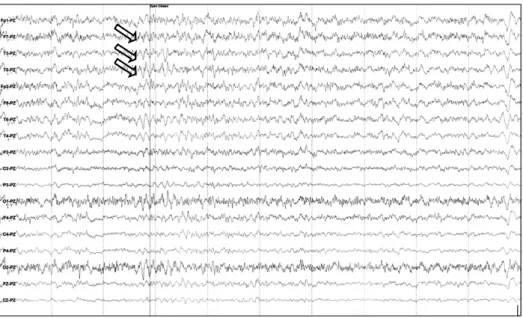
Figure 1. Electroencephalogram was characterized excessive beta wave (arrows) at the anterior and temporal zone.
total protein 26 mg/dL, and glucose 60 mg were detected. CSF was negative for cryptococcal antigen, herpes simplex virus, enterococcus, and Mycobacterium tuberculosis. Electroence- phalogram showed excessive beta waves (Figure 1). Brain and spine MRI showed nonspecific high signal intensity in both subcortical white matters of the front lobes without abnormal- ities in the spine. Two days after, she developed acute confusion. Based on malar rash, hematologic disorder, im- munologic disorder, and ANA positivity, diagnosis of SLE was made. Because of no evidence of infection was found, acute confusional state was assessed as a symptom of NPSLE.
SLEDAI score was assessed as 14. 125 mg/day of methyl- prednisolone was administered promptly. Clonazepam 0.5 mg/day was administered to control the myoclonus. In 3 days, confusion abated and she recovered clear consciousness with full orientation. However, myoclonus continued for 10 days al- though its severity was lessened. At the time of discharge, the patient was fully conscious, oriented, without any neurologic deficit. Six months later, the patient was doing well without any symptoms of SLE or NPSLE.
Discussion
NPSLE should be considered as a possible differential diag- nosis for rapid onset neuropsychiatric symptoms even in pre- viously healthy individuals. Before making the diagnosis of NPSLE, secondary causes of neuropsychiatric symptoms such as infections, metabolic or endocrine disturbances should be vigorously excluded. However, timely diagnosis of NPSLE is important, since treatment delay is associated with poor prog- nosis (1). The patient we described presented with rapidly evolving neuropsychiatric symptoms of myoclonus and acute
confusional state. Although she had symptoms and signs sug- gestive of active SLE, the diagnosis of NPSLE was difficult, since she was not previously diagnosed with SLE. CSF exami- nation and MRI findings were non-specific, but no definite evidence for CNS infection such as herpes viral infection or tuberculosis was found. Treatment with high dose cortico- steroids promptly reversed the symptoms without neurologic damage.
Myoclonus is a movement disorder characterized by sudden, brief, involuntary muscle jerks. Myoclonus most often occurs in context of variety of neurological and nonneurological dis- orders including autoimmune diseases such as SLE (2).
However, movement disorders other than chorea have been less well characterized in SLE (3). Only one case of my- oclonus as a symptom of NPSLE has been described pre- viously (4). Recent advances in understanding of immune mediated mechanisms suggest a common pathogenetic mecha- nisms mediated by autoantibodies. Anti-glutamate-receptor an- tibodies was suggested to play an important role in movement disorders as well as NPSLE (5). N-Methyl-D-aspartate re- ceptors (NMDARs) are receptors for the neurotransmitter glu- tamate, the major excitatory neurotransmitter of the brain crit- ically important for many brain functions. Antibodies to NMDAR modulate NMDAR activation by synergizing with the natural agonist glutamate to increase excitatory post- synaptic potentials, causing overexcitability of the neurons (6).
Alterations in NMDAR activity can produce uncontrolled movement, seizures, coma, and hallucinations (7). In SLE, presence of anti-NMDAR antibodies in the CSF were shown to correlate with acute, diffuse central nervous system (CNS) manifestations of NPSLE that include seizure disorders, acute
58 Min Jin Lee et al.
confusional state, and psychosis (8). The patient we described developed acute confusional state shortly after the develop- ment of myoclonus. Close clustering of two NPSLE symp- toms of myoclonus and acute confusion may implicate com- mon pathogenic mechanisms.
Summary
Although not included in the ACR case definition of NPSLE, myoclonus can occur as a part of diffuse CNS involvement and may be associated with diffuse neurologic dysfunction of NPSLE.
References
1. Bertsias GK, Ioannidis JP, Aringer M, Bollen E, Bombardieri S, Bruce IN, et al. EULAR recom- mendations for the management of systemic lupus eryth- ematosus with neuropsychiatric manifestations: report of a task force of the EULAR standing committee for clin- ical affairs. Ann Rheum Dis 2010;69:2074-82.
2. Kojovic M, Cordivari C, Bhatia K. Myoclonic disorders:
a practical approach for diagnosis and treatment. Ther Adv Neurol Disord 2011;4:47-62.
3. The American College of Rheumatology nomenclature and case definitions for neuropsychiatric lupus synd- romes. Arthritis Rheum 1999;42:599-608.
4. Joseph FG, Lammie GA, Scolding NJ. CNS lupus: a study of 41 patients. Neurology 2007;69:644-54.
5. Panzer J, Dalmau J. Movement disorders in paraneo- plastic and autoimmune disease. Curr Opin Neurol 2011;24:346-53.
6. DeGiorgio LA, Konstantinov KN, Lee SC, Hardin JA, Volpe BT, Diamond B. A subset of lupus anti-DNA anti- bodies cross-reacts with the NR2 glutamate receptor in systemic lupus erythematosus. Nat Med 2001;7:1189-93.
7. Chen HS, Lipton SA. The chemical biology of clinically tolerated NMDA receptor antagonists. J Neurochem 2006;97:1611-26.
8. Arinuma Y, Yanagida T, Hirohata S. Association of cere- brospinal fluidanti-NR2 glutamate receptor antibodies with diffuse neuropsychiatric systemic lupus erythema- tosus. Arthritis Rheum 2008;58:1130-5.