www.krspine.org
Cervical Intervertebral Disc Calcification in Children - A Case Report-
Dong-Eun Shin, M.D., Chang-Soo Ahn, M.D., Yong-Suk Cho, M.D.
J Korean Soc Spine Surg 2011 Dec;18(4):254-258.
Originally published online December 31, 2011;
http://dx.doi.org/10.4184/jkss.2011.18.4.254
Korean Society of Spine Surgery
Department of Orthopedic Surgery, Inha University School of Medicine
#7-206, 3rd ST. Sinheung-Dong, Jung-Gu, Incheon, 400-711, Korea Tel: 82-32-890-3044 Fax: 82-32-890-3467
©Copyright 2011 Korean Society of Spine Surgery pISSN 2093-4378 eISSN 2093-4386
The online version of this article, along with updated information and services, is located on the World Wide Web at:
http://www.krspine.org/DOIx.php?id=10.4184/jkss.2011.18.4.254
This is an Open Access article distributed under the terms of the Creative Commons Attribution Non-Commercial License (http://
creativecommons.org/licenses/by-nc/3.0) which permits unrestricted non-commercial use, distribution, and reproduction in any medium, provided the original work is properly cited.
Journal of Korean Society of
Spine Surgery
J Korean Soc Spine Surg. 2011 Dec;18(4):254-258.
http://dx.doi.org/10.4184/jkss.2011.18.4.254
Case Report
pISSN 2093-4378 eISSN 2093-4386Cervical Intervertebral Disc Calcification in Children - A Case Report-
Dong-Eun Shin, M.D., Chang-Soo Ahn, M.D.
*, Yong-Suk Cho, M.D.
Department of Orthopaedic Surgery, CHA Bundang Medical Center, CHA University, Korea Department of Orthopaedic Surgery, CHA Gumi Medical Center, CHA Hospital, Korea*
Study Design: A case report.
Objectives: This case report presents a child who was treated conservatively after having being diagnosed with cervical intervertebral disc calcification.
Summary of Literature Review: Cervical intervertebral disc calcification is considered as a degenerative change of spine. It is common in adults and in most cases, no symptoms are observed. In children, by contrast, it is a rare condition and frequently accompanies symptoms such as severe neck pain and dysphagia.
Materials and Methods: A 7-year-old male patient who suffered from neck pain and torticollis without trauma had been diagnosed with cervical intervertebral disc calcification and was treated conservatively. He was discharged after symptom relief, and has been followed up and observed in our outpatient department.
Results: The improvements of symptom and radiographic findings were found in the month follow up.
Conclusions: Cervical intervertebral disc calcification shows similar symptoms to laryngopharyngeal abscess, traumatic injury and infective spondylitis, but through careful physical examination and radiologic evaluation, differential diagnosis is possible. After diagnosis, conservative treatment alone is sufficient. Antibiotic usage and surgical treatment are avoidable.
Key Words: Children, Cervical spine, Intervertebral disc, Calcification
Received: September 3, 2011 Revised: November 28, 2011 Accepted: December 1, 2011 Published Online: December 31, 2011 Corresponding author: Chang-Soo Ahn, M.D.
Department of Orthopaedic Surgery, CHA Gumi Medical Center, CHA University, 855 Hyungkok-dong, Gumi-si, Gyongsangbuk-do 730-040, Korea TEL: 82-31-780-5289, FAX: 82-31-708-3578
E-mail: [email protected]
“This is an Open Access article distributed under the terms of the Creative Commons Attribution Non-Commercial License (http://
creativecommons.org/licenses/by-nc/3.0/) which permits unrestricted non-commercial use, distribution, and reproduction in any medium, provided the original work is properly cited.”
서 론
소아에서의 추간판 석회화증은 1924년 처음 보고된 이래 현 재까지 약 200 례 정도 보고된 드문 질환이며, 퇴행성 변환의 일 환으로 나타나는 성인에서의 추간판 석회화증과는 다른 원인 과 경과를 취하는 것으로 알려져 있다. 대부분 상부 경추에 호발 하며, 경추부 통증, 사경 및 연하곤란과 같은 비특이적인 증상을 나타내기에 농양이나 감염성 척수염, 그리고 외상성 손상과 혼 동될 수 있어 항생제의 남용 및 수술적 치료 등의 과도한 치료가 시행될 가능성이 있어 정확한 문진, 이학적 검사 및 영상학적 검 사를 통한 진단과 안정 및 보존적 치료를 통하여 호전된 증례를 보고하고자 한다.
증례보고
환자는 7세된 여아로 내원 2주일전부터 갑자기 발생한 좌 경부 동통 및 운동장해를 주소로 본원 외래로 내원하였다. 과 거력상 특이 할 만한 병력이나 외상은 발견할 수 없었고, 이학 적 검사상, 경추부 통증 및 경도의 사경과 함께 목의 신전 시
심해지는 통증으로 경추부 운동이 제한되는 소견이 관찰되었 다. 그 이외의 견갑부 및 상지로 방사되는 신경학적 이상은 동 반되지 않았다. 혈액 검사 및 기타 임상 병리 검사상 백혈구가 13830/mm2 (정상치 4000~10000/mm2), 단핵구 72% (정상치 43~73%), ESR 33mm/hr (정상치 1~20mm/hr), CRP 5.16mg/
dL (정상치 0~0.3mg/dL)로 상승된 것 이외의 다른 특이 소견은 보이지 않았다.
Cervical Intervertebral Disc Calcification Journal of Korean Society of Spine Surgery
www.krspine.org
255
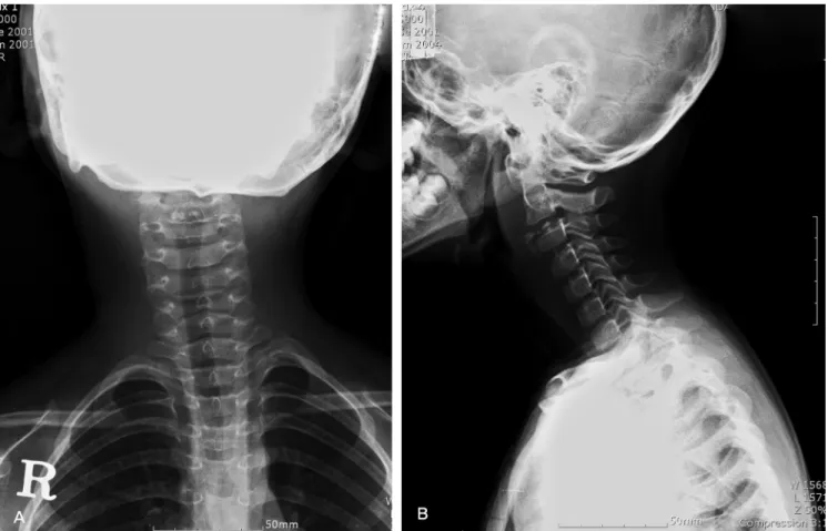
Fig. 1. Initial X-ray. Intervertebral disc calcification at C2-3 with right anterior hernication with inferior migration and associated retropharyngeal widening (A) AP view (B) Lateral viewFig. 2. Initial MRI. Acute symptomatic intervertebral disc calcification at C2-3 with right sided prevertebral hermiation of the calcification migrating inferiorly (A) T2WI Axial view (B) T1WI Sagittal view
Dong-Eun Shin et al Volume 18 • Number 4 • December 2011
단순 방사선 소견상(Fig. 1) 제 2, 3 경추간판이 전하방으로 탈 출되어 있는 명확한 음영증가 소견이 관찰되었다. 입원 시 37.2 도의 미열과 혈액학 검사상에서의 미약한 염증 소견을 보여 추 간판 농양의 감별 위해 초기에 시행한 자기공명영상 소견 상 (Fig. 2) 농양 등의 감염성 질환 소견은 보이지 않았으며, T1 및 T2 강조영상 모두에서 저신호 강도를 보이는 석회화 음영을 관 찰할수 있었다. 또한 척추강내 수핵탈출 및 척수압박소견도 보 이지 않아 보존적 치료로 침상 안정 및 NSAID(Ibuprofen 20mg/
kg/day) 사용만을 유지하며 증상을 관찰하기로 했다.
내원 3일째 경부의 통증이 완화되었으며, 혈액학적 소견 또 한 ESR 16 mm/hr (정상치 1~20mm/hr), CRP 0.18 mg/dL (정 상치 0~0.3mg/dL)로 감소하여 석회화에 대한 정밀 관찰 위하여 시행한 전산화 단층 촬영상(Fig. 3) 제 2, 3번 경추간판의 우측에 전하방으로 탈출되는 모양의 다발성 석회화 음영이 관찰되었다.
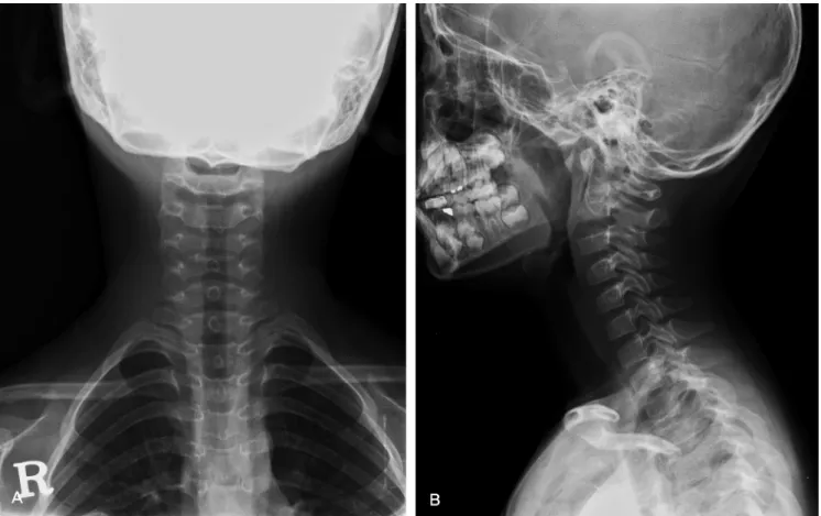
보존적 치료 후 통증, 경부의 운동 제한, 및 사경 등의 모든 증 상이 사라져 입원 5일째 퇴원하였으며 2개월 추시관찰 결과 증 상의 재발 및 이학적 검사상의 이상은 발견되지 않았으며 단순 방사선 소견상(Fig. 4.) 석회화 음영이 감소되었음을 관찰할 수 있었다.
고 찰
소아에서 특별한 외상력 없이 생긴 갑작스런 경부 통증과 운 동제한이 있는 경우, 가장 먼저 염증성 질환 및 종양에 대하여 의심해보아야 하며, 세균성 추간판염, 골수염, 농양, 및 척추염 등의 염증성 질환과 유골성 골종 및 골모세포종 등의 종양성 질 환의 감별 진단을 위해 방사선학적 검사 와 혈액검사를 시행해 야 한다. 1)
추간판 석회화증은 퇴행성 변화의 일환으로 증상 없이 성인 에 있어서 흔하게 나타나는 반면 소아에서 나타나는 추간판의 석회화증은 비교적 드물어 지금까지 대략 200례 정도가 보고 되 어있다. 신생아에서부터 청소년 시기까지 모두 나타날 수 있지 만 6~10세에 가장 흔하게 나타난다고 알려져 있으며,2,3) 발병 원 인에 대해서는 이전에 있었던 외상과 본 례에서와 같이 혈역학 적 이상 소견을 들어 염증성 질환이 제기 되었지만 뚜렷한 연관 성을 찾아내지 못하였다.4) 그 이외에도 연골 석회화증이나 부갑 상선 항진증 및 비타민 D 과잉과 같은 대사성 질환과의 연관성 에 대해서도 연구 되었으나 밝혀내지 못하였다.5) Swischuk 등은 외상이나 감염, 혈관염 등의 여러 요인에 의해 유발된 이차적인 추간판내 수핵으로의 혈액 공급 장애가 일시적인 석회화를 발생 Fig. 3. Spinal CT. Acute symptomatic intervertebral disccalcification at C2-3 with right sided prevertebral hermiation of the calcification migrating inferiorly (A) Axial view (B) Sagittal view
Cervical Intervertebral Disc Calcification Journal of Korean Society of Spine Surgery
www.krspine.org
257
시키고 혈액공급의 재개에 의해서 소실된다고 주장하였다.6)주 증상은 경추부 운동제한이나 사경 등을 동반한 경부 동통 이며 드물게는 경부의 상지통을 나타내기도 한다. 경우에 따라 서는 본 례에서와 같이 ESR/CRP 및 백혈구 등의 염증을 나타내 는 지표의 증가와 함께 미열을 동반하는 경우가 흔하게 보고되 나7) 신경관의 침범에 의한 방사통, 척추증 등의 신경학적 증상 을 나타내는 경우는 거의 없다.8) Dai 등은 증상이 평균 한달 정 도 지속되며 단순 방사선상 석회화는 5개월 가량 지속된다고 보 고했다.4) 또한 Sonnabend 등은 90% 정도에서 방사선상 석회화 소견이 없어지나 약 8% 가량에서는 석회화 음영이 소실되지 않 았고 다발성으로 나타난 군에서는 각각의 추간판 석회화 음영이 서로 다른 속도로 소실됨을 보고하였다.7) 성인과는 달리 소아에 있어서는 경추에 가장 호발하며 증상 또한 흉추나 요추에 발병 하는 경우에 비해 경우에 발병하는 경우에 비해 급성 증상을 동 반하는 경우가 많다.5,9)
척추 단순 방사선 소견상 대부분 수핵 안에 석회화가 관찰되 며, 척추체의 형태 변화가 동반되나 이러한 방사선학적 소견은 증상의 정도와는 관련이 없고,4,10) 약 30%의 환아에서 추간판 돌 출(Protrusion)이 관찰되나 이 또한 증상과는 관련이 없었다고 보고하였다.7)
결 론
소아에 있어서 경추 추간판 석회화증은 매우 드문 질환으로 써 이환된 환아들은 흔히 사경 및 운동 제한을 동반한 갑작스런 경부 통증 호소한다. 이러한 증상들은 보존적 치료 만으로 보통 수주 안에 회복되며 또한 좋은 예후를 보인다. 다만 감염성 질환 및 종양에 대한 감별을 위한 최소한의 검사는 필요할 것으로 사 료된다.
REFERENCES
1. Herring JA, Hensinger RN. Cervical disc calcification. J Pediatr Orthop. 1988:8:613-6.
2. Gerlach R, Zimmermann M, Kellermann S, Lietz R, Raabe A, Seifert V. Intervertebral disc calcification in childhood—
a case report and review of the literature. Acta Neurochir (Wien). 2001:143:89-93.
3. Calderone M, Severino M, Pluchinotta FR, Zangardi T, Martini G. Idiopathic intervertebral disc calcification in childhood. Arch Dis Child. 2009:94:233-4.
Fig. 4. F/U X-ray after 2 months. Detcreased intervertebral disc calcification at C2-3 (A) AP view (B) Lateral view
Dong-Eun Shin et al Volume 18 • Number 4 • December 2011
4. Dai LY, Ye H, Qian QR. The natural history of cervical disc calcification in children. J Bone Joint Surg Am.
2004:86:1467-72.
5. Girodias JB, Azouz EM, Marton D. Intervertebral disk space calcification. A report of 51 children with a review of the literature. Pediatr Radiol. 1991:21:541-6.
6. Swischuk LE, Jubang M, Jadhav SP. Calcific discitis in children: vertebral body involvement (possible insight into etiology). Emerg Radiol. 2008:15:427-30.
7. Sonnabend DH, Taylor TK, Chapman GK. Intervertebral
disc calcification syndromes in children. J Bone Joint Surg Br. 1982:64:25-31.
8. Bagatur AE, Zorer G, Centel T. Natural history of paediatric intervertebral disc calcification. Arch Orthop Trauma Surg.
2001:121:601-3.
9. Silverman FN. Calcification of the intervertebral disks in childhood. Radiology. 1954:62:801-16.
10. Dhammi IK, Arora A, Monga J. Calcified thoracic intervertebral disc at two levels as a cause of mid-back pain in a child: a case report. J Orthop Sci. 2002:7:587-9.
소아에서의 경추 추간판 석회화증 -증례보고-
신동은 • 안창수* • 조용석
차의과학대학교 분당차병원 정형외과학교실, 차의과학대학교 구미차병원 정형외과학교실*
연구 계획: 증례보고
목적: 본 연구의 목적은 소아의 경추 추간판에 생긴 석회화증에 대해 보존적인 치료로 완치된 증례를 보고하고자 함이다.
선행문헌의 요약: 경추 추간판의 석회화증은 척추의 퇴행성 변화의 일종으로 성인에게 있어서는 흔하게 볼 수 있으며, 대부분 특별한 증상을 나타내지 않는다. 반면 소아에 있어서는 매우 드물게 나타나며, 심한 경추부 통증과 연하곤란 등의 증상을 동반하는 경우가 흔하다.
대상 및 방법: 특별한 외상력 없이 경부 통증과 사경을 주소로 외래로 내원한 7세 남아는 경추 추간판 석회화증을 진단받고 보존적인 치료 후 증상 호전 되어 퇴원하였으며 이후 2개월 후 추시관찰 하였다.
결과: 본 환자는 정확한 이학적 검사 및 자기공명영상에 의해 경추 추간판 석회화증으로 진단 후 보존적일 치료만을 시행 증상 호전되어 5일째 퇴원 하 였으며, 2개월 추시 관찰상 증상 및 영상의학적 소견이 호전되는 양상을 보였다.
결론: 경추 추간판 석회화증은 인후두 농양이나 외상성 손상 또는 감염성 척추염과 감별해 하지만, 진단 후에는 보존적 치료만으로 효과적이다.
색인 단어: 소아, 경추, 추간판, 석회화 약칭 제목: 경추 추간판 석회화증