© 2012 Korean Breast Cancer Society. All rights reserved. http://ejbc.kr | pISSN 1738-6756
INTRODUCTION
Traditionally, breast cancer was classified by morphology, and hormonal receptors were thought to be an important prognostic factor. However, among breast cancer patients with homogenous morphology and hormonal receptor, it was known that clinical prognosis can be heterogeneous [1]. The prognosis of breast cancer has been known to be associated with tumor size, nodal status, hormonal receptor status, histo- logic grade, nuclear grade, human epidermal growth factor receptor 2 (HER2) expression, Ki-67 expression, etc. [2]. How- ever, the numerous studies for gene molecular profiles of breast cancer replaced the traditional prognostic factors these days.
The molecular difference was researched by the gene micro- array of breast cancer, and different gene expression profiles
were found, which were classified into basal-like type, HER2 type, normal breast-like type and luminal-type. Luminal-type breast cancer was characterized by profiles of estrogen receptor (ER) positive or progesterone receptor (PR) positive; it showed better prognosis than other subtypes, such as basal-like type and HER2 type. Recently, luminal-type breast cancer was classified into types A and B by Ki-67 or HER2 expression [3].
Also, in recent studies, luminal-type A breast cancer has shown better prognosis than luminal-type B [4]. In this study, we analyzed luminal-type breast cancer in order to find out the clinicopathological factors affecting prognosis in luminal-type breast cancer.
METHODS
We reviewed the clinical data of breast cancer patients who underwent surgery from February 2005 to November 2007. A total of 159 patients with profiles of ER positive or PR positive were classified as having luminal-type breast cancer. They were divided into two subtypes A and B by using a cutoff of 14%
for Ki-67. The cutoff value of Ki-67 was determined such that it was an optimal value relevant to disease-free survival (DFS) of luminal-type breast cancer in other previous studies [5-7].
Analysis of the Potent Prognostic Factors in Luminal-Type Breast Cancer
Han-Sung Kim, Inseok Park, Hyun Jin Cho, Geumhee Gwak, Keunho Yang, Byung Noe Bae, Ki Whan Kim, Sehwan Han, Hong-Joo Kim, Young-Duck Kim
Department of Surgery, Inje University Sanggye Paik Hospital, Inje University College of Medicine, Seoul, Korea ORIGINAL ARTICLE
Purpose: Luminal-type breast cancer has a good prognosis compared to other types, such as human epidermal growth factor receptor 2 and triple negative types. Luminal-type breast cancer is classified into luminal A and B, according to the prolif- eration index. We investigated the clinicopathological factors that affect the prognosis of the luminal-type subgroups. Methods:
We reviewed the medical records and the pathologic reports of 159 luminal-type breast cancer patients who were treated be- tween February 2005 and November 2007. We divided luminal- type breast cancer into luminal A and B, according to Ki-67 (cut- off value, 14%) and analyzed the clinicopathologic factors, such as age at diagnosis, intensity score of estrogen receptor and progesterone receptor, histologic grade, and Bcl-2. Moreover,
we compared the disease-free survival (DFS) of each group.
Results: In the univariate analysis, age (p=0.004), tumor size (p=
0.010), lymph node metastasis (p=0.001), and Bcl-2 (p=0.002) were statistically significant factors in luminal-type breast cancer.
In the multivariate analysis, lymph node (p=0.049) and Bcl-2 (p=
0.034) were significant relevant factors in luminal-type breast cancer. In the subgroup analysis, the increased Bcl-2 (cutoff value, 33%) was related with a longer DFS in the luminal B group (p=
0.004). Conclusion: In our study, luminal A breast cancer showed a longer DFS than luminal B breast cancer, further, Bcl-2 may be a potent prognostic factor in luminal-type breast cancer.
Key Words: Bcl-2, Breast neoplasms, Prognosis
Correspondence to: Geumhee Gwak
Department of Surgery, Inje University Sanggye Paik Hospital, Inje University College of Medicine, 1342 Dongil-ro, Nowon-gu, Seoul 139-707, Korea Tel: +82-2-950-1017, Fax: +82-2-933-9389
E-mail: [email protected]
This research was supported by the Inje University research grants in 2012.
Received: September 3, 2012 Accepted: November 15, 2012
Cancer
We analyzed the clinicopathological factors affecting the prognosis of both groups. Approval of the present study was obtained from the Institutional Review Board (SP IRB 12-076).
Clinicopathologic parameters
The clinicopathological factors contained age at diagnosis, tumor size, lymph node metastasis, histologic grade, ER, PR, Bcl-2, adjuvant chemotherapy, radiotherapy, and endocrine therapy. Age at diagnosis was set at 35 years and tumor size was set at 2 cm. Lymph node metastasis was classified by being either positive or negative. Histologic grade was classified into stages I, II, and III by the Modified Bloom and Richard- son’s histologic grade. ER and PR were classified by the inten- sity (0, 1+, 2+, 3+) and proportion (%), and Bcl-2 was classified by the cutoff value of 33% expression. Prognosis of patients was determined by the DFS. According to these standards, we analyzed the differences of DFS and the associated clinico- pathological factors between subtypes A and B.
Immunohistochemical staining (IHC) for ER, PR, Bcl-2, and Ki-67 Liquid mouse monoclonal antibody ER NCL-1-ER-6F11 (Leica Microsystems Inc., Newcastle Upon Tyne, UK) and PR NCL-L-PGR-312 (Leica Microsystems Inc.) diluted 1:80 with normal goat serum (NGS diluted 1 in 5 TBS) were used as the primary antibody for the ER and PR assay. The secondary antibody was goat anti-mouse peroxidase conjugated immu- noglobulines, we used the 3, 3´-diaminobenzidine tetrahydro- chloride (DAB) as the chromogen. ER and PR were scored as 0, 1+, 2+, and 3+ according to the intensity with the descrip- tion of the percentage in regards to the proportion of stained nuclei in 10 high power fields. The ER and PR positivities were defined as any positive scores or percentages over zero.
IHC for Bcl-2 and Ki-67 was performed by the avidin-bio- tin peroxidase complex method with aminoethylcarbazole as a chromogen using the Vectastain ABC Elite kit (Vector Labo- ratories, Burlingame, USA). The sections were counterstained with Mayer’s hematoxylin. For the Bcl-2 measurement, the sections were incubated in monoclonal mouse anti-human Bcl-2 oncoprotein (1:100; Dako, Glostrup, Denmark), and brown nuclear immunostaining was examined. For the Ki-67 measurement, the sections were incubated with monoclonal mouse anti-human Ki-67 antigen (1:100; Dako), and brown nuclear immunostaining was examined. Ki-67 of >14% and Bcl-2 of >33% were considered as positive expressions.
IHC and fluorescence in situ hybridization for HER2/neu IHC of HER2/neu protein were performed on 4 μm thick paraffin embedded tissue sections placed on poly-L-Lysine coated slides. After deparaffinization and blocking of the
endogenous peroxidase, HER2/neu immunostaining was per- formed using rabbit anti-human c-erbB-2 oncoprotein as the primary antibody (Dako, Copenhagen, Denmark) at 1:100 dilution. Binding of the primary antibody was checked by Dako Quick-Staining, Labelled Streptavidin-Biotin System (LSAB; Dako, Carpentaria, USA), followed by the addition of diaminobenzidine (DAB) as a chromogen. Each slide was scored in a blinded fashion by two pathologists according to the manufacturer’s recommended criteria. The immunostain- ing was read in a semiquantitative manner and was graded as follows: 0, 1+, 2+, and 3+. Intensity scores of 0 or 1+ were designated as negative expression and 3+ were designated as positive expression for HER2/neu. Scores of 2+ were taken as equivocal cases, which were further recommended for the fluorescence in situ hybridization (FISH) analysis.
Two-color FISH was conducted on 3.5 μm consecutive sections of the TMA paraffin blocks using 20 μL of LSI HER2/
CEP17 probes (Vysis Inc., Downers Grove, USA). At least a 2-fold increase of the HER2 signals over the CEP 17 signals using the LSI HER2 probe in the tumor cell was considered as the criterion for gene amplification.
Statistical methods
Pearson’s chi-square test was used for the univariate analysis in order to analyze the clinicopathological factors affecting the prognosis between subtypes A and B. Logistic regression was used for the multivariate analysis. The difference of DFS between subtypes A and B was analyzed by the Kaplan-Meier method. In each luminal A and B breast cancer groups, the univariate analysis of clinicopathological factors affecting the prognosis was conducted by the Log-rank test; moreover, the multivariate analysis was administered by the Cox multivariate regression model. The SAS software (SAS 9.0; SAS Institute Inc., Cary, USA) was used for the statistical analysis. Results that had p-value <0.05 were statistically significant.
RESULTS
In a total of 159 patients, 99 patients were diagnosed with luminal-type A breast cancer and 60 patients were diagnosed with luminal-type B breast cancer. Breast-conserving surgery (BCS) was performed on 90 patients (56.7%) and mastectomy was performed on 69 patients (43.3%) (Table 1). Further, the median follow-up period was 65.9 months (range, 7-95 months). Adjuvant management contained chemotherapy, radiotherapy, endocrine therapy. Chemotherapy was performed on 121 patients (76.1%) and not on 38 patients (33.9%). Radio- therapy was conducted on 80 patients (50.3%) and not on 79 patients (49.7%). Endocrine therapy was performed on 143
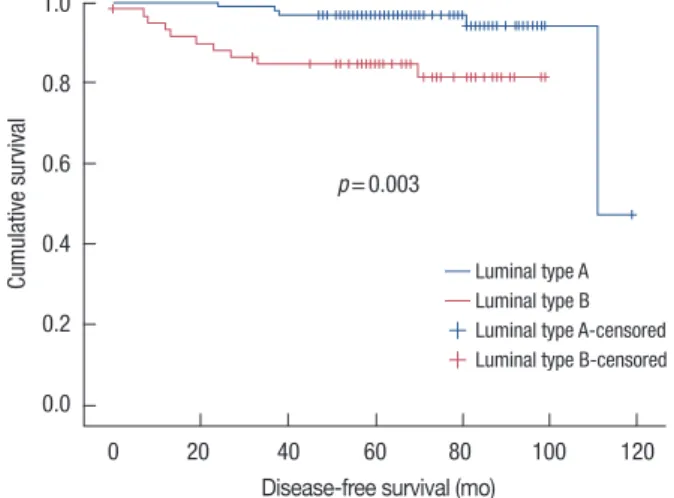
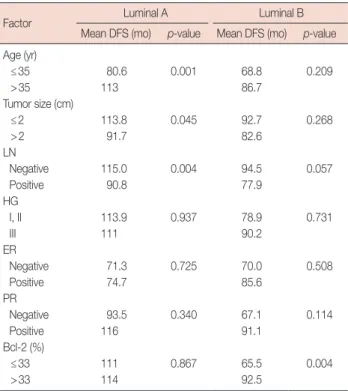
patients (89.9%) and not on 16 patients (10.1%). Luminal-type A breast cancer group had longer DFS than luminal-type B breast cancer group with statistical significance (p=0.003) (Figure 1). The receiver operating characteristic curve of Ki-67 suggested the cutoff value was at 10%, 14%, and 20%.
Also, the optimal cutoff value was 14% because the widest area under the curve was seen at 14% (Figure 2).
When we compared the luminal-type A group and the luminal-type B group, the proportion of the Bcl-2 high expres- sion (>33%) (p=0.013) rate and the small size tumor (≤2 cm) (p=0.001) increased significantly in the luminal-type A Table 1. Clinicopathologic factors of luminal type A and B breast cancer
Factor Luminal A
No. (%) Luminal B
No. (%) p-value
Age (yr)
≤35 8 (8.1) 5 (8.3) 1.000
>35 91 (91.9) 55 (91.7)
Operation methods
BCS 57 (57.6) 33 (55.0) 0.752
Mastectomy 42 (42.4) 27 (45.0)
Tumor size (cm)
≤2 69 (71.1) 24 (42.1) 0.001
>2 28 (28.9) 33 (57.9)
LN
Negative 66 (71.7) 30 (53.6) 0.033
Positive 26 (28.3) 26 (46.4)
HG
I, II 70 (87.5) 21 (38.9) <0.001
III 10 (12.5) 33 (61.1)
ER
Negative 3 (3.0) 3 (5.0) 0.673
Positive 96 (97.0) 57 (95.0)
PR
Negative 7 (7.5) 11 (21.2) 0.033
Positive 86 (92.5) 41 (78.8)
HER2/neu
No amplication 95 (96) 51 (85.0) 0.014
Amplication 4 (4.0) 9 (15.0)
Bcl-2 (%)
≤33 13 (13.1) 18 (30) 0.013
>33 86 (86.9) 42 (70)
Adjuvant treatment RTx
Done 54 (54.5) 26 (43.3) <0.001
None 45 (45.5) 34 (56.7)
CTx
Done 81 (81.8) 40 (66.7) 0.025
None 18 (18.2) 20 (33.3)
ETx
Done 95 (96.0) 48 (80.0) 0.002
None 4 (4.0) 12 (20.0)
BCS=breast-conserving surgery; LN=lymph node; HG=histologic grade;
ER=estrogen receptor; PR=progesterone receptor; Bcl-2=B-cell lymphoma 2 protein; RTx=radiotherapy; CTx=chemotherapy; ETx=endrocrine therapy.
group. The proportion of lymph node (LN) metastasis posi- tive (p=0.033) and histologic high grade (p<0.001) rate in- creased significantly in the luminal-type B group (Table 1).
In the luminal-type breast cancer group, patients in the age group of over 35 years showed longer DFS than the younger age group (p=0.004) and the small tumor size group (≤2 cm) also showed longer DFS than the group with a tumor size over 2 cm (p=0.010). The lymph node metastasis negative group showed longer DFS than the node positive group. Moreover, the high Bcl-2 expression group (>33%) showed longer DFS than the low expression group (p=0.002) (Table 2). In the multivariate analysis, nodal status and Bcl-2 expression were statistically significant relevant factors in the luminal-type breast cancer group (Table 3).
In the subgroup analysis with luminal A group, age at diag-
Figure 1. Disease-free survival curves of luminal type A and B breast cancer.
Cumulative survival
0 20 40 60 80 100 120 Disease-free survival (mo)
1.0
0.8
0.6
0.4
0.2
0.0
p=0.003
Luminal type A Luminal type B Luminal type A-censored Luminal type B-censored
Table 2. Analysis of clinicopathologic factors affecting to disease-free survival (DFS) in luminal type breast cancer
Factor Mean DFS (mo) p-value
Age (yr)
≤35 77.4 0.004
>35 108.3
Tumor size (cm)
≤2 112.2 0.010
>2 87.2
LN
Negative 113.4 0.001
Positive 84.9
HG
I, II 111.3 0.586
III 103.2
Bcl-2 (%)
≤33 91.3 0.002
>33 113.4
LN=lymph node; HG=histologic grade; Bcl-2=B-cell lymphoma 2 protein.
nosis (p=0.001), tumor size (p=0.045) and LN metastasis (p=0.004) were statistically significant factors to affect DFS in the univariate analysis, but not in the multivariate analysis (p=0.240) (Table 4). The younger age group (age ≤35 years)
Table 3. Multivariate analysis of clinicopathologic factors in luminal type breast cancer
Factor HR 95% CI
p-value
Lower Upper
Age 0.708 0.143 3.519 0.708
Tumor size 2.004 0.482 8.332 0.339
LN 5.316 1.008 28.023 0.049
Bcl-2 0.259 0.080 0.905 0.034
HR=hazard ratio; CI=confidence interval; LN=lymph node; Bcl-2=B-cell lymphoma 2 protein.
Table 4. Analysis of clinicopathologic factors affecting to disease-free survival (DFS) in luminal type A and B breast cancer
Factor Luminal A Luminal B
Mean DFS (mo) p-value Mean DFS (mo) p-value Age (yr)
≤35 80.6 0.001 68.8 0.209
>35 113 86.7
Tumor size (cm)
≤2 113.8 0.045 92.7 0.268
>2 91.7 82.6
LN
Negative 115.0 0.004 94.5 0.057
Positive 90.8 77.9
HG
I, II 113.9 0.937 78.9 0.731
III 111 90.2
ER
Negative 71.3 0.725 70.0 0.508
Positive 74.7 85.6
PR
Negative 93.5 0.340 67.1 0.114
Positive 116 91.1
Bcl-2 (%)
≤33 111 0.867 65.5 0.004
>33 114 92.5
LN=lymph node; HG=histologic grade; ER=estrogen receptor; PR=proges- terone receptor; Bcl-2=B-cell lymphoma 2 protein.
showed longer DFS (p=0.001). The smaller tumor size group (≤2 cm) (p=0.045) and the LN metastasis negative group (p=0.004) also showed longer DFS (Table 4).
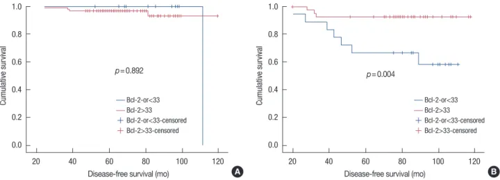
In the luminal B group, Bcl-2 expression (p=0.004) was the only statistically significant factor affecting DFS in the univar- iate analysis (Table 5). Additionally, in the multivariate analy- sis to the luminal B group, Bcl-2 expression was a statistically significant independent factor (p=0.012). Further, Bcl-2 expression over 33% (p=0.004) showed longer DFS curve (Figure 1). In the luminal A group, Bcl-2 expression (p=0.867) was not a statistically significant factor (Table 4) and thus, had no statistically difference in DFS (p=0.89) (Figure 3).
Figure 2. Receiver operating characteristic (ROC) curve of Ki-67 with cutoff value 14% (A), 10% (B), and 20% (C).
AUC=area under the curve.
A
Sensitivity
0.0 0.2 0.4 0.6 0.8 1.0 1-Specificity
1.0
0.8
0.6
0.4
0.2
0.0
AUC=0.684 ROC curve
B
Sensitivity
0.0 0.2 0.4 0.6 0.8 1.0 1-Specificity
1.0
0.8
0.6
0.4
0.2
0.0
AUC=0.659 ROC curve
C
Sensitivity
0.0 0.2 0.4 0.6 0.8 1.0 1-Specificity
1.0
0.8
0.6
0.4
0.2
0.0
AUC=0.544 ROC curve
Table 5. Multivariate analysis of clinicopathologic factors in luminal type B breast cancer
Factor HR 95% CI
p-value
Lower Upper
Age 0.005 0.544 5.128 0.993
Tumor size 0.913 0.097 8.602 0.937
LN 4.150 0.349 49.408 0.260
HG 1.175 0.092 15.050 0.902
ER 0.451 0.384 5.156 0.360
PR 0.283 0.023 3.431 0.321
Bcl-2 0.177 0.131 2.448 0.012
HR=hazard ratio; CI=confidence interval; LN=lymph node; HG=histologic grade; ER =estrogen receptor; PR =progesterone receptor; Bcl-2 =B-cell lymphoma 2 protein.
DISCUSSION
Breast cancer is a heterogeneous disease and shows various clinical courses within the same molecular subtype. Perou et al. [8] classified breast cancer into several subgroups, such as normal-like breast type, luminal-type, HER2 type and basal- like type, according to gene profile. In addition, Brenton et al.
[4] also reported that the prognosis of breast cancer differs according to its genotype. Luminal-type breast cancer showed better prognosis than other subtypes, and additionally, when we divide luminal-type breast cancer to types A and B by Ki-67 expression (cutoff value, 14%), luminal-type A (Ki-67
≤14%) breast cancer showed a lower relapse risk and a longer survival period than luminal-type B (Ki-67 >14%) breast cancer. Additionally, luminal-type B breast cancer showed poor prognosis than luminal-type A breast cancer, but showed better response to systemic chemotherapy. Also, ER-positive tumors responded better to endocrine therapy. Furthermore,
up to 80% of patients with ER and PR-positive tumors, whose profile of luminal type breast cancer responding to endocrine therapy, compared to less than 10% of patients with ER and PR-negative tumors [9-11]. In our study, luminal-type A breast cancer showed longer DFS than luminal-type B breast cancer and was statistically significant; it also increased the proportion of the low histologic grade.
Bcl-2 protein is known to be an anti-apoptotic factor in the cell cycle and it also related to poor prognosis in many other malignancies. Target therapy to Bcl-2 has been attempted consistently in order to apply for clinical use in many areas such as chronic myeloid leukemia, malignant melanoma, multiple myeloma, etc. [12,13]. However, in breast cancer, the expression of Bcl-2 has been reported to be a good prognostic factor related with ER positive, high nuclear grade, low histo- logic grade and HER2 negative [2,14,15].
Elledge et al. [16] and Kim et al. [17] reported that Bcl-2 expression is associated with the prognosis and expression of ER and PR in breast cancer patients undergoing hormonal therapy. Ali et al. [18] investigated the expression of Ki-67 and Bcl-2 in ER positive breast cancer. They reported that Ki-67/
Bcl-2 index is associated with breast cancer-specific survival as well as with the significant prognostic factor in ER positive breast cancer, and also has a strong relationship to luminal- type B breast cancer. In our study, we found that the low Bcl-2 expression group is associated with shorter DFS in luminal- type A breast cancer and luminal-type B breast cancer. Our study suggested that Bcl-2 expression is related to DFS in lumi- nal-type A breast cancer and luminal-type B breast cancer.
Histologic grade was known as the independent prognostic factor in breast cancer [19-23]. In this study, luminal-type B breast cancer showed an increased proportion of high histologic grade and a shorter DFS than luminal-type A breast cancer.
Figure 3. Disease-free survival curves of luminal-type A (A) and B (B) breast cancer according to Bcl-2.
Cumulative survival
20 40 60 80 100 120 Disease-free survival (mo)
p=0.892 1.0
0.8
0.6
0.4
0.2
0.0
Bcl-2-or<33 Bcl-2>33 Bcl-2-or<33-censored Bcl-2>33-censored
A
Cumulative survival
20 40 60 80 100 120 Disease-free survival (mo)
p=0.004 1.0
0.8
0.6
0.4
0.2
0.0
Bcl-2-or<33 Bcl-2>33 Bcl-2-or<33-censored Bcl-2>33-censored
B
However, there is no significant difference in DFS according to histologic grade in the total group and the subgroup analysis.
Further study should be performed for the analysis of progno- sis according to histologic grade in luminal-type breast cancer.
This study has some limitations, however. First, selection bias was observed in this study. The numbers of patients should be similar for both luminal-type A and luminal-type B groups.
Also, the overall survival comparison was not performed due to the limitation in the number of patients and the follow-up period. Therefore, further study with more polished design, such as patient selection, larger patient group, more variant factors and longer follow-up period should be performed.
In summary, luminal-type A breast cancer showed longer DFS than luminal-type B breast cancer, which was related with high grade tumor. Bcl-2 expression was significantly related with DFS in luminal-type breast cancer and its subgroup, such as type B. Therefore, Bcl-2 expression may be a potent prog- nostic factor in luminal-type breast cancer.
CONFLICT OF INTEREST
The authors declare that they have no competing interests.
REFERENCES
1. Zaha DC, Lazăr E, Lăzureanu C. Clinicopathologic features and five years survival analysis in molecular subtypes of breast cancer. Rom J Morphol Embryol 2010;51:85-9.
2. Kröger N, Milde-Langosch K, Riethdorf S, Schmoor C, Schumacher M, Zander AR, et al. Prognostic and predictive effects of immunohisto- chemical factors in high-risk primary breast cancer patients. Clin Can- cer Res 2006;12:159-68.
3. Cheang MC, Chia SK, Voduc D, Gao D, Leung S, Snider J, et al. Ki67 index, HER2 status, and prognosis of patients with luminal B breast cancer. J Natl Cancer Inst 2009;101:736-50.
4. Brenton JD, Carey LA, Ahmed AA, Caldas C. Molecular classification and molecular forecasting of breast cancer: ready for clinical applica- tion? J Clin Oncol 2005;23:7350-60.
5. Goldhirsch A, Ingle JN, Gelber RD, Coates AS, Thürlimann B, Senn HJ; Panel members. Thresholds for therapies: highlights of the St Gallen International Expert Consensus on the primary therapy of early breast cancer 2009. Ann Oncol 2009;20:1319-29.
6. Ahlin C, Aaltonen K, Amini RM, Nevanlinna H, Fjällskog ML, Blomqvist C. Ki67 and cyclin A as prognostic factors in early breast cancer. What are the optimal cut-off values? Histopathology 2007;51:491-8.
7. Neri A, Marrelli D, Pedrazzani C, Caruso S, De Stefano A, Mariani F, et al. Prognostic relevance of proliferative activity evaluated by Mib-1 im- munostaining in node negative breast cancer. Eur J Surg Oncol 2008;
34:1299-303.
8. Perou CM, Sørlie T, Eisen MB, van de Rijn M, Jeffrey SS, Rees CA, et al.
Molecular portraits of human breast tumours. Nature 2000;406:747-52.
9. Osborne CK. Steroid hormone receptors in breast cancer management.
Breast Cancer Res Treat 1998;51:227-38.
10. Hess KR, Pusztai L, Buzdar AU, Hortobagyi GN. Estrogen receptors and distinct patterns of breast cancer relapse. Breast Cancer Res Treat 2003;78:105-18.
11. Steroid receptors in breast cancer: an NIH Consensus Development Conference, Bethesda, Maryland, June 27-29, 1979. Cancer 1980;46:
2759-963.
12. O’Brien S, Moore JO, Boyd TE, Larratt LM, Skotnicki AB, Koziner B, et al. 5-year survival in patients with relapsed or refractory chronic lym- phocytic leukemia in a randomized, phase III trial of fludarabine plus cyclophosphamide with or without oblimersen. J Clin Oncol 2009;
27:5208-12.
13. Bedikian AY, Millward M, Pehamberger H, Conry R, Gore M, Trefzer U, et al. Bcl-2 antisense (oblimersen sodium) plus dacarbazine in patients with advanced melanoma: the Oblimersen Melanoma Study Group. J Clin Oncol 2006;24:4738-45.
14. Hellemans P, van Dam PA, Weyler J, van Oosterom AT, Buytaert P, Van Marck E. Prognostic value of bcl-2 expression in invasive breast cancer.
Br J Cancer 1995;72:354-60.
15. Chen HH, Su WC, Guo HR, Chang TW, Lee WY. p53 and c-erbB-2 but not bcl-2 are predictive of metastasis-free survival in breast cancer patients receiving post-mastectomy adjuvant radiotherapy in Taiwan.
Jpn J Clin Oncol 2002;32:332-9.
16. Elledge RM, Green S, Howes L, Clark GM, Berardo M, Allred DC, et al.
bcl-2, p53, and response to tamoxifen in estrogen receptor-positive metastatic breast cancer: a Southwest Oncology Group study. J Clin Oncol 1997;15:1916-22.
17. Kim K, Chie EK, Han W, Noh DY, Park IA, Oh DY, et al. Prognostic val- ue of p53 and bcl-2 expression in patients treated with breast conserva- tive therapy. J Korean Med Sci 2010;25:235-9.
18. Ali HR, Dawson SJ, Blows FM, Provenzano E, Leung S, Nielsen T, et al.
A Ki67/BCL2 index based on immunohistochemistry is highly prog- nostic in ER-positive breast cancer. J Pathol 2012;226:97-107.
19. Lundin J, Lundin M, Holli K, Kataja V, Elomaa L, Pylkkänen L, et al.
Omission of histologic grading from clinical decision making may re- sult in overuse of adjuvant therapies in breast cancer: results from a na- tionwide study. J Clin Oncol 2001;19:28-36.
20. Trudeau ME, Pritchard KI, Chapman JA, Hanna WM, Kahn HJ, Mur- ray D, et al. Prognostic factors affecting the natural history of node-neg- ative breast cancer. Breast Cancer Res Treat 2005;89:35-45.
21. Ivshina AV, George J, Senko O, Mow B, Putti TC, Smeds J, et al. Genetic reclassification of histologic grade delineates new clinical subtypes of breast cancer. Cancer Res 2006;66:10292-301.
22. Schumacher M, Schmoor C, Sauerbrei W, Schauer A, Ummenhofer L, Gatzemeier W, et al. The prognostic effect of histological tumor grade in node-negative breast cancer patients. Breast Cancer Res Treat 1993;25:
235-45.
23. Roberti NE. The role of histologic grading in the prognosis of patients with carcinoma of the breast: is this a neglected opportunity? Cancer 1997;80:1708-16.