Original Article
Plasma levels of insulin-like growth factor-1 and insulin-like growth factor binding protein-3 in
women with cervical neoplasia
Si Won Lee1,*, Soo Yoon Lee2,3,*, Sa Ra Lee2,3, Woong Ju2,3, Seung Cheol Kim2,3
1Department of Obstetrics and Gynecology, Cheil General Hospital and Women's Healthcare Center, Kwandong University College of Medicine,
2Department of Obstetrics and Gynecology, 3Medical Research Institute, Ewha Womans University School of Medicine, Seoul, Korea
Objective: The goal of this study was to investigate the relationship between plasma levels of insulin-like growth factors-1 (IGF-1) and IGF-binding protein-3 (IGFBP-3) and the risk for cervical intraepithelial neoplasia (CIN) and cervical cancer.
Methods: Plasma levels of IGF-1 and IGFBP-3 of 44 cervical cancer patients, 82 CIN patients and 40 neoplasm-free patients were investigated. Then the associations of the plasma levels of IGF-1 and IGFBP-3 with cervical neoplasm or its clinicopathologic parameters were analyzed.
Results: The mean IGF-1 concentrations were significantly different among the control, CIN, and cervical cancer groups;
the levels were higher in the CIN group compared to the controls. According to the quartile category, the plasma IGF-1 level was significantly higher (p=0.0015) in the CIN group than in the controls. The IGFBP-3 level showed no association between the controls and CIN groups (p=0.842). Although the mean IGF-1/IGFBP-3 molar ratio had borderline significance (p=0.08) among the study population, the quartile comparison showed a significantly higher IGF-1/IGFBP-3 molar ratio in the CIN group compared to the control group (p=0.041).
Conclusion: Plasma levels of IGF-1 and the IGF-1/IGFBP-3 molar ratio might be useful for the development early detection of cervical lesions and used as an adjuvant diagnostic tool for cervical neoplasia after more larger scale research.
Key Words: IGF-1, IGFBP-3, IGF-1/IGFBP-3 molar ratio, Cervical neoplasia
Received December 3, 2009, Revised June 30, 2010, Accepted August 5, 2010
Correspondence to Seung Cheol Kim
Department of Obstetrics and Gynecology, Ewha Womans University School of Medicine, 911-1 Mok-dong, Yangcheon-gu, Seoul 158-710, Korea
Tel: 82-2-2650-6101, Fax: 82-2-2647-9860 E-mail: [email protected]
*These two authors contributed equally.
INTRODUCTION
It is well known that cells with accelerated rates of division and proliferation are predisposed to develop into cancer cells.1,2 Cell proliferation and apoptosis are especially important for the formation of certain neoplasia. The insulin-like growth factors (IGF) are multifunctional peptides that play a pivotal role in cell regulation. The IGF family consists of IGF-1, IGF-2 and several IGF-binding proteins (IGFBP). IGF-1 is a broad- spectrum growth factor that has been shown to play an im- portant role in regulation of cell proliferation, differentiation and apoptosis, and may thus be involved in the development of cancer;3,4 it acts in an endocrine, paracrine, and autocrine
manner in many tissues, and has acute anabolic effects on pro- tein and carbohydrate metabolism by increasing the cellular uptake of amino acids, and by stimulating glycogen and pro- tein synthesis.5 It acts as a mitogen by increasing DNA syn- thesis and stimulating the expression of cyclin D1.6 IGF-1 also can stimulate the expression of Bcl proteins and the sup- pression of Bax, which results in blocking the initiation of the apoptotic pathway.7
The action of IGF-1 and -2 is mediated by the IGF-1 receptor (IGF-1R). The type IGF-1R is a tyrosine kinase similar to the insulin receptor; it mediates the promotion of growth by binding to both IGF-1 and IGF-2.8,9 The IGF-1R affects several growth promoting functions. It stimulates mitogenesis in many differ- ent cell types and protects the cells from apoptosis.10,11 The re- ceptor also plays an important role in the transformation of cells induced by viral proteins and oncogene proteins.12 Previous studies have reported that overexpression of IGF-1R may be as- sociated with certain aggressive tumors.13,14
IGF-1 interacts with its cell membrane receptor to influence various cellular activities. The bioavailability of the IGF family is regulated by IGFBPs. The IGFBPs are composed of six pro- teins; all IGFBPs have growth inhibitory effects.15 The in- hibitory effects are caused by competitive binding of the IGFs,
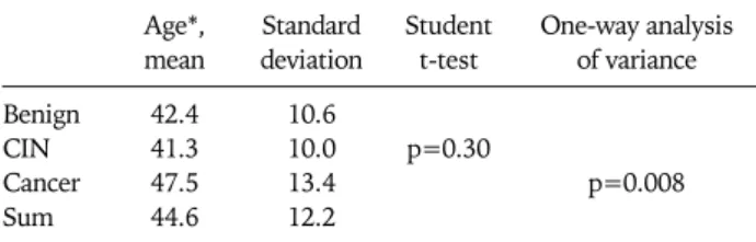
Table 1. Comparison of age between the benign controls, the cer- vical intraepithelial neoplasia (CIN) and the cancer patients
Age*, mean
Standard deviation
Student t-test
One-way analysis of variance Benign
CIN Cancer Sum
42.4 41.3 47.5 44.6
10.6 10.0 13.4 12.2
p=0.30
p=0.008
No differences were noted between the benign controls and the CIN group, but significant difference in age was noted in the cancer group compared to the controls.
*Patient's age at diagnosis.
and prevention of their binding to the receptor. More than 90%
of the plasma IGF-1 is bound to IGFBP-3, the most abundant of the six different IGFBPs identified (IGFBP 1-6). IGFBP-3 has been shown to play a major role in the regulation of the in- teraction between IGF and IGF-1R;16,17 it modulates free IGF and inhibits its transfer from the circulation to tissues. The in- hibitory effects have been shown to be both IGF-dependent and independent.
There are several lines of evidence that support a role for the IGF family in the development of neoplasia. The results from previous studies have shown an increased risk of cancer asso- ciated with high circulating IGF-1 levels, especially in cancers of epithelial and glandular origin such as the prostate, colon, breasts, and lungs.18-21 The finding of high IGFBP-3 levels ap- pears to be associated with a decreased risk.22-24
With regard to cervical cancer, it is a cancer of epithelial ori- gin similar to other cancers with high plasma IGF levels. Thus, IGF may be involved in carcinogenesis of the cervix. There is some evidence that the IGF receptor is overexpressed in cer- vical cancers, and reports suggest that cervical cancer might be sensitive to IGF-1.25 One study reported a positive correla- tion between high plasma IGF levels and squamous intra- epithelial lesions.26
Cervical cytology has been considered the primary screening method for early detection of cervical intraepithelial neoplasia (CIN) and cervical cancer. The measurement of the plasma concentration of IGF-1 and IGFBP-3 may be relatively easier and more convenient for patients that commonly have blood drawn at routine health examinations; this would likely be preferred to obtaining specimens for cervical cytology. IGF levels may be useful as biomarkers for the assessment of risk for CIN and/or cervical cancer. However, there is little re- search on the relationship between the IGF family and cervical neoplasias.
This study was performed to investigate the relationship be- tween plasma levels of IGF-1 and IGFBP-3 and the risk for CIN and/or cervical cancer.
MATERIALS AND METHODS 1. Study population
One hundred and sixty six women that underwent surgery from November 2004 to May 2007, at the department of Gynecology at Ewha Womans University, Mokdong hospital, were included in this retrospective study. A control group consisted of 40 patients that were diagnosed with benign gy- necological disease by pathology. The group with CIN con- sisted of 82 cases (40 cases of CIN II and 42 cases of CIN III), and the cancer group consisted of 44 cases of cervical cancer at a variety of different stages. Among all surgery cases during the study period, CIN cases and controls were selected if the patient agreed to sample collection during the study period.
All participants signed an informed consent that explained the further use of blood samples and cervical tissues before
donating the blood samples. The clinical and pathological da- ta used for this study were collected by retrospective medical chart review. This study was exempted from approval of the Institutional Review Board, at the Ewha Womans University, Mokdong Hospital, because of its retrospective design.
2. Blood sample collection and analysis
From all participating women, a 10-mL blood sample was col- lected by venipuncture and added to a heparinized Vacutainer tube. The blood samples were centrifuged at 1,500 × g for 20 minutes. The plasma and Buffy coat were aspirated and stored separately at −70oC. Plasma levels of IGF-1 and IGFBP-3 were measured using an ELISA kit from Diagnostics Systems Laboratories Inc. (Webster, TX, USA). All samples were tested in duplicate and the mean values of the patient’s plasma IGF-1 and IGFBP-3 levels were determined. If differences between the two results were more than 10%, the test was repeated. In addi- tion, the effects of HPV infection on the levels of the IGF family were studied by dividing the patients into HPV (+) and HPV (−) groups and evaluating the levels of IGF-1 and IGFBP-3.
3. Statistical analysis
The ANOVA test was used to compare the variables between cases and controls. We used the plasma levels of IGF-1 and IGFBP-3 for the statistical analysis. The Spearman correlation coefficient was used to examine the relationship between the IGF levels and age. For evaluation of the IGF levels in cancer patients, we used the Mann-Whitney U test. We categorized their plasma levels into quartiles based on the distribution in control subjects and used the results for the analysis. The IGF-1/
IGFBP-3 molar ratio that was calculated from [(IGF-1×0.13)/
(IGFBP-3×0.035)] was also evaluated. The chi-square test was used to compare the variables between cases and control patients.
RESULTS 1. Characteristics of the subjects
The ages of the three groups are listed in Table 1. The differ- ence in age between the control and the CIN group was not significant (p=0.30); however, the patients were significantly
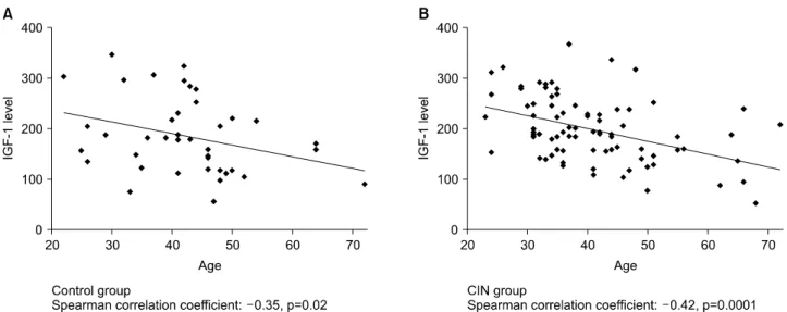
Fig. 1. Scatter plots showing inverse correlation of serum concentrations of insulin-like growth factors-1 (IGF-1) with age, separately among the control group (A) and the cervical intraepithelial neoplasia (CIN) group (B).
Fig. 2. Serum levels of insulin-like growth factors-1 (IGF-1), IGF-binding protein-3 (IGFBP-3), and IGF-1/IGFBP-3 molar ra- tio in the study population showing significantly increased lev- els of IGF-1 and IGF-1/IGFBP-3 molar ratio in the cervical intra- epithelial neoplasia (CIN) group.
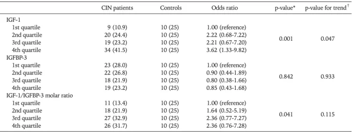
Table 2. Levels of IGF-1, IGFBP-3 and IGF-1/IGFBP-3 molar ratio according to the quartile category between CIN and the control group
CIN patients Controls Odds ratio p-value* p-value for trend†
IGF-1
1st quartile 9 (10.9) 10 (25) 1.00 (reference)
2nd quartile 20 (24.4) 10 (25) 2.22 (0.68-7.22)
0.001 0.047
3rd quartile 19 (23.2) 10 (25) 2.21 (0.67-7.20)
4th quartile 34 (41.5) 10 (25) 3.62 (1.33-9.82)
IGFBP-3
1st quartile 23 (28.0) 10 (25) 1.00 (reference)
2nd quartile 22 (26.8) 10 (25) 0.90 (0.44-1.89)
0.842 0.933
3rd quartile 18 (21.9) 10 (25) 0.80 (0.38-1.66)
4th quartile 19 (23.2) 10 (25) 0.85 (0.43-1.68)
IGF-1/IGFBP-3 molar ratio
1st quartile 11 (13.4) 10 (25) 1.00 (reference)
2nd quartile 18 (21.9) 10 (25) 1.64 (0.52-5.19)
0.041 0.115
3rd quartile 27 (32.9) 10 (25) 2.36 (0.77-7.27)
4th quartile 26 (31.7) 10 (25) 2.36 (0.76-7.28)
IGF-1: insulin-like growth factors-1, IGFBP-3: IGF-binding protein-3, CIN: cervical intraepithelial neoplasia.
*chi-square test. †Weighted linear regression was performed to model the natural logarithm of odds ratios for the risk of cervical neoplasm as a function of qualitatively classified IGF-1, IGFBP-3, and IGF-1/IGFBP-3 molar ratio (1st quartile, 1; 2nd quartile, 2; 3rd quartile, 3; 4th quar- tile, 4) using the inverse variance calculated from confidence intervals of each category; standard error=[LN (upper limit)−LN (lower lim- older in the cancer group (p=0.008). The parity was 1.5±0.87,
1.69±1.09, and 1.97±1.42 in the controls, CIN and the cancer groups respectively; there was no significant difference among the three groups. Other factors such as smoking and the num- ber of intimate partners were not evaluated in this study.
2. IGF-1 and IGFBP-3 levels among the study groups IGF-1 and IGFBP-3 levels showed an inverse correlation with age both in the control group and the CIN group (Fig. 1).
The mean plasma IGF-1 levels were 184.9±74.6 ng/mL in the control group, 200±65.5 ng/mL in the CIN group, and 168.1±
72.4 ng/mL in the cancer group (p=0.048). However since the mean age of the cancer group was significantly greater than the control or CIN groups, the lower plasma IGF-1 level may be affected by age factor. The levels of IGFBP-3 were 3074.9±602.0 ng/mL, 3096.9±618.4 ng/mL and 2881.5±
699.9 ng/mL respectively (p=0.17). The results demonstrated a significant increase in the IGF-1 in the CIN group; however, there was no significant difference noted in the IGFBP-3 levels among the study groups (Fig. 2).
3. IGF-1/IGFBP-3 molar ratio between the controls and CIN cases
The IGF-1/IGFBP-3 molar ratio was 0.059±0.018, 0.064±
0.017, and 0.057±0.019 in the controls, CIN and cancer groups (p=0.08), respectively; there was no significant asso- ciation noted among the three groups (Fig. 2). However, ac- cording to the quartile category, the molar ratio was relatively high in the CIN group (p=0.041). There were 64.6% (53 cas- es) of the CIN group above the 3rd quartile of the controls.
4. IGF-1 and IGFBP-3 levels among the study groups according to the quartile category
In the quartile category, the plasma IGF-1 level was sig- nificantly higher (p=0.0015) in the CIN group than the con- trols (Table 2). About 41.5% (34 patients) of the CIN group was in the 4th quartile group of the controls. The IGFBP-3 level showed no association between the controls and the CIN cases (p=0.842). As mentioned above, the age was significantly higher in the cancer group compared to the control and CIN groups. Therefore, the analysis based on the quartile category was performed only between the control and CIN groups.
5. IGF-1 and HPV infection
HPV testing was not performed in all patients, especially among the controls. Only 71 cases out of 82 patients in the CIN group and 34 cases out of 44 patients in the cancer group had HPV testing. We used Hybrid Capture 2® (Digene Corporation, Gaithersburg, MD, USA) assay method for HPV detection. In the CIN group, 54 patients had positive results, and their mean circulating IGF-1 level was 195.42±8.0 ng/mL. In the negative cases, the mean IGF-1 level was 220.21±18.4 ng/mL and the p-value was 0.16.
In the cancer group, 26 patients were positive for HPV in- fections, and their mean plasma IGF level was 167.0±67.8 ng/mL; eight cases had negative results and their mean IGF-1 level was 170.6±52.8 ng/mL. There was no significant corre- lation identified (p=0.92) between the HPV infection status and IGF-1 levels.
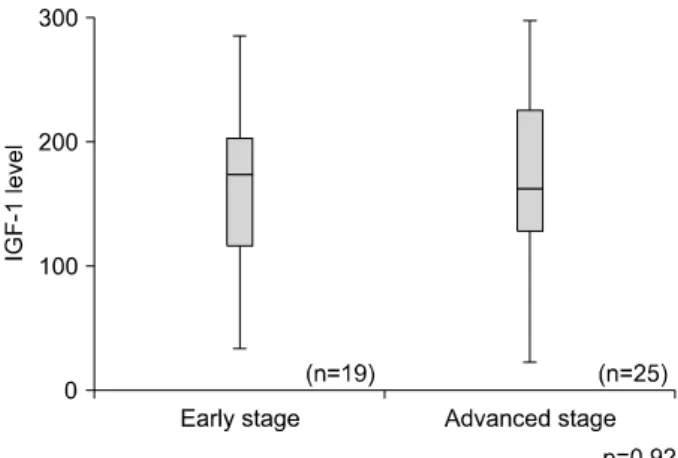
Fig. 3. Levels of insulin-like growth factors-1 (IGF-1) according to cancer stage (p-value=0.92). There was no statistical difference in serum IGF-1 between early and advanced stage cervical cancer.
6. IGF-1 levels and cancer stages
The levels of IGF-1 were evaluated according to the cervical cancer stage and no correlation was noted between IGF-1 lev- els and the cancer stage (p=0.92) (Fig. 3). During early stage disease (CIS-IA), the mean level of IGF-1 was 169.9±17.3 ng/mL (95% CI, 133.6 to 206.3), and for advanced stage dis- ease (over stage IB), the mean level was 166.6±13.9 ng/mL (95% CI, 137.8 to 195.3).
DISCUSSION
The results of the current study showed that the plasma IGF-1 level and IGF-1/IGFBP-3 molar ratio were significantly associated with CIN, but had no significant association with cervical cancer. Elevated plasma IGF-1 and IGF-1/IHFBP-3 ratios were associated with the development of CIN; although it is not clear whether increased plasma levels of IGF-1 and IGF-1/IGFBP-3 were the cause or the result of CIN.
The insulin-like growth factor family was first isolated and se- quenced in 1978. The "Insulin-like growth factors" were named after their primary structure that resembled proinsulin.27 These factors play an important role in human growth and main- tenance of homeostasis, and recently their involvement in carci- nogenesis has been recognized. IGF presumably plays an im- portant role in carcinogenesis by regulating the growth of can- cer cells and by interacting with cancer related molecules; this has been confirmed by several cell culture experiments.28 Many epidemiological studies have shown that IGF-1 levels were higher in cancer patients with sarcoma, leukemia, prostate, col- orectal, breast, liver, and uterine cancers. Animal experiments have shown that over-expression of IGF-1 increases the ten- dency for tumor development.29
Previous studies have demonstrated some relationship be- tween IGF-1 and cervical cancer; however, such results have been inconsistent. One study proposed that IGF-1 was asso- ciated with cervical cancer growth in vitro; however, another
study found no association between high plasma IGF-1 levels and cervical cancer.30 Another study reported that low values of IGF-1 might be associated with cervical cancer.31 However, Wu et al.26 found a strong dose-dependent correlation with the IGF-1 level and squamous intraepithelial lesions of the cervix including cervical cancer.
The results of this study showed that circulating IGF-1 levels were significantly correlated with CIN. As previously re- ported, IGF-1 stimulates cell proliferation by influencing DNA synthesis and cyclin D production; it can suppress apop- tosis by interacting with Bax and Bcl genes. In addition, IGF-1 might promote angiogenesis by effecting vascular endothelial growth factor production.32 Therefore, the circulating IGF-1 level was higher in the CIN group than in the control cases.
Our study showed that IGF-1 was not associated with cervical cancer. These results can be explained by the following. First, age was an important factor associated with the IGF-1 levels.
The levels of circulating IGF-1 change with age.33 They in- crease from birth to puberty, and decrease after puberty. The results of this study were consistent with these prior findings.
Since the mean age was significantly higher in the cervical can- cer group compared to controls and CIN cases, age may have affected the plasma IGF-1 levels. Second, there are likely to be many alterations not yet revealed in the microenvironment during carcinogenesis, thus these alterations may affect the circulating IGF-1 levels in patients with cervical cancer.
Our study has limitations in that plasma IGF levels may be af- fected by body mass index, diet, physical activity, and menstru- al status, which are potential confounders of the case control study comparing plasma IGF levels. A large cohort of cases and controls should be recruited to be able to demonstrate a clear relationship between IGF variables and cervical neoplasia.
IGFBP-3 appears to have a protective effect against carcino- genesis in several cancers. One study reported that plasma lev- els of IGFBP-3 in patients with cervical cancer were sig- nificantly lower than in the control group; however, the levels returned to normal following cancer treatment. In addition, IGFBP-3 down-regulated growth factors in vitro in cervical cancer.34 A former study revealed that the plasma IGFBP-3 was significantly higher in groups with squamous intraepithelial lesions than in controls; however, after adjustment for IGF-1, no correlation was observed with the IGFBP-3 levels.26
In the present study, we could not find any relationship be- tween cervical neoplasia and the plasma IGFBP-3 levels. IGFBP-3 is a binding protein, and can play a dual role. As stated above, IGFBP-3 inhibits cell growth and accelerates apoptosis in both an IGF-dependent and IGF-independent manner. However, in an environment with a high IGF-1 concentration, IGFBP-3 can enhance the effect of IGF by presenting and releasing IGF-1 for interaction with the receptor, while protecting the receptor from down-regulation by exposure to a high IGF-1 concentration.35
It is well known that HPVs are involved in the carcinogenesis of cervical cancer.36 Persistent infection with HPVs has been identified as a major risk factor for cervical cancer and CIN.
However, only a small portion of persistently infected women will develop cervical cancer. Therefore, there are likely several interacting factors that influence carcinogenesis leading to cancer of the cervix. Both the viral factors, as associated with HPV infection and the humoral factors, similar to those asso- ciated with the IGF family influence cell proliferation, are re- sistant to apoptosis and cancer development. Immortalization of HPV-infected cervical squamous epithelial cells is a major factor in the carcinogenesis of cancer of the cervix. It may be initiated by inactivation of cell cycle regulatory p53 and reti- noblastoma genes by HPV E6 and E7 genes. Thus, this may trigger the up-regulation of growth factors, such as IGF or the epidermal growth factor.37
The results of this study showed no relationship between IGF levels and HPV infections. The association between HPV in- fections and IGF levels has not been extensively explored in cer- vical neoplasia. One study found no association with the mRNA expression of IGF-1 and HPV infection status;38 how- ever, another study revealed that cervical cells transduced with high-risk HPV E6 and E7 genes in cell cultures had an 85-fold increase in IGFBP-3 levels.39 Theoretically, high levels of IGF-1 may affect HPV infection and vice versa; however, there are no studies that have found a positive correlation between HPV in- fection and plasma IGF-1 and IGFBP-3 levels, including our study. Therefore, further study will be needed to determine the relationship between HPV infections and the IGF family.
According to our results, plasma levels of IGF-1 and the IGF-1/IGFBP-3 molar ratio were not associated with cervical cancer, but demonstrated significantly higher levels in the CIN group compared to the control group. Therefore, these factors may be involved in the progression from normal to pre- cancerous lesions of the cervix. In the future, plasma levels of IGF-1 and the IGF-1/IGFBP-3 molar ratio may be useful as early detection markers, or as an adjuvant diagnostic tool or prognostic marker in patients with cervical neoplasia.
In conclusion, the results of this study show a positive corre- lation between IGF-1 and IGFBP-3 plasma levels and CIN.
Larger studies are needed to assess the role of the IGF family in the development of cervical neoplasia.
CONFLICT OF INTEREST
No potential conflict of interest relevant to this article was reported.
ACKNOWLEDGEMENTS
This study was supported by a grant of the Korea Healthcare Technology R&D Project, Ministry for Health, Welfare &
Family Affairs, Korea (A080884).
REFERENCES
Increased cell division as a cause of human cancer. Cancer Res 1990; 50: 7415-21.
2. Kim K, Ryu SY. Major clinical research advances in gynecologic cancer 2009. J Gynecol Oncol 2009; 20: 203-9.
3. Sara VR, Hall K. Insulin-like growth factors and their binding proteins. Physiol Rev 1990; 70: 591-614.
4. Rosenfeld RG, Lamson G, Pham H, Oh Y, Conover C, De Leon DD, et al. Insulinlike growth factor-binding proteins. Recent Prog Horm Res 1990; 46: 99-159.
5. Baxter RC, Saunders H. Radioimmunoassay of insulin-like growth factor-binding protein-6 in human serum and other body fluids. J Endocrinol 1992; 134: 133-9.
6. Furlanetto RW, Harwell SE, Frick KK. Insulin-like growth fac- tor-I induces cyclin-D1 expression in MG63 human osteosarco- ma cells in vitro. Mol Endocrinol 1994; 8: 510-7.
7. Minshall C, Arkins S, Straza J, Conners J, Dantzer R, Freund GG, et al. IL-4 and insulin-like growth factor-I inhibit the de- cline in Bcl-2 and promote the survival of IL-3-deprived mye- loid progenitors. J Immunol 1997; 159: 1225-32.
8. LeRoith D, Werner H, Beitner-Johnson D, Roberts CT Jr.
Molecular and cellular aspects of the insulin-like growth factor I receptor. Endocr Rev 1995; 16: 143-63.
9. Stewart CE, Rotwein P. Growth, differentiation, and survival:
multiple physiological functions for insulin-like growth factors.
Physiol Rev 1996; 76: 1005-26.
10. Esposito DL, Blakesley VA, Koval AP, Scrimgeour AG, LeRoith D. Tyrosine residues in the C-terminal domain of the in- sulin-like growth factor-I receptor mediate mitogenic and tu- morigenic signals. Endocrinology 1997; 138: 2979-88.
11. Hongo A, D'Ambrosio C, Miura M, Morrione A, Baserga R.
Mutational analysis of the mitogenic and transforming activ- ities of the insulin-like growth factor I receptor. Oncogene 1996; 12: 1231-8.
12. LeRoith D, Baserga R, Helman L, Roberts CT Jr. Insulin-like growth factors and cancer. Ann Intern Med 1995; 122: 54-9.
13. Hakam A, Yeatman TJ, Lu L, Mora L, Marcet G, Nicosia SV, et al. Expression of insulin-like growth factor-1 receptor in hu- man colorectal cancer. Hum Pathol 1999; 30: 1128-33.
14. Xie Y, Skytting B, Nilsson G, Brodin B, Larsson O. Expression of insulin-like growth factor-1 receptor in synovial sarcoma: as- sociation with an aggressive phenotype. Cancer Res 1999; 59:
3588-91.
15. Ferry RJ Jr, Katz LE, Grimberg A, Cohen P, Weinzimer SA.
Cellular actions of insulin-like growth factor binding proteins.
Horm Metab Res 1999; 31: 192-202.
16. Arany E, Afford S, Strain AJ, Winwood PJ, Arthur MJ, Hill DJ.
Differential cellular synthesis of insulin-like growth factor binding protein-1 (IGFBP-1) and IGFBP-3 within human liver. J Clin Endocrinol Metab 1994; 79: 1871-6.
17. Ballard FJ, Knowles SE, Walton PE, Edson K, Owens PC, Mohler MA, et al. Plasma clearance and tissue distribution of labelled insulin-like growth factor-I (IGF-I), IGF-II and des (1-3) IGF-I in rats. J Endocrinol 1991; 128: 197-204.
18. Pollak M. Insulin-like growth factor physiology and cancer risk.
Eur J Cancer 2000; 36: 1224-8.
19. Giovannucci E. Insulin, insulin-like growth factors and colon cancer: a review of the evidence. J Nutr 2001; 131(Suppl 11):
3109S-20S.
20. Yu H, Rohan T. Role of the insulin-like growth factor family in cancer development and progression. J Natl Cancer Inst 2000;
92: 1472-89.
21. Sandhu MS, Dunger DB, Giovannucci EL. Insulin, insulin-like growth factor-I (IGF-I), IGF binding proteins, their biologic in- teractions, and colorectal cancer. J Natl Cancer Inst 2002; 94:
22. Chan JM, Stampfer MJ, Giovannucci E, Gann PH, Ma J, Wilkinson P, et al. Plasma insulin-like growth factor-I and pros- tate cancer risk: a prospective study. Science 1998; 279: 563-6.
23. Wolk A, Mantzoros CS, Andersson SO, Bergstrom R, Signorello LB, Lagiou P, et al. Insulin-like growth factor 1 and prostate cancer risk: a population-based, case-control study. J Natl Cancer Inst 1998; 90: 911-5.
24. Yu H, Spitz MR, Mistry J, Gu J, Hong WK, Wu X. Plasma levels of insulin-like growth factor-I and lung cancer risk: a case-con- trol analysis. J Natl Cancer Inst 1999; 91: 151-6.
25. Steller MA, Delgado CH, Zou Z. Insulin-like growth factor II mediates epidermal growth factor-induced mitogenesis in cer- vical cancer cells. Proc Natl Acad Sci U S A 1995; 92: 11970-4.
26. Wu X, Tortolero-Luna G, Zhao H, Phatak D, Spitz MR, Follen M. Serum levels of insulin-like growth factor I and risk of squ- amous intraepithelial lesions of the cervix. Clin Cancer Res 2003; 9: 3356-61.
27. Rinderknecht E, Humbel RE. The amino acid sequence of hu- man insulin-like growth factor I and its structural homology with proinsulin. J Biol Chem 1978; 253: 2769-76.
28. Macaulay VM. Insulin-like growth factors and cancer. Br J Cancer 1992; 65: 311-20.
29. Rogler CE, Yang D, Rossetti L, Donohoe J, Alt E, Chang CJ, et al. Altered body composition and increased frequency of di- verse malignancies in insulin-like growth factor-II transgenic mice. J Biol Chem 1994; 269: 13779-84.
30. Steller MA, Delgado CH, Bartels CJ, Woodworth CD, Zou Z.
Overexpression of the insulin-like growth factor-1 receptor and autocrine stimulation in human cervical cancer cells. Cancer Res 1996; 56: 1761-5.
31. Serrano ML, Romero A, Cendales R, Sanchez-Gomez M, Bravo MM. Serum levels of insulin-like growth factor-I and -II and in- sulin-like growth factor binding protein 3 in women with squ-
amous intraepithelial lesions and cervical cancer. Biomedica 2006; 26: 258-68.
32. Bustin SA, Jenkins PJ. The growth hormone-insulin-like growth factor-I axis and colorectal cancer. Trends Mol Med 2001; 7: 447-54.
33. Gomez JM, Mourot B, Fostier A, Le Gac F. Growth hormone re- ceptors in ovary and liver during gametogenesis in female rain- bow trout (Oncorhynchus mykiss). J Reprod Fertil 1999; 115:
275-85.
34. Mathur RS, Mathur SP. In vitro downregulation of growth fac- tors by insulin-like growth factor binding protein-3 in cervical cancer. Gynecol Oncol 2003; 91: 410-5.
35. Conover CA, Powell DR. Insulin-like growth factor (IGF)- binding protein-3 blocks IGF-I-induced receptor down-regu- lation and cell desensitization in cultured bovine fibroblasts.
Endocrinology 1991; 129: 710-6.
36. Walboomers JM, Jacobs MV, Manos MM, Bosch FX, Kummer JA, Shah KV, et al. Human papillomavirus is a necessary cause of invasive cervical cancer worldwide. J Pathol 1999; 189: 12-9.
37. Dassonville O, Formento JL, Francoual M, Ramaioli A, Santini J, Schneider M, et al. Expression of epidermal growth factor re- ceptor and survival in upper aerodigestive tract cancer. J Clin Oncol 1993; 11: 1873-8.
38. Nakamura K, Hongo A, Kodama J, Miyagi Y, Yoshinouchi M, Kudo T. Down-regulation of the insulin-like growth factor I re- ceptor by antisense RNA can reverse the transformed pheno- type of human cervical cancer cell lines. Cancer Res 2000; 60:
760-5.
39. Berger AJ, Baege A, Guillemette T, Deeds J, Meyer R, Disbrow G, et al. Insulin-like growth factor-binding protein 3 expression increases during immortalization of cervical keratinocytes by human papillomavirus type 16 E6 and E7 proteins. Am J Pathol 2002; 161: 603-10.