Corresponding author: Sang-Won Park
Department of Prosthodontics, School of Dentistry, Chonnam National University 300 Yongbong-dong, Buk-ku, Kwangju, 500-757, Korea
Tel, +82 62 530 5842: e-mail, [email protected]
Received January 28, 2009 / Last Revison February 25, 2009 / Accepted July 15, 2009,
INTRODUCTION
Osseointegration is the essential biological basis of the cur- rent dental implants.1Many researchers have found that the responses of cell and tissue to implant is affected not only by the chemical properties, but also the surface topography or roughness of the implant surfaces.2So, there were many efforts to modify titanium implant surface to achieve better tis- sue responses.
The implant surfaces characteristics most frequently described in the literature may be subdivided into implants with rough- ened surfaces by coated (titanium plasma spray, hydroxyapatite, etc), implants with roughened surfaces with the electro- chemical modifications (anodic oxidation) of the commer- cially pure titanium implants, and implants with roughened surfaces without coated (sand-blasted, acid-etched etc). These surfaces were known to promote initial healing capacity by roughness.10The bone formation that occurs during osseoin- tegration may show the osteoblast activities which are affect- ed by the implant surfaces.13A number of in vitro and in vivo studies have been conducted to compare the effect of implant surfaces on the bone formation. Novaes and colleagues14
compared hydroxyapatite (HA), titanium plasma-sprayed (TPS), sandblasted, and machined implants. They found that in the relation to bone implant contact (BIC), the sandblasted surfaces were statistically superior to the turned surfaces and showed greater BIC than the HA and TPS surfaces after 90 days in place without loading. Human histologic find- ings demonstrated the improved BIC on rough surfaced implants compared to turned surfaced implants.15When the surface topography of an implant is altered, its surface chem- istry is also altered. Cell behavior is not dependent on topog- raphy alone; surface topography and chemistry are inseparable.
In a study investigating bone tissue reactions to various sur- face oxide properties, Sul16concluded that, either separately or altogether, surface chemistry and topography play important roles of bone responses to the implant surfaces. Recently, a num- ber of studies about nano-treated surfaces of implants have been conducted. Oh et al. cited that28TiO2nanotube arrays and associated nanostructures could be useful as the well-adhered bioactive surface layers on Ti implant metals and alloys for ortho- pedic and dental applications. Karlsson et al.29suggested that the anodized nano-porous alumina membranes seem to pro- vide better surface for osteoblastic cell growth, with cells
Histologic evaluation and removal torque analysis of nano- and microtreated titanium implants in the dogs
Seok Ahn1, DDS,MSD, Mong-Sook Vang2, DDS, MSD, PhD, Hong-So Yang2, DDS, MSD, PhD, Sang-Won Park2, DDS, MSD, PhD, Hyun-Pil Lim3, DDS, MSD
1Graduate student,2Professor, 3Clinical professor, Department of Prosthodontics, Graduate school, Chonnam National University, Gwangju, Korea
STATEMENT OF PROBLEM. A number of studies about the nano-treated surfaces of implants have been conducting along with micro-treat- ed surfaces of implants. PURPOSE. The purpose of this study was to get information for the clinical use of nano-treated surfaces compared with micro-treated surfaces by measuring removal torque and analyzing histological characteristics after the placement of various surface-treat- ed implants on femurs of dogs. MATERIAL AND METHODS. Machined surface implants were used as a control group. 4 nano-treated sur- face implants and 3 micro-treated surface implants [resorbable blast media surface (RBM), sandblast and acid-etched surface (SAE), anodized RBM surface] were used as experimental groups. Removal torque values of implants were measured respectively and the histological analyses were conducted on both 4weeks and 8weeks after implant surgery. The surfaces of removed implants after measuring removal torque values were observed by scanning electron microscopy (SEM) at 8 weeks. RESULTS. 1. Removal torque values of the nano-treated groups were lower than those of micro-treated groups. 2. Removal torque values were similar in the anodized RBM surface groups. 3. On the histological views, there was much of bone formation at 8 weeks, but there was no difference between 4 and 8 weeks, and between the types of implant surfaces as well. CONCLUSION. it is suggested that implant topography is more effective in removal torque test than surface chemistry. To get better clinical result, further studies should be fulfilled on the combined effect of surface topography and chemistry for the implant surface treatments. KEYWORDS. removal torque, implant, surfaces characteristics, dog, histology, SEM [J Adv Prosthodont 2009;1:75-84]
ⓒ 2009 The Korean Academy of Prosthodontics
This is an Open Access article distributed under the terms of the Creative Commons Attribution Non-Commercial License (http://creativecommons.org/licenses/by- nc/3.0) which permits unrestricted non-commercial use, distribution, and reproduction in any medium, provided the original work is properly cited.
treated by sputtering method in laboratory.
For group 2 and 4, the sputtering parameters were as follows:
300 W, (1.0 - 1.2) × 10-2torr, for 3 h. Group 4 is heat-treated at 600℃ for 1 hour after deposition.
For group 3 and 5, the sputtering parameters were as follows:
dental implants were coated by sputter-coated using a CMS- 18 radiofrequency magnetron sputtering system (Kurt J.
Lesker Company, Clairton, PA, USA). The machine was oper- ated at 300 W, 1.0 - 1.2 × 10⁻²torr, for 7 h. Pins were rotated 120 degree between each of three coated periods to cover the entire 360�surface of the specimens. CaP pins were subjected to a post coated heat treatment of 600℃ for 1 h to achieve 60%
crystallinity.
Implants surfaces in micro-treated surface groups were produced by a company (ExFeel�, MEGAGEN, Kyungsan, Korea)
For group 6, 7 and 8, sanding conditions are as follows:
to 20 kg were used in this study. The dogs were anesthetized with the combinations of Ketamine (5 mg/kg, Yu-han, Gunpo, Korea) and Rompun (0.3 mg/kg, Bayer Korea, Ansan, Korea) intramuscularly.
Surgical procedures and implant placement
Both hind legs were prepared in the standard sterile fashions.
The flat surface on the lateral aspect of the proximal femur was selected for the implant placement. The skin incision was performed to expose the whole lateral aspect of femur, mus- cles were dissected to allow elevation of the periosteum and then implant sites were prepared. All implants were 3.75 mm in diameter, which were larger than the final drill sizes, which were 3.3 mm in diameter. Eight implants sites were drilled on each leg using specially designed acrylic stent (Fig. 2).
Postsurgical teratment
The surgical site was closed in layers with resorbable suture materials (SURGIFIT, AILEE Co Ltd, Korea). All animals received 50 injection of 0.15 ml/kg Baytril�50 (Bielkorea, Seoul, South Korea) for 7 days and 0.1 ml/kg Pirin�(Green Cross Veterinary Products Co, Seoul, Korea) for 2 days intramuscularly.
Animals sacrifice
Two animals were sacrificed at 4 weeks and another two at 8 weeks after surgery by the injection of overdose of thiopen- tal sodium (T6023, Sigma, MO, USA).
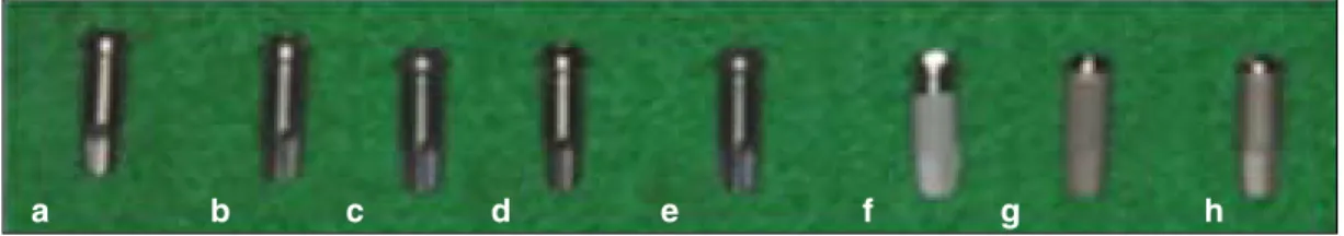
Fig. 1.Eight fixtures with differently treated implant surface. From left; a. Machined surface, b. 20 nm TiO2coated sur- face, c. CaP coated surface, d. Heat treated 80 nm TiO2coated surface, e. Heat treated CaP coated surface, f. Resorbable blast media (RBM) surface, g. Sandblast and acid-etched (SAE) surface, h. Anodized RBM surface.
a b c d e f g h Table I.Surface characteristics
Group Surface characteristics n
Control 1 Machined surface 8
Nano-treated 2 20 nm TiO2coated surface 8
3 CaP coated surface 8
4 Heat treated 80 nm TiO2coated surface 8 5 Heat treated CaP coated surface 8 Micro-treated 6 Resorbable blast media surface 8 7 Sandblast and acid-etched surface 8
8 Anodized RBM surface 8
Removal torque values measurement
The implants and adjacent tissues were removed en bloc.
Removal torque values were measured with a torque mea- surement device (MGT12, ELECTROMATIC Equipment, NY, USA) (Fig. 3). When the ruptures between bones and implants occurred, the peak force values fell quickly.
Scanning electron microscopy (SEM)
After the removal torque test at 8 weeks, the implants were fixed in the neutral buffered formalin. After dehydration in a graded series of alcohol, the implants were dried and mount- ed on metallic stubs using double side tapes. All samples were placed in the vacuum chamber of the SEM. Thereafter, the implant surfaces were observed by SEM (S-4700, Hitachi, Tokyo, Japan).
Preparation of specimens and the histological analyses The implants, after measuring removal torque value, were removed from bones in the 8-th week experimental group to see the SEM of removed implant surfaces, but the implants after measuring removal torque value were replaced with the original site to see better bone implant relationships in the 4- th week experiment group. The implants and surrounding bones were fixed in the neutral buffered formalin, dehydrated with ascending concentrations of ethanol for 24 h at each stage.
Following transitional acetone immersion, the samples were immersed in 100% polymethylmethacrylate monomer for 24 h, followed by immersion in a 1 : 1 ratio of polymethyl- methacrylate to methylmethacrylate monomer for 24 h. The sam- ples were placed in the embedding molds containing poly- methylmethacrylate resin for 24 h. Thereafter, the samples were Fig. 3.a. The jig was especially designed for removal torque measurement. The upper part of the metal jig was able to move from the middle into the space where the arrow was directed and the lower part can move back and forward. This kind of movement was necessary to place the hole of the upper part of the zig that can position on the right surface of the implant fixtures. b. Removal torque measurement device (MGT12, ELECTROMATIC Equipment, USA). c. Connect the fixture mount with implants. d. Mount holder was attached with the measured appliance and measured the removal torque.
Fig. 2.a. Implant placement diagram. b. Acrylic stent which has guide holes 10 mm apart. c. Implants were placed in order. d. Mounts were removed.
a b
c d
a b c d
Statistics analyses
The differences of removal torque values were analyzed by two-way ANOVA and for post hoc comparison Duncan’
s test was executed. All calculations were made using SPSS Version 12 for Windows.
RESULTS
Removal torque values
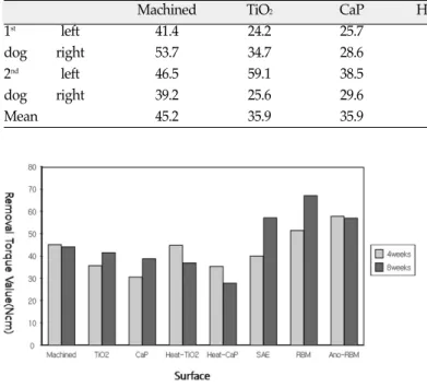
The removal torque values, measured after a 4-week and 8- week healing period, were summarized in Table II. The mean values of the removal torque and diagram were found (Fig. 4).
The removal torque value of one implant (heat treated CaP coat- ed surface) of a dog sacrificed at 8 week was not measured because the femur was fractured and the implant at frac-
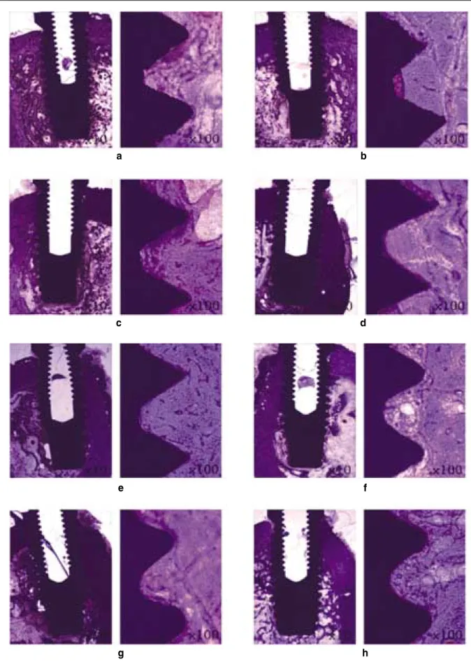
the intimate contact with implants in the cortex area. In medulla area, much endosteal bone formation was observed on adjacent area to cortex and had thin trabecular patterns. There was no difference between 4 weeks (Fig. 7) and 8 weeks (Fig.
8), and between the types of implant surfaces as well.
DISCUSSION
Many studies showed that surface roughness has a positive influence on the resistance to shear and tensile forces.18-19 However, the degree of surface roughness may not be the only aspect of surface topography that effects osseointegration.
The intimacy of bone contact with the implant surface may be important as may the ionic charge, surface energy and surface tension or other still undefined properties of the surface.
Fig. 4.A diagram of removal torque value at 4-th and 8-th week. It showed the mean values of removal torque value between 4 weeks and 8 weeks and there was no statistic significance (P 〉.05).
Fig. 5.A fracture site of femur. one femur of a dog sacrificed at 8 week was fractured at midline and one implant at fractured site failed to be osseointegrated.
Table II.Removal torque values (Ncm) data after 4-week of healing time
Machined TiO2 CaP Heat-TiO2 Heat-CaP SAE RBM Ano-RBM
1st left 41.4 24.2 25.7 40.7 31.5 40.7 58.1 43.5
dog right 53.7 34.7 28.6 34.9 36.4 35.6 56.8 65.0
2nd left 46.5 59.1 38.5 72.8 54.6 37.9 32.8 50.6
dog right 39.2 25.6 29.6 31.3 19.3 46.3 58.2 72.7
Mean 45.2 35.9 35.9 44.9 35.5 40.1 51.5 58.0
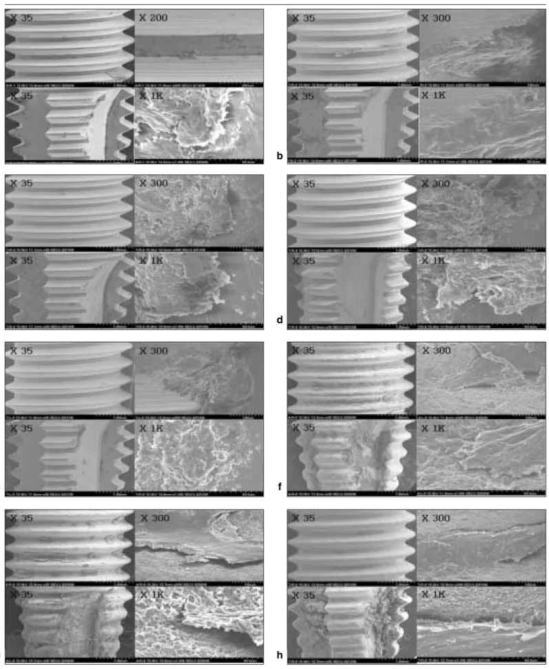
Fig. 6.Scanning electron microscopy of removed implant surfaces. a. Machined surface. b. 20 nm TiO2coated surface. c. CaP coated surface. d. Heat treated 80 nm TiO2coated surface. e. Heat treated CaP coated surface. f. Sandblast and acid-etched (SAE) surface. g. Resorbable blast media (RBM) sur- face. h. Anodized RBM surface.
a b
c d
e f
g h
Fig. 7.The histological section in the buccolingual direction at 4 weeks. a. Machined surface, b. 20 nm TiO2coated surface, c. CaP coat- ed surface, d. Heat-TiO2coated surface, e. Heat CaP coated surface, f. SAE, g. RBM, h. Anodized RBM. All implants osseointegrat- ed well. Bone quality of the original sites was different dependent on the installed sites of implants.
c d
e f
g h
Fig. 8.The histological sections in the buccolingual direction at 8 weeks. a. Machined surface, b. 20 nm TiO2coated surface, c. CaP coat- ed surface, d. Heat-TiO2coated surface, e. Heat CaP coated surface, f. SAE, g. RBM, h. Anodized RBM. All implants were removed after measuring the removal torque values.
a b
c d
e f
g h
experiments were done. The bone quality has similar pat- tern in the radiographs, so the problem of the places of implants was excepted. However, the implants were not placed at the mid-sagittal of the femur and the cortical-cancellous ratio was not constantly maintained. A rough estimate of comparative healing rates between dogs and humans would suggest that the events of wound healing and bone remodeling happened approximately 1.5 times sooner in dogs than would occur in the human, 4 weeks for dog means 6 weeks for human.
The process of osseointegration is affected by many fac- tors, including surgical techniques and the conditions of the implant bed.23Clinical observations have also indicated that the final healing time is affected by individual differences and oper- ation conditions.24 In this study, same clinician installed implants and the implants were planted always the same place with the same sequences. All implants except one that installed at the fracture site of femur healed well. It means that the suitable implant number according to the different bone conditions should be considered avoiding the fracture of bones under the different loading circumstances.
A greater removal force can be generally interpreted as an increase in bone healing around the implants and improvement in osseointegration. In this study, removal torque values of nano- treated group were lower than those of micro-treated group at both 4 weeks and 8 weeks. This result implies that micro-treat- ed surface have better conditions to satisfy the osseointegra- tion than nano-treated surface and surface roughness is more important than surface composition for the resistance of removal torque. In RBM surface and SAE surface group, removal torque value was increased at 8 weeks compared to at 4 weeks. This means that osseointegration is influenced more by the bone maturity than the bone strength. But in the anodized RBM surface group which was additionally treated with anodizing oxidation on sandblasted micro-roughness sur- face, the removal torque values at 4 weeks & 8 weeks were sim- ilar. It seems that when anodized treatment was added to micro- roughness, osseointegration was done at the early stage.
may all influence implant osseointegration.25Consequently, mod- ifications in implant body design and implant surfaces have been suggested to increase the success in poor quality bone by hypothetically, gaining better anchorage and providing more surface area of the load to decrease stress to the softer bone types.26Salata and coworkers27have suggested that faster development of implant stability for oxidized implants than turned components when placed in bone defects. Bone formation towards the rough surfaced implant is facilitated by a more sta- ble connection between bone matrix and the implant sur- face, which can be explained by the degree of protein adsorp- tion on the anodic-oxidized implants. Glauser et al.26con- cluded that the applied immediate loading protocols in the com- bination with a slightly tapered implant and a modified implant surface texture was shown to be a successful treatment alternative even in regions exhibiting soft bones. The results from the study by Olsson et al.27indicated that early loading can be applied to the cross-arch dental bridges supported by six to eight oxidized implants in the maxilla. From this study, it seems that the surface composition may be least effective factor in clinical loading conditions.
In the SEM view of removed implants at 8 weeks, the surfaces of removed implants in nano-treated groups showed relatively clean surfaces with a little bone tissue but the surfaces of removed implants in micro-treated groups showed rough surface with much bone tissue or organic residues. It seems that micro-roughness surface would be more resistant to removal torque than nano-surface, because mechanical locking effect could be achieved on rotational force applied to implants.
This study has showed no statistical significances between the surfaces of implants. This meant that each surface of the implants had a few number of it and the differences between the dogs were so huge that the measurements of the standard deviation gave large numbers. Between the 4 weeks and 8 weeks, it had no statistical significance because of the initial formation of osseointegration at the early stage.
The tissue responses may not depend on only one specific sur- face property but rather on a number of different alterations.
However, it is not fully understood whether these properties influence the bone tissue response separately or synergistically.
In this study the experimental condition may not always be extrapolated the clinical situations. This may be due to differences in bone anatomical, physiological, and unloaded conditions.
In this study, micro-treated groups have showed higher ability of removal than nano-treated groups and these revealed different results than the review of Webster. In addition, both nano- and micro-treated surface implants showed no sta- tistical significance. However, between the 4 and 8 weeks, the results showed no differences and it meant initial osseointe- gration had occurred at the early stage.
From this experiment, we can suggest that the nano-treated surface that newly start to be investigated need to be studied in combination with micro-treated surfaces for better clinical results rather than it is studied by itself.
CONCLUSION
From this study, following results were obtained: Removal torque values of the nano-treated groups were lower than those of micro-treated groups at both 4 week and 8 week. There were no statistically significant difference between the groups, the machined group and nano-treated groups with smooth surfaces relatively had similar removal torque values at both 4 weeks and 8 weeks, but micro-treated groups showed higher removal torque values at 8 weeks than at 4 weeks. Removal torque val- ues at both 4 weeks and 8 weeks were similar in the anodized RBM surface groups and this suggested that osseointegration was done at the early stage compared to RBM surface and SAE surface groups. On the histological views, there was much of bone formation at 8 weeks, but there was no difference between 4 and 8 weeks, and between the types of implant sur- faces as well. On the SEM views, the surfaces of removed implants in nano-treated groups showed relatively clean sur- faces with few bone tissue while the surfaces of removed micro-treated implants showed rough surface with much bone tissues.
From this experiment, it is suggested that implant topography is more effective in removal torque test than surface chemistry.
To get better clinical result, further studies should be ful- filled on the combined effect of surface topography and chemistry for the implant surface treatments.
REFERENCES
1. Li DH, Liu BL, Zou JC, Xu KW. Improvement of osseointegration of titanium dental implants by a modified sandblasting sur- face treatment: an in vivo interfacial biomechanics study. Implant Dent 1999;8:289-94.
2. Buser D, Schenk RK, Steinemann S, Fiorellini JP, Fox CH, Stich H. Influence of surface characteristics on bone integration of ti- tanium implants. A histomorphometric study in miniature pigs.
J Biomed Mater Res 1991;25:889-902.
3. Bowers KT, Keller JC, Randolph BA, Wick DG, Michaels CM.
Optimization of surface micromorphology for enhanced os- teoblast responses in vitro. Int J Oral Maxillofac Implants 1992;7:302-10.
4. Brunette DM. The effects of implant surface topography on the behavior of cells. Int J Oral Maxillofac Implants 1988;3:231-46.
5. Mustafa K, Silva Lopez B, Hultenby K, Wennerberg A, Arvidson K. Attachment and proliferation of human oral fibroblasts to ti- tanium surfaces blasted with TiO2particles. A scanning electron microscopic and histomorphometric analysis. Clin Oral Implants Res 1998;9:195-207.
6. Johansson C, Albrektsson T. Integration of screw implants in the rabbit: a 1-year follow-up of removal torque of titanium im- plants. Int J Oral Maxillofac Implants 1987;2:69-75.
7. Han CH, Johansson CB, Wennerberg A, Albrektsson T. Quantitative and qualitative investigations of surface enlarged titanium and titanium alloy implants. Clin Oral Implants Res 1998;9:1-10.
8. Wennerberg A, Albrektsson T, Johansson C, Andersson B.
Experimental study of turned and grit-blasted screw-shaped implants with special emphasis on effects of blasting material and surface topography. Biomaterials 1996;17:15-22.
9. Ericsson I, Johansson CB, Bystedt H, Norton MR. A histomor- phometric evaluation of bone-to-implant contact on machine-pre- pared and roughened titanium dental implants. A pilot study in the dog. Clin Oral Implants Res 1994;5:202-6.
10. Larsson C, Thomsen P, Aronsson BO, Rodahl M, Lausmaa J, Kasemo B, Ericson LE. Bone response to surface-modified tita- nium implants: studies on the early tissue response to ma- chined and electropolished implants with different oxide thick- nesses. Biomaterials 1996;17:605-16.
11. Sul YT, Johansson CB, Jeong Y, Wennerberg A, Albrektsson T.
Resonance frequency and removal torque analysis of implants with turned and anodized surface oxides. Clin Oral Implants Res 2002;13:252-9.
12. Letic、-Gavrilovic、A, Scandurra R, Abe K. Genetic potential of in- terfacial guided osteogenesis in implant devices. Dent Mater J 2000;19:99-132.
13. Novaes AB Jr, Souza SL, de Oliveira PT, Souza AM.
Histomorphometric analysis of the bone-implant contact ob- tained with 4 different implant surface treatments placed side by side in the dog mandible. Int J Oral Maxillofac Implants 2002;17:377-83.
14. Ivanoff CJ, Hallgren C, Widmark G, Sennerby L, Wennerberg A.
Histologic evaluation of the bone integration of TiO(2) blasted and turned titanium microimplants in humans. Clin Oral Implants Res 2001;12:128-34.
15. Ivanoff CJ, Widmark G, Johansson C, Wennerberg A. Histologic evaluation of bone response to oxidized and turned titanium mi- cro-implants in human jawbone. Int J Oral Maxillofac Implants 2003;18:341-8.
16. Sul YT. The significance of the surface properties of oxidized ti- tanium to the bone response: special emphasis on potential biochemical bonding of oxidized titanium implant. Biomaterials 2003;24:3893-907.
17. Thomas KA, Kay JF, Cook SD, Jarcho M. The effect of surface macrotexture and hydroxylapatite coating on the mechanical strengths and histologic profiles of titanium implant materi- als. J Biomed Mater Res 1987;21:1395-414.
18. Carlsson L, Ro¨stlund T, Albrektsson B, Albrektsson T. Removal torques for polished and rough titanium implants. Int J Oral Maxillofac Implants 1988;3:21-4.
19. Webster TJ, Ergun C, Doremus RH, Siegel RW, Bizios R. Enhanced functions of osteoblasts on nanophase ceramics. Biomaterials 2000;21:1803-10.
20. Webster TJ, Schadler LS, Siegel RW, Bizios R. Mechanisms of en- hanced osteoblast adhesion on nanophase alumina involve vit- ronectin. Tissue Eng 2001;7:291-301.
21. Berglundh T, Abrahamsson I, Lang NP, Lindhe J. De novo alve-