Conventional arthrography has proved to be a useful imag- ing method to detect full-thickness rotator cuff tear, in spite of their invasiveness. But there is a potential risk of misin- terpreting a full-thickness rotator cuff tear as normal.
We report a case of a surgically confirmed medium sized full-thickness rotator cuff tear with false negative arthrog- raphy, which demonstrates pitfalls of conventional arthrog- raphy.
CASE REPORT
A 42 year old man visited our department suffering from a non-dominant left shoulder pain, which started after lift- ing a heavy object 1 year before presentation. The pain was more severe at night. A clinical examination showed positive impingement sign and abduction weakness.
Plain radiography showed type III acromion with a large spur according to the classifications of Bigliani et al.1)and Park et al.10).
Before visiting our hospital the patient had already received MRI at a private radiologic clinic, but the MRI films were not available at the time of presentation. Therefore, arthrog- raphy was performed at our hospital after injecting 14 cc
Urografin�(Schering, Germany). The MRI then became available to us. It had been carried out using a 0.5-T system (Signa; GE Medical Systems, Milwaukee, WI). Oblique coro- nal and axial scans of the shoulder joint showed about a 12×
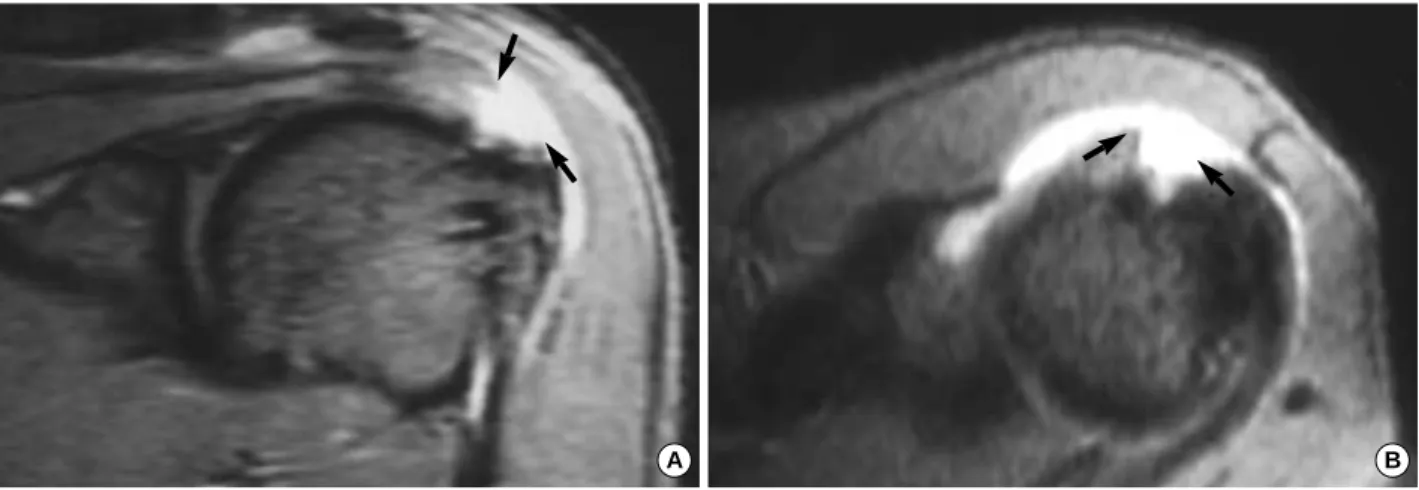
10 mm sized high signal intensity lesion at the insertion site of the supraspinatus tendon on the T2-weighted gradient echo image (flip angle 20°), which suggested a full-thick- ness rotator cuff tear (Fig. 1). In contrast to the MRI find- ings, single-contrast arthrography showed no abnormal find- ings without any leakage of contrast media (Fig. 2). There- fore, there was significant discrepancy between MRI and arthrography.
On arthroscopic examination of the glenohumeral joint, there seemed to be no partial or full-thickness rotator cuff tear (Fig. 3). But bursoscopy of the subacromial space showed a U-shaped full-thickness tear of the supraspinatus tendon at the attachment site on the greater tuberosity measuring 3.0 cm in diameter, which had an intact joint capsule attached to the bone. These findings were confirmed by mini-open skin incision (Fig. 4), through which the torn supraspina- tus tendon was repaired using 2 suture anchors.
At 4 months after surgery, his symptoms were relieved completely, and shoulder strength was regained. Shoulder function was normal with a full range of motion.
DISCUSSION
Rotator cuff tears are common sources of chronic shoul-
498 J. of Korean Orthop. Assoc.
2004; 39: 498-501
498
Arthrographic Pitfalls in the Diagnosis of Full-Thickness Tears of the Rotator Cuff
- A Case Report -
Tae-Soo Park, M.D., and Young-Sun Kim, M.D.*
Department of Orthopaedic Surgery and Diagnostic Radiology*, Guri Hospital, Hanyang University College of Medicine, Guri, Korea
498 498 Address reprint requests to
Tae-Soo Park, M.D.
Department of Orthopaedic Surgery, Guri Hospital, Hanyang University College of Medicine, 249-1 Gyomoon-dong, Guri 471-020, Korea Tel: +82.31-560-2314, Fax: +82.31-557-8781
E-mail: [email protected]
We report a case of full-thickness rotator cuff tear with operative confirmation, which was arthrographi- cally intact. In this case, there was a discrepancy between conventional arthrography and magnetic res- onance imaging (MRI), which suggested a full-thickness tear of the rotator cuff. The cause was confirmed to be an intact synovial capsule of the shoulder joint with a torn rotator cuff.
Key Words: Rotator cuff, Full-thickness tear, Arthrography, Pitfall, MRI
Arthrographic Pitfalls in the Diagnosis of Full-Thickness Tears of the Rotator Cuff 499
der pain and loss of function in middle-aged people. Accord- ing to Neer9), the end stage of impingement syndrome is rotator cuff tear with biceps brachii rupture and bony changes.
The detection of full-thickness rotator cuff tear is very impor- tant because its treatment approaches differ from that of ten- dinitis of the rotator cuff. Arthrography is one of the useful imaging tools for diagnosing full-thickness rotator cuff tear despite its invasiveness. Controversy remains regarding the accuracy and utility of shoulder arthrography13). The surgi- cally proven accuracy of arthrography is 98% in our hospi- tal, which is comparable with reported data ranging from 91% to 99%3,4,8).
In spite of the high accuracy of arthrography for detecting full-thickness rotator cuff tear, several pitfalls of arthrogra-
phy have been reported2,5-8,11,12,14). One such cause is a small full-thickness tear filled with clot, scar tissue or a thin syn- ovial covering. In the present case, the tear size was 3.0 cm in diameter, which was much different from the cases cited in the previous literatures. However, no evidence of either a partial or a full-thickness tear was observed by arthrography although a full-thickness tear of medium size was confirmed at surgery (Fig. 2). In operative finding, the supraspinatus tendon was found to have torn from the greater tuberosity in a U shaped flap fashion, but the glenohumeral joint cap- sule was intact (Fig. 4A). Therefore, we believed that the intact capsule prevented contrast media from leaking into the subacromial space in arthrography.
This report alerts the possibility of a rare but potentially serious pitfall of arthrography for the diagnosis of rotator
A B
Fig. 1.Oblique coronal (A) and axial MRI scans (B) showed a 12×10 mm-sized defect at the insertion site of the supraspinatus ten- don, which was observed as a high signal intensity lesion (arrows) on T2-weighted gradient echo image. The MRI diagnosis was of a full-thickness rotator cuff tear.
Fig. 2.Anteroposterior projection of a conventional arthrogram of the left shoulder in the neutral position of the arm showed no leakage of contrast media at all, although this examination was performed after a full range of motion exercise.
Fig. 3.Arthroscopic picture of the glenohumeral joint showed the intact rotator cuff (R). H, humeral head; R, rotator cuff.
H R
500 Tae-Soo Park∙Young-Sun Kim
cuff tear. This pitfall could also be applied to MR arthrog- raphy of the shoulder joint in the same manner. No leakage of contrast media from the shoulder joint capsule on con- ventional or MR arthrography does not exclude the possi- bility of a full-thickness rotator cuff tear, especially in case with discrepant arthrography and MRI findings.
REFERENCES
1. Bigliani LU, Morrison DS and April EW: The morphology of the acromion and its relationship to rotator cuff tears. Orthop Trans, 10: 228, 1986.
2. Blanchard TK, Constant CR, Bearcroft PW, Marshall TJ and Dixon AK:Imaging of the rotator cuff: an arthrographic pitfall.
Eur J Radiol, 8: 817-819, 1998.
3. Burk DL Jr, Karasick D and Kurtz AB: Rotator cuff tears:
Prospective comparison of MR imaging with arthrography, sonog- raphy, and surgery. Am J Roentgenol, 153: 87-92, 1989.
4. Furtschegger A and Resch H: Value of ultrasonography in pre- operative diagnosis of rotator cuff tears and postoperative follow-up.
Eur J Radiol, 8: 69-75, 1988.
5. Grant LB: Full thickness supraspinatus tendon tears with intact
superior glenohumeral capsule. Arthroscopy, 9: 186-189, 1993.
6. Hawkins RJ, Misamore GW and Hobeika PE: Surgery of full thickness rotator cuff tears. J Bone Joint Surg, 67-A: 1349-1355, 1985.
7. Hazlett JW: Tears of the rotator cuff. In Proceedings of the Dewar Orthopaedic Club. J Bone Joint Surg, 53-B: 772, 1971.
8. Mink JH, Harris E and Rappaport M: Rotator cuff tears: Eval- uation using double-contrast shoulder arthrography. Radiology, 157: 621-623, 1985.
9. Neer CS II: Impingement lesions. Clin Orthop, 173: 70-77, 1983.
10. Park TS, Park DW, Kim SI and Kweon TH: Roentgenograph- ic assessment of acromial morphology using supraspinatus outlet radiographs. Arthroscopy, 17: 496-501, 2001.
11. Post M, Silver R and Singh M: Rotator cuff tear: Diagnosis and treatment. Clin Orthop, 173: 78-92, 1983.
12. Resnick D: Shoulder arthrography. Radiol Clin North Am, 19:
243-252, 1981.
13. Stiles RG: Imaging of the shoulder. Radiology, 188: 603-613, 1993.
14. Wolfgang GL: Rupture of the musculotendinous cuff of the shoul- der. Clin Orthop, 134: 230-243, 1978.
Fig. 4.Operative finding (A) and its sche- matic drawing (B) showed a full-thickness tear of the supraspinatus tendon (S) from the greater tuberosity, of 3.0 cm in diame- ter, and an intact glenohumeral joint cap- sule (C). S, full-thickness tear of the supra- spinatus tendon; C, glenohumeral joint capsule.
A B
S C
Arthrographic Pitfalls in the Diagnosis of Full-Thickness Tears of the Rotator Cuff 501
견관절 조영술에서는 정상으로 판단되었으나 수술 당시 전층 파열로 확인되었던 회전근 개 파열 1예를 체험하였기에 보고하고자 한다. 회전근 개가 전층 파열되었으나 관절 낭이 찢어지지 않고 정상적으로 보존된 경우였으며 이로 인하여 자기 공명 영상에서는 전층 파열 소견을 보이는 반면, 관절 조영술에서는 정상 소견을 나타낸 것으로 사료된다.
색인 단어: 회전근 개, 전층 파열, 관절 조영술, 함정, 자기 공명 영상
회전근 개 전층 파열 진단 시 관절 조영술의 함정 - 1예 보고 -
박태수ㆍ김영선*
한양대학교 의과대학 구리병원 정형외과학교실, 진단방사선과학교실*