지방육종은 성인에서 생기는 연부 조직 육종 중 악성 섬유 성 조직구종 다음으로 흔하며 전체 악성 연부 조직 종양의 1 6 - 18 %를 차지한다 (1-4). 지방육종의 조직학적 분류는 저자 들마다 상이한데 Enzinger 등은 (3) 지방육종을 고분화형, 점 액형, 원형세포형, 다형성형으로 분류하고 고분화형을 다시 지방종성 (lipoma-like), 경화성 (sclerosing), 염증성 ( i n f l a m- matory), 역분화성 ( d e d i f f e r e n t i a t e d )으로 세분하였다. Evans는 (5) 지방육종을 고분화형, 점액형, 역분화형, 다형성형, 그리고 다형성형 부분을 동반한 고분화형으로 분류하였다. 지방육종 은 조직학적 아형에 따라 예후와 외과적 치료 방향이 영향을 받으므로 정확한 수술전 평가가 중요하며 (1,6,7), 또한 자기 공명영상 ( M R )이 연부 조직 종양을 발견하고 종양의 특징을 알아보는 데 있어서는 가장 우수한 방법이다 (8-10). 이에 저 자들은 지방육종의 아형에 따른 자기공명영상 소견을 분석하 여 각각의 특징을 알아보고자 하였다.
대상 및 방법
1 9 9 2년 5월부터 1 9 9 8년 9월까지 지방육종으로 진단 받은 2 1 명의 MR 소견을 후향적으로 분석하였다. 환자의 연령 분포 는 2 6세에서 7 7세까지로 평균 연령은 5 5세였고 남자가 5명, 여 자가 1 6명이었다. 발생 부위는 대퇴부 1 3예, 둔부 3예, 배부와 하퇴부에 각각 2예, 그리고 액와부에 1예이었다. 조직학적 아 형은 Enzinger 등의 (3) 분류에 따랐으며 고분화형 7예, 점액 형 7예, 다형성형 2예, 원형세포형 1예, 2가지 이상의 형으로 구성된 혼합형이 3예 (고분화형과 점액형 2예, 점액형과 다형 성형 1예) 그리고 분류 불가능한 경우가 1예 있었다. 각각의 아형을 T1 및 T2 강조영상에서 근육과 비교한 신호 강도 ( S I )와 종양 전체의 균질도(homogeneity), 종양 내 격막( s e p- t a )의 유무와 신호 강도, 종양의 조영 증강 정도와 조영 증강 의 균질도에 따라 분석하였다. MR 영상은 1.0T unit ( SMT - 100X, Shimadzu, Kyoto) 또는 1.5T unit (Signa Horizon; GE Medical System, Milwaukee, WI)를 사용하였고 스핀 에코 기 법으로 횡단면 T1 강조영상 (450-650msec/9-20msec/2-4, repe- tition time/echo time/excitations)과 T2 강조영상 ( 1 5 0 0 -
목적 : 지방육종의 아형에 따른 MR 소견을 알아보고자 하였다.
대상 및 방법 : 1 9 9 2년 5월부터 1 9 9 8년 9월까지 지방육종으로 진단 받은 2 1명 (남자 5명, 여자 1 6명, 평균나이 5 5세)의 환자를 대상으로 하였다. 고분화형 7예, 점액형 7예, 다형성형 2예, 원형세포형 1예, 혼합형이 3예, 그리고 분류가 불가능한 경우가 1예 있었다. 각각의 아형을 MR 신호 강도 및 조영 증강 정도에 따라 분석하였고 조직학적 소견과 비교하였다.
결과 : 7예의 고분화형 중 6예는 종양 전체가 T1 및 T2 강조영상에서 지방과 같은 신호강도를
보였고 T1 및 T2 강조영상에서 모두 내부에 저신호강도의 격막이 관찰되었다. 1예에서는 T 1 강조영상에서 저신호강도, T2 강조영상에서는 비균질한 고신호강도, 조영 후 비균질한 조영증 강을 보였으며 조직학적으로 역분화형이었다. 7예의 점액형 중 2예에서 각각 2개의 종괴가 있 었고 9개의 종괴 모두 T1 강조영상에서 저신호강도를 보였으며 6개에서는 내부에 그물망 모 양의 고신호강도가 관찰되었다. T2 강조영상에서는 모두 균질한 고신호강도로 보였고 조영증 강을 한 7개의 종괴 중 5개에서 비균질한 조영증강을 보였다. 3예의 혼합형 중 2예는 점액형 과 고분화형의 혼합형이었는데 각각의 아형이 분리되어 보였다. 다형성형, 원형세포형, 점액형 과 다형성형의 혼합형, 그리고 분류가 불가능한 예에서는 T1 강조영상에서 모두 저신호강도, T2 강조영상에서는 비균질한 고신호강도로 관찰되었고 조영 후 불규칙한 조영증강을 보였다.
결론 : 고분화형과 점액형은 특징적인 M R소견을 보여 다른 아형과 감별할 수 있었다.
1원자력병원 진단방사선과
이 논문은 1 9 9 8년 1 0월 2 7일 접수하여 1 9 9 8년 1 2월 2 6일에 채택되었음.
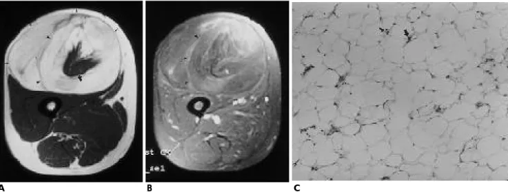
Fig. 1. A 59-year-old woman with well differentiated liposarcoma in the thigh.
A. T1-weighted axial image shows well defined mass (small arrows) in anterior compartment of the thigh with same signal intensity as that of fat. Intratumoral septa (arrowheads) and entrapped rectus femoris muscle (large arrow) are noted.
B. Enhanced fat suppression axial image shows septal enhancement (arrowheads).
C. Photomicrograph (H&E, ×100) shows lipoblasts with deeply staining nuclei (arrows).
A B C
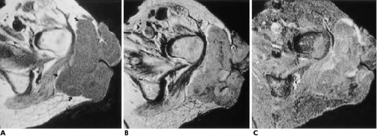
Fig. 2. A 71-year-old man with recurrent mass in the buttock.
A-C. Round mass (arrowheads) in the buttock shows low signal intensity on T1-weighted image (A), heterogeneous enhance- ment on enhanced image (B), and heterogeneous high signal in- tensity on T2-weighted image (C).
D. Microphotograph (H&E, ×200) represents dedifferentiated type. Primary mass was well differentiated liposarcoma, scle- rosing type.
A B C
D
3 0 0 0 / 6 0 - 8 0 / 2 - 4 )을 얻었고 Gd-DTPA (Magnevist, Schering, Germany, 0.1 mmol/Kg )를 이용하여 조영 증강 후 축상 및 종상 영상을 얻었다. 절편 두께는 5 - 1 0 m m였고 간격은 2 - 5 m m 였으며 m a t r i x는 2 5 6×256, FOV는 1 5 - 2 0 m m로 하였다. 모든 예 에서 수술을 시행하였으며 MR 소견을 조직학적 소견과 비교 분석하였다.
결 과
모든 종괴는 근육층 사이에 있었으며 주위 근육과 뚜렷한 경계를 보였다. 7예의 고분화형 중 6예는 종양 전체가 T1 및 T2 강조영상에서 지방과 같은 신호강도를 보였고 T1 및 T 2 강조영상에서 모두 내부에 저신호강도의 격막이 관찰되었다.
조직학적으로는 다양한 크기의 지방세포와 진하게 염색되는 핵을 가진 지방아세포들이 관찰되었다 (Fig. 1). 나머지 1예는 재발한 종괴로서 T1 강조영상에서 저신호강도, T2 강조영상에 서는 비균질한 고신호강도, 조영 후 비균질한 조영증강을 보였 으며 조직학적으로 역분화형 (dedifferentiated type)으로 확인 되었고 (Fig. 2) 재발 전 종괴의 아형은 고분화형 중 경화성 (sclerosing type)이었다. 7예의 점액형중 2예에서 각각 2개의 종 괴가 있었는데 1예는 완전히 분리된 서로 다른 종괴였고 다른 1예에서는 종괴가 장골( i l i u m )을 경계로 내측과 외측에 있었
다. 9개의 종괴 모두 T1 강조영상에서 저신호강도를 보였고, 6 개에서는 내부에 그물망 모양의 고신호강도가 관찰되었으며 T2 강조영상에서는 모두 고신호강도로 보였다. T1 강조영상에 서 고신호강도로 나타난 그물망 모양은 T2 강조영상에서는 저 신호강도로 관찰되었으며 조직학적으로 점액형 기질 내에 작 은 방추형 세포들과 모세혈관들이 관찰되었다 (Fig. 3) 조영증 강을 한 7개의 종괴 중 5개에서 비균질한 조영증강을 보였다.
3예의 혼합형 중 2예는 점액형과 고분화형의 혼합형이었는데 모든 신호강도에서 지방과 같은 신호강도를 보이는 부분 외에 그것과 경계가 잘 지어지는 앞에서 기술한 점액형과 같은 소 견을 보이는 부분이 있었으며 조영 후 비균질한 조영증강을 보였다. 조직학적으로 앞에서 기술한 고분화형과 점액형의 소 견을 보이는 두 부분이 뚜렷하게 구분되어 보였다 (Fig. 4). 1 예는 점액형과 다형성형의 혼합형이었고 두 부분이 뚜렷이 나 누어지지 않았으며 T1 및 T2 강조영상에서 각각 저신호강도 와 비균질한 고신호강도로, 그리고 조영 후 불규칙한 조영증강 을 보였다. 2예의 다형성형 (Fig. 5)과 1예의 원형세포형 ( F i g . 6) 그리고 분류가 불가능한 1예에서는 T1 강조영상에서 모두 저신호강도, T2 강조영상에서는 비균질한 고신호강도로 관찰 되었고 조영 후 불규칙한 조영증강을 보여 비특이적인 MR 소 견을 나타냈다. 조직학적으로는 다형성형과 원형세포형에서 각각 다형태의 종양세포와 둥근 종양세포들이 관찰되었다.
Fig. 3. A 58-year-old woman with myxoid liposarcoma in the but- t o c k .
A. T1-weighted axial image shows isointense masses (arrow- heads) to the muscle containing septa (arrows) with high signal intensity.
B. T2-weighted axial image shows homogeneous high signal in- tensity of the masses (arrowheads).
C. The mass shows heterogeneous enhancement (arrow) after ad- ministration of Gd-DTPA.
D. Photomicrograph (H&E, ×100) shows small spindle cells (ar- rows) in the myxoid background.
A B C
D
고 찰
지방육종은 악성 지방아세포에서 기원하는 연부 조직 육종 으로 주로 대퇴부(25-30 %)와 후복막강 (20 %)에 생기며 상 지와 어깨, 하지와 오금부, 둔부 그리고 배부에도 생길 수 있 다 (1, 3-4).
지방육종의 예후는 조직학적 분화 정도와 밀접한 관련이 있는데 분화가 좋은 형 (고분화형, 점액형)은 악성도가 낮아 국소 재발하는 경우는 있지만 전이 경향은 없고 분화가 좋지 않은 형 (원형세포형, 다형성형)은 국소 재발과 전이율이 높 다 (6, 7). 그러므로, 조직학적 아형에 따라 예후와 외과적 치 료 방향이 달라진다.
고분화형은 육안적으로 지방종과 매우 유사하며 조직학적 으로 지방아세포, 지방세포, 섬유성 격막으로 구성된다 ( 4 , 13). MR 신호강도도 지방과 같으나 지방종과 다른 점은 격막 이 두껍고 지방 성분과 다른 결절이 있다는 것이다 (11, 12).
본 연구에서도 MR 신호 강도가 지방과 같았으나, 종양 내 격 막이 T2 강조 영상에서 고신호강도로 보였던 Arkun 등의 (11) 연구 결과와는 달리 T2 강조 영상에서 저신호강도의 격 막이 관찰되었다. 통상 역분화형은 고분화형의 일부분이 악성 도가 높은 다형성 육종 (pleomorphic sarcoma)의 형태로 발견 될 때를 말하나 (secondary dedifferentiation) 일부는 처음부터
악성도가 높은 형태로 나오며 (de novo 또는 primary dediffer- entiation) 재발 또는 전이암이 원발암의 조직소견과 매우 다 를 때를 의미한다 (14, 15). 저자들의 경우는 후자에 속하며 원 발암은 경화성의 고분화형이었고 MR 소견은 대부분의 육종 과 같이 비특이적이었다. 점액형은 가장 흔한 아형으로 3가지 조직 성분 - 지방아세포 (lipoblast), 미세한 모세혈관 망, 그리 고 점액성 기질 - 으로 구성된다. 점액성 기질때문에 T1 강조 영상에서 내부 구조가 근육과 동등신호강도로 비교적 균질하 게 보이며 T2 강조영상에서 균질하게 고신호강도로 보인다고 하며 (11-13) 본 연구에서도 9개의 종괴가 모두 이와 같은 MR 소견을 보였다. 조영증강은 비균질하게 되는 것으로 알려 져 있으며 조영이 되지 않는 부분은 조직학적으로 모세혈관 망이 없는, 점액 성분이 축적된 부분에, 조영되는 부분은 좀 더 세포들이 많은 부분에 해당한다 (11). 본 연구에서도 조영 증강을 한 7개의 종괴 중 5개에서 비균질한 조영증강을 보였 는데 조영증강이 되었던 종괴가 그렇지 않은 종괴보다 세포밀 도가 높았다. Sundaram등 ( 1 3 )에 의하면 7 1 %에서 T1 강조영 상에서 그물망 형태의 고신호강도가 보였고 이는 지방조직에 의한 것이라 하였고 A r k u n등 ( 1 1 )에 의하면 이 부분이 T2 강 조영상에서 저신호강도로 보인다고 하였는데 본 연구에서도 6예 ( 6 7 % )에서 이러한 소견이 보였으나 조직학적으로 지방 성분을 확인하지는 못했다. 다형성형은 종양의 악성도가 가장 높은 형으로 전이율이 가장 높고 조직학적으로 특징적인 지방 Fig. 4. A 43-year-old woman with mixed liposarcoma in the but- t o c k .
A, B. T1-(A) and T2-weighted (B) axial images show mass with two components; lateral one (▲) with signal intensity equal to the fat, medial one (open arrow) with low signal intensity on T1- weighted image and homogeneous high signal intensity on T2- weighted image. Hyperintense septa in the mass is noted on T1- weighted image (arrows).
C. Enhanced scan shows heterogeneous enhancement of medial o n e .
D. Microphotograph (H&E, ×100) demonstrates sharply defined border (arrowheads) between well differentiated type (left side) and myxoid type (right side).
A B C
D
Fig. 5. A 58-year-old woman with pleomorphic liposarcoma in the buttock.
A, B. Lobular contoured mass (arrows) in subcutaneous layer of the buttock shows low signal intensity on T1-weighted image (A) and mixed signal intensity on T2 weighted image (B).
C. Enhanced fat suppression image shows heterogeneous en- hancement of the mass.
D. Microphotograph (H&E, ×200) shows pleomorphism of the tumor cells (arrows).
A B C
D
Fig. 6. A 74-year-old woman with round cell liposarcoma in the infrapatellar area.
A. T1-weighted sagittal image shows infrapatellar mass (arrows) with low signal intensity.
B. T2-weighted image shows mixed high and low signal intensity on the mass.
C. Microphotograph (H &E, ×200) shows round shaped tumor cells (arrows).
A B C
을 보여 지방육종의 다른 형과 감별진단이 가능하였다.
참 고 문 헌
1 . Springfield D. Liposarcoma. Clin Orthop1 9 9 3 ; 2 8 9 : 5 0 - 5 7
2 . Reszel PA, Soule EH, Coventry MB. Liposarcoma of the extremi- ties and limb girdles: a study of 222 cases. J Bone Joint Surg [A m]
1 9 6 6 ; 4 8 : 2 2 9 - 2 4 4
3 . Enzinger FM, Weiss SW. Soft tissue tumors. 2nd ed. St Louis:
Mosby, 1988:346-382
4 . Kransdorf MJ, Moser RP Jr., Meis JM, Meyer CA. Fat-containing soft-tissue masses of the extremities. R a d i o G r a p h i c s 1 9 9 1 ; 1 1 : 8 1 - 1 0 6 5 . Evans HL. Liposarcoma: a study of 55 cases with a reassessment of
its classification. Am J Surg pathol 1 9 7 9 ; 3 : 5 0 7 - 5 2 3
6 . Reitan JB, Kaalhus O, Brennhovd IO, Sager EM, Stenwig AE, Talle
1 1 . Arkun R, Memis A, Akalin T, Ustun E, Sabah D, Kandioglu G.
Liposarcoma of soft tissue: MRI findings with pathologic correla- tion. Skeletal Radiol 1997;26:167-172
1 2 . Jelinek JS, Kransdorf MJ, Shmookler BM, Aboulafia AJ, Malawer MM. Liposarcoma of the extremities: MR and CT findings in the histologic subtypes. R a d i o l o g y 1 9 9 3 ; 1 8 6 : 4 5 5 - 4 5 9
1 3 . Sundaram M, Baran G, Merenda G, McDonald DJ. Myxoid li- posarcoma: magnetic resonance imaging appearance with clinical and histologic correlation. Skeletal Radiol 1990;19:359-362 1 4 . Nascimento AG, Kurtin PJ, Guillou L, Fletcher CDM. Dedifferen-
tiated liposarcoma a report of 9 cases with a peculiar neurallike whorling pattern associated with metaplastic bone formation. Am J Surg Pathol 1 9 9 8 ; 2 2 : 9 4 5 - 9 5 5
1 5 . Henricks WH, Chu YC, Goldblum JR, Weiss SW. Dedifferentiated liposarcoma a clinicopathological analysis of 155 cases with a pro- posal for an expanded definition of dedifferentiation. Am J Surg P a t h o l 1997;21:271-281
Address reprint requests to : Jeong Hoon Lee, M.D., Department of Diagnostic Radiology Korea Cancer Center Hospital
#215-4 -Dong, -Ku, Seoul, 139-706, Korea.
Tel. 82-2-974-2501 Fax. 82-2-972-3093
Purpose : To evaluate the MR imaging findings of liposarcomas of different histologic subtypes.
Materials and Methods : We evaluated MR images of 21 patients (5 men and 16 women, mean age, 55 years) with liposarcoma and correlated the findings with the results of histopathology. In the study group seven liposarcomas were well-differentiated, seven were myxoid, three were mixed, two were pleomorphic, and one was round cell.
Results : On T1 -and T2 - weighted images, six of seven well-differentiated liposarcomas showed signal intensity equal to the fat and hypointense septa, while the other showed low signal intensity on a T1 - weighted image, heterogeneous high signal intensity on a T2- weighted image, heterogeneous enhancement after the administration of contrast media and was dedifferentiate. Nine masses in seven patients with myxoid liposarcoma showed low signal intensity on T1-weighted images, six of the nine showed lace-like foci of high signal intensity. On T2 -weighted images, all masses showed homogeneous high signal intensity.
After administration of contrast media, five of seven masses showed heterogeneous enhancement. Two of three mixed form were well-differentiated and myxoid types, and two subtypes were separable on MR.
Pleomorphic, round cell, mixed type myxoid and pleomorphic and unclassified cases showed low signal intensity on T1-weighted images, heterogeneous high signal intensity on T2-weighted and heterogeneous e n h a n c e m e n t .
Conclusion : Using MR imaging, well-differentiated and myxoid liposcarcomas may be differentiated from other types.
Index words : L i p o s a r c o m a
Magnetic resonance(MR), tissue characterization Soft tissues, MR
Soft tissues, neoplasms