Biomedical Science Letters 2019, 25(1): 23~31 https://doi.org/10.15616/BSL.2019.25.1.23 eISSN : 2288-7415
Estrogen Induces CK2α Activation via Generation of Reactive Oxygen Species
Soo-Yeon Jeong* and Suhn-Young Im†,* *
Department of Biological Sciences, College of Natural Sciences, Chonnam National University, Gwangju 61186, Korea
The protein kinase CK2α (formerly Casein Kinase II) is implicated in tumorigenesis and transformation. However, the mechanisms of CK2α activation in breast cancer have yet to be elucidated. This study investigated the mechanisms of CK2α activation in estrogen signaling. Estrogen increased reactive oxygen species (ROS) production, CK2α activity, and protein expression in estrogen receptor positive (ER+) MCF-7 human breast cancer cells, which were inhibited by the antioxidant N-acetyl-L-cysteine. H2O2 enhanced CK2α activity and protein expression. Human epidermal growth factor (EGF) increased ROS production, CK2α activity and protein expression in EGF receptor 2 (HER2)-overexpressing MCF-7 (MCF-7 HER2) cells, but not in MCF-7 cells. Estrogen induced the phosphorylation of p38 mitogen-activated protein kinase (MAPK). The p38 inhibitor, SB202190, blocked estrogen-induced increases in ROS production, CK2α activity and CK2α protein expression. The data suggest that ROS/p38 MAPK is the key inducer of CK2α activation in response to estrogen or EGF.
Key Words: Estrogen, CK2α, ROS, P38 MAPK
INTRODUCTION
Breast cancer is the most common cause of cancer death among women (Ferlay et al., 2010), and is characterized by molecular and cellular heterogeneity (Perou et al., 2000).
The etiology of breast cancer is multifactorial, with major risk factors including age, early menarche, delayed meno- pause, use of contraceptives or oral medications, hormone therapy, family history, history of benign breast disease, and obesity; estrogen is a major risk factor for breast carcino- genesis and growth of breast cancer cells (Yager and Liehr, 1996; Clemons and Goss, 2001).
Reactive oxygen species (ROS) act as potent mutagens,
increasing genomic instability and initiation as well as the progression of cancer (Carew and Huang, 2002). Oxidative stress is one of the mechanisms underlying the carcinogenic effect of estrogen in breast cancer. Genotoxic estrogen metab- olites such as catechols and estrogen-semi-quinones gene- rated during estrogen metabolism are known to produce ROS (Roy et al., 2007; Okoh et al., 2011). Mitochondria are the main source of estrogen-induced ROS in breast cancer cells (Felty et al., 2005; Parkash et al., 2006).
Protein kinase CK2α (formerly Casein Kinase 2 or II) is a highly conserved and ubiquitous serine/threonine kinase, which is localized in the cell nucleus and cytoplasm. CK2α is a tetrameric complex composed of catalytic (α or α') and regulatory (β) subunits with a α2β2, αα'β2, or α'2β2 structure
Original Article
Received: October 22, 2018 / Revised: February 8, 2019 / Accepted: February 12, 2019
*Graduate student, **Professor.
†Corresponding author: Suhn-Young Im. Department of Biological Sciences, College of Natural Sciences, Chonnam National University, Gwangju 61186, Korea.
Tel: +82-62-530-3414, Fax: +82-62-530-3409, e-mail: [email protected]
○CThe Korean Society for Biomedical Laboratory Sciences. All rights reserved.
○CCThis is an Open Access article distributed under the terms of the Creative Commons Attribution Non-Commercial License (http://creativecommons.org/licenses/by-nc/3.0/) which permits unrestricted non-commercial use, distribution, and reproduction in any medium, provided the original work is properly cited.
(Allende and Allende, 1995). CK2α modulates 300 potential substrates in the cell, as well as a wide range of pathways related to cell growth, proliferation and apoptosis (Litchfield, 2003; Duncan and Litchfield, 2008). CK2α is implicated in tumorigenesis and transformation. CK2α inhibits the activity and stability of tumor suppressor proteins (p53 and phospha- tase and tensin homolog deleted on chromosome 10 (PTEN)), promotes cell survival signaling via up-regulation of proto- oncogenic products (c-Myc, c-Myb, and c-Jun) and transcri- ptional activators (α-catenin and Max) (Singh and Ramji, 2008). Overexpression of CK2α has been observed in many human cancers, including breast cancers (Filhol et al., 2015;
Chua et al., 2017). However, the underlying mechanisms of CK2α expression in breast cancers remain elusive.
The molecular mechanism underlying the activation of CK2α is mediated via ROS (Kim et al., 2014). Therefore, it is of interest to investigate whether a similar mechanism occurs in estrogen signaling. In this study, we found that estrogen increased CK2α activation and expression via ROS /p38 mitogen activated protein kinase (MAPK) in estrogen receptor-positive (ER+) cell lines.
MATERIALS AND METHODS Cell cultures
MCF-7 and T47D, MBA-MB-231 human breast cancer cells (American Type Culture Collection, ATCC, Manassas, VA, USA Rockville, MD, USA) were cultured in Dulbecco's Modified Eagle's Medium (DMEM) or RPMI1640 medium (GIBCO Invitrogen, Carlsbad, CA, USA) supplemented with 10% (volume/volume) fetal bovine serum (GIBCO Invitro- gen, Carlsbad, CA, USA) at 37℃ in a humidified atmos- phere of 5% CO2 and 95% air. MCF-7 overexpressing HER2 (MCF-7 HER2+) and vector control (MCF-7 vec) cells were kindly provided by professor Ko, and were described else- where (Seo et al., 2012).
Antibodies and reagents
The following antibodies were used for Western blot analysis. Anti-phospho-CK2 (Thr306, Ser362) (Sigma-Aldrich, St Louis, MO, USA), anti-CK2α, anti-phospho-p38 mitogen activated protein kinase (MAPK) (Thr180, Tyr182), anti-
phospho-JNK anti-phospho-ERK were purchased from Cell Signaling Technology (Beverly, MA, USA). Anti-GAPDH, anti-normal rabbit IgG were from Santa Cruz Biotechnology (Santa Cruz, CA, USA). The antioxidant, N-Acetyl-L-Cysteine (Sigma-Aldrich), p38 inhibitor (SB202190, 5 mg/kg), JNK inhibitor (SP600125, 12.5 mg/kg) or ERK inhibitor (PD- 98059, 12.5 mg/kg) were purchased from Calbiochem Merck (Darmstadt, Germany).
Measurement of intracellular ROS
The oxidation-sensitive fluorescent probes CM-H2DCF- DA were used to monitor the production of hydrogen per- oxide. Cells in each well were treated with estrogen and/or reagents for 3 hr, after which they were incubated with 5 μM CM-H2DCF-DA (DCF) (Sigma-Aldrich) for the detection of cytosolic ROS for 30 min at 37℃ in the dark. Fluorescence was read with excitation at 485 nm and emission at 530±
30 nm with an automated microplate reader (IniniteTM M200 (Tecan, AG, Switzerland).
CK2α activity assay
Cells were homogenized in lysis buffer (50 mM Tris-HCl, pH 7.5, 150 mM NaCl, 1% Triton X-100, 0.5% sodium deoxycholate, 0.1% SDS, 1 mM EGTA, 1 mM EDTA, 10 mM sodium fluoride, 1 mM sodium orthovanadate, 1 mM dithiothreitol and 1 mM PMSF). The phosphotransferase activity of CK2α was measured using a CK2α assay kit (Millipore, Temecula, CA, USA) in accordance with the manufacturer's recommendations. Briefly, cell lysates, sub- strate peptide, protein kinase A inhibitor cocktail, and γ- [32P] ATP in the assay dilution buffer were incubated for 10 min at 30℃. The phosphorylated substrate was then separ- ated on P81 phosphocellulose paper and quantified with a scintillation counter.
Western blot analysis
Whole cell extracts were prepared using PhosphoSafe Extraction Reagent (Novagen Merck, Darmstadt, Germany) with phenylmethylsulfonyl fluoride protease inhibitor (Sigma- Aldrich, St Louis, MO, USA). Equal amounts of cell lysates were separated on 10% SDS-polyacrylamide gel under re- ducing conditions, and electrophoretically transferred onto
Protran Nitrocellulose Transfer Membranes (Whatman, Dassel, Germany). The membranes were blocked by 30 min of in- cubation at room temperature (RT) in 5% BSA in Tris- buffered saline (TBS)-0.1% Tween 20, followed by 2 hr in- cubation at 4℃ with primary antibodies in 5% BSA in TBS- 0.1% Tween 20. The blots were washed for 30 min with three changes of TBS-0.1% Tween 20 solution, followed by incubation for 1 hr at RT with horseradish peroxidase- conjugated anti-rabbit IgG antibody (Santa Cruz Biotech- nology, Santa Cruz, CA, USA). The blots were again washed three times for 30 min each, and finally developed in Lumi- GLO reagent (ECL, GE Health Care UK Limited Cell Sig-
naling Technology, Beverly, MA, USA). GAPDH was used as a loading control. The density of each band was deter- mined using the densitometry Fluor-STM Imager (Bio-Rad, Muncher, Germany).
Statistical analysis
Data are expressed as means ± SE Statistical significance was determined via one-way analysis of variance (StatView;
Abacus Concepts Inc., Berkeley, CA, USA). All experiments were conducted at least twice. Reproducible results were obtained and representative data are, therefore, provided in the figures.
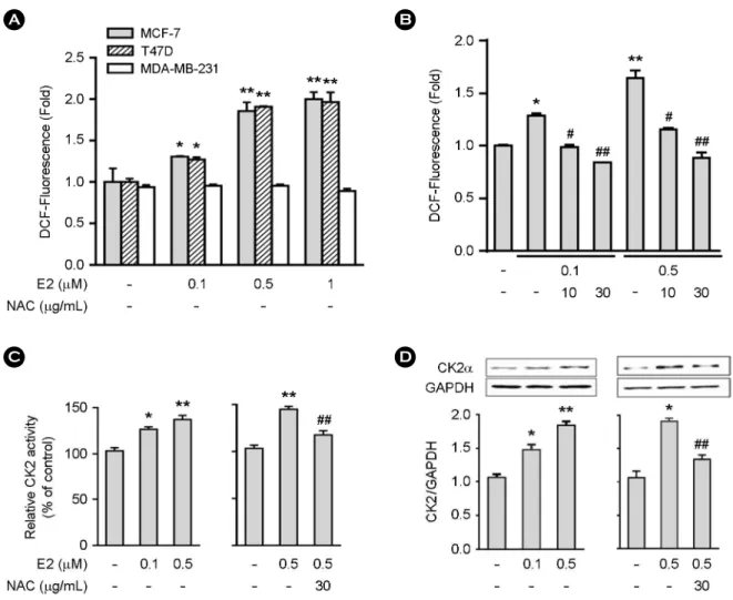
Fig. 1. Estrogen increases ROS production in ER+ MCF-7 and T47D cells. A and B, Cells (4 × 10³) were treated for 3 hr with varying concentrations of estrogen for the measurement of ROS. B, MCF-7 cells (4 × 10³) were treated for 3 hr with varying concentrations of estrogen NAC (10 or 30 μg/mL) was added 30 min before estrogen (0.1 or 0.5 μM) treatment (B). C and D, Cells (1 × 10⁶) were treated with NAC and/or estrogen for the measurement of CK2 activity (C) and protein expression (D). Data represent the mean ± SE of two to three independent experiments with three to five samples per experiment. *P < 0.01 versus control group. **P < 0.001 versus control group. #P < 0.01 versus estrogen-treated group. ##P < 0.001 versus estrogen-treated group.
A
C
B
D
RESULTS
Estrogen increases ROS production and CK2α expression
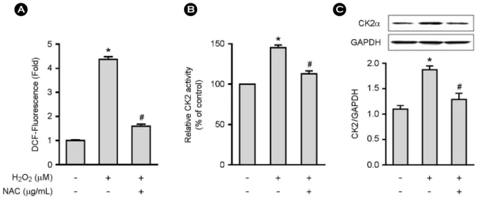
We first assessed the effect of estrogen on ROS production and CK2α expression using estrogen receptor (ER)-positive MCF-7 and T47D and ER-negative MDA-MB-231 breast cancer cells. ROS production was increased about 1.7-fold in ER+ MCF-7 and T47D cells upon estrogen stimulation, but such increase was not observed in ER-negative MDA- MB-231 cells (Fig. 1A). The presence of NAC, a commonly used ROS scavenger, blocked the estrogen-induced ROS production (Fig. 1B). CK2α activity was increased in a dose- dependent manner in MCF-7 cells upon estrogen stimulation (Fig. 1C). We also investigated the effect of estrogen on CK2α protein expression by Western blot analysis, which revealed that estrogen increased CK2α protein expression in MCF-7 cells in dose-dependent manners (Fig. 1D). Both estrogen-induced CK2α activity and protein expression were blocked by NAC (Fig. 1C and D). We analyzed the direct role of ROS in CK2α activity and protein expression. The addition of H2O2 to MCF-7 cells resulted in increases in ROS production (Fig. 2A), CK2α activity (Fig. 2B) and protein expression (Fig. 2C), which were blocked by NAC. These data indicate that estrogen upregulates CK2α activity and
protein expression via ROS production in ER+ breast cancer cells.
Comparison of ROS production, p38 phosphorylation, and CK2α activity and protein expression: MCF-7 versus MCF-7 HER2 cells
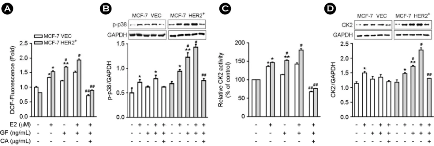
MCF-7 vec and MCF-7 HER2+ cells were stimulated either estrogen alone or estrogen combined with EGF. When the cells were exposed to estrogen alone, ROS production (Fig. 3A), p38 phosphorylation (Fig. 3B), CK2α activity (Fig. 3C), and protein expression (Fig. 3D) were increased similarly in both cell lines. When the cells were stimulated with EGF alone, ROS production (Fig. 3A), p38 phospho- rylation (Fig. 3B), CK2α activity (Fig. 3C), and protein expression (Fig. 3D) were only increased in MCF-7 HER2+ cells. When the cells were treated with estrogen + EGF, more higher increases in ROS production (Fig. 3A), p38 phos- phorylation (Fig. 3B), CK2α activity (Fig. 3C) and protein expression (Fig. 3D) were observed in MCF-7 HER2+ cells compared to MCF-7 vec cells. All the increases of ROS pro- duction and CK2α activity, and protein expression induced by exposure to estrogen combined with EGF in MCF-7 HER2+ cells were blocked by pre-treatment with NAC (Fig.
3A-D). These data indicate that the presence of HER2 in breast cells contributes to higher levels of ROS production
A B C
Fig. 2. H2O2 increases CK2 activity and protein expression in MCF-7 cells. A, Cells (4 × 10³/well for A; 2 × 10⁶/well for B and C) were pretreated with NAC (30 μg/mL) for 30 min followed by H2O2 (30 μM) for 3 hr to quantify ROS levels (A), CK2 activity (B) and protein expression (C). Data represent the mean ± SE of two to three independent experiments with three to five samples per experiment. *P
< 0.001 versus control group. #P < 0.001 versus H2O2-treated group.
and CK2α activity and expression.
ROS-dependent p38 mediates CK2α activation
We next investigated the roles of p38, JNK, and ERK MAPK in estrogen-induced CK2α activation. Estrogen in- duced phosphorylation of the three MAPK in MCF-7 cells, all of which were blocked by NAC (Fig. 4). Pre-treatment of cells with the p38 inhibitor, SB202190, blocked estrogen- induced increases in CK2α activity (Fig. 5A) and protein expression (Fig. 5B). JNK inhibitor or ERK inhibitor failed to inhibit estrogen-induced CK2α expression (data not shown), suggesting that JNK and ERK do not involved importantly
in this context. Further, SB202190 inhibited H2O2-induced CK2α activation (Fig. 5C) and protein expression (Fig. 5D), suggesting that p38 mediated CK2α activity and protein expression.
DISCUSSION
The mechanism of CK2α overexpression in breast cancers is poorly understood. In this study, we showed that estrogen induced CK2α activation and protein expression via ROS/
p38 signaling. It is well known that estrogen increases ROS production via several mechanisms including estrogen me- Fig. 3. EGF increases ROS production, CK2 activity and protein expression in MCF-7 HER2+ cells. MCF-7 vec or MCF-7 HER2+ cells (4 × 10³/well for A; 1 × 10⁶/well for B and C) were pretreated with NAC (30 μg/mL) for 30 min followed by estrogen (0.5 μM) or EGF (50 ng/mL) for the measurement of ROS (A), p38 phosphorylation (B), CK2 activity (C) and protein expression (D). Data represent the mean ± SE of two to three independent experiments with three to five samples each. *P < 0.01 versus control group. **P < 0.001 versus control group. #P < 0.01 versus EGF-treated MCF-7 vec group. ##P < 0.001 versus estrogen + EGF-treated group.
A B C D
Fig. 4. Estrogen induces MAPK phosphorylation in MCF-7 cells. Cells (1 × 10⁶) were treated with estrogen (0.5 μM) for the time indicated to measure the phosphorylation of p38, JNK, and ERK. To see the effect of NAC, cells were pretreated with NAC (30 μg/mL) for 30 min, and were cultured for another 30 min with estrogen. Data represent the mean ± SE of three independent experiments with three samples each. *P < 0.005 versus control group. #P < 0.001 versus estrogen-treated group.
tabolism and generation of genotoxic estrogen metabolites such as catechol estrogen and estrogen-semi-quinones (Roy et al., 2007; Okoh et al., 2011). We found that estrogen pro- duced ROS in ER+ MCF-7 and T47D cells, but not in ER- MDA-MB-231 cells. ROS can be generated intracellularly by a variety of enzymes (i.e., NADPH oxidase, xanthine oxi- dase, lipoxygenase) and through the mitochondrial respira- tory chain (Sauer et al., 2001). Mitochondria however, are the largest source of ROS in epithelial cells. Studies have shown that mitochondria is the major source of intracellular ROS
in response to estrogen (Felty et al., 2005).
The ROS generated by estrogen induced CK2α activation and protein expression via CK2α activation and protein expression, which were blocked by NAC. The ROS (H2O2) also directly increased the CK2α activity and protein ex- pression.
The members of the epidermal growth factor receptor (EGFR)/ErbB family: EGFR (also known as ErbB1 and HER1), HER2 (also known as HER2/neu and ErbB2), ErbB3 (also known as HER3), and ErbB4 (also known as
A B
D C
Fig. 5. The p38 mediates estrogen-induced CK2 activation in MCF-7 cells. Cells (1 × 10⁶) were pretreated with SB202190 (20 μM) for 30 min followed by estrogen (0.5 μM, A and B) or H2O2 (30 μM, C and D) for the measurement of CK2 activity (A, C) and protein expression (B, D). Data represent the mean ± SE of two to three independent experiments with three to five samples each. *P < 0.01 versus control group. #P < 0.01 versus estrogen-treated group. ##P < 0.01 versus H2O2-treated group.
HER4) are the most notable cancer molecular targets identi- fied to date (Mendelsohn and Baselga, 2003). Overexpres- sion of HER2, which is a transmembrane receptor tyrosine kinase (Groenen et al., 1994; Lemke, 1996) is seen in 30%
of breast cancer patients and is associated with poor survival, increased metastasis, and resistance to chemotherapy (Slamon et al., 1987; Toikkanen et al., 1992). As a result, HER2 is the most well established therapeutic target in breast cancer.
EGFR overexpression is observed in all subtypes of breast cancer, EGFR is more frequently overexpressed in triple- negative breast cancer (TNBC) and inflammatory breast cancer, which are especially aggressive (Rakha et al., 2007;
Burness et al., 2010).
In addition to estrogen, we observed that EGF increased ROS levels and CK2 activation as well in MCF-7 HER 2 cells. Treatment of the cells with the combinations of estro- gen and EGF led to enhanced ROS and CK2α activation compared with stimulation with either estrogen or EGF alone.
What is the mechanism of up-regulation of ROS/ CK2α in MCF-7 HER 2 cells? NADPH oxidase was specifically in- volved in the generation of ROS by growth factors, such as transforming growth factor-β1 (Ohba et al., 1994; Thannickal and Fanburg, 1995), insulin (Mahadev et al., 2001), PDGF- and EGF (Sundaresan et al., 1995; Bae et al., 1997). Add- itionally, we previously reported that exogenous stimuli such as platelet activating factor, LPS, TNF-α, and interleukin-1 triggering ROS generation were found to enhance CK2α activity (Kim et al., 2014). Therefore, up-regulation of CK2α in MCF-7 HER 2 cells is probably attributed to EGF- dependent generation of higher levels of ROS, which may explain the aggressive features of HER2-positive breast car- cinomas and their rapid proliferation compared with other types of breast cancer (Me´nard et al., 2003).
The p38 MAPK signaling pathway mediates the inflam- matory response via proteins, such as proinflammatory cyto- kines, chemokines, degradative enzymes, growth factors, and adhesion molecules (Zhang et al., 2007). P38 is an oxidative stress-sensitive kinase. During the ROS-mediated p38 activa- tion, a few protein kinases, such as apoptosis signal-regulated kinase 1, an upstream MAPK kinase kinases (Saitoh et al., 1998; Fujino et al., 2007), cGMP-dependent protein kinase (Li et al., 2006; Hofmann et al., 2006), protein kinase A
(Brennan et al., 2006), and protein kinase C (Giorgi et al., 2010) that are directly regulated by ROS, are known to phos- phorylate and activate p38 MAPK. We have previously reported that p38 is involved in ROS-mediated CK2α modu- lation (Kim et al., 2014). Consistent with these findings, we demonstrated that p38 phosphorylation occurred in response to estrogen, which was abrogated by pre-treatment with NAC, indicating that ROS generated by estrogen induced p38 phosphorylation. Indeed, the p38 inhibitor, SB202190, was shown to inhibit CK2α activation and protein expression (Fig. 5), indicating that p38 kinase catalyzed ROS-mediated CK2α activation and protein expression. Therefore, appar- ently ROS/p38 is a key common upstream pathway under- lying the enhanced CK2α activity, suggesting that the ROS/
p38 leading to CK2α activation mediated estrogen signaling.
Classification of breast cancer has been historically based on both analysis of tumor morphology and histological detection of three marker proteins: the ER, the progesterone receptor (PR), and the HER2 or ErbB2. Tumors which ex- press none of these three markers are collectively referred to as triple-negative breast cancer (TNBC; ER-, PR-, HER2-).
More recent gene expression analyses showed that breast cancer is a more heterogeneous disease than previously assumed. The current classification includes five major trans- criptional subtypes: basal-like, HER2-enriched, luminal A, luminal B, and normal breast-like (Perou et al., 2000; Sorlie et al., 2003). Recently, the aberrant expression of CK2α and its subtypes in breast cancers has been reported (Giusiano et al., 2011). Therefore, it is of interest to investigate the linkage between oxidative stress and p38/CK2α expression in various types of breast cancer in the future.
In summary, we demonstrated that ROS/p38 acts as a key inducer of CK2α activation in response to estrogen or growth factor (EGF) stimulation, i.e., estrogen or EGF → ROS↑→ p38 MAPK↑ → CK2α activation↑. Therefore, this study adds an important element to our understanding of the mechanisms underlying CK2α activation in breast cancers.
ACKNOWLEDGEMENT
This work was supported by the Mid-Career Researcher Program through the National Research Foundation of Korea
(NRF) grant (No. 2015-1397, 2016-1093).
CONFLICT OF INTEREST
No conflicts of interest are involved in this manuscript.
REFERENCES
Allende JE, Allende CC. Protein kinases. 4. Protein kinase CK2:
an enzyme with multiple substrates and a puzzling regulation.
FASEB Journal. 1995. 9: 313-323.
Bae YS, Kang SW, Seo MS, Baines IC, Tekle E, Chock PB, Rhee SG. Epidermal growth factor (EGF)-induced generation of hydrogen peroxide. Role in EGF receptor-mediated tyrosine phosphorylation. Journal of Biological Chemistry. 1997. 272:
217-221.
Brennan JP, Bardswell SC, Burgoyne JR, Fuller W, Schröder E, Wait R, Begum S, Kentish JC, Eaton P. Oxidant-induced acti- vation of type I protein kinase A is mediated by RI subunit interprotein disulfide bond formation. Journal of Biological Chemistry. 2006. 281: 21827-21836.
Burness ML, Grushko TA, Olopade OI. Epidermal growth factor receptor in triplenegative and basal-like breast cancer: pro- mising clinical target or only a marker? Cancer Journal. 2010.
16: 23-32.
Carew JS, Huang P. Mitochondrial defects in cancer. Molecular Cancer. 2002. 1: 15.
Chua MM, Ortega CE, Sheikh A, Lee M, Abdul-Rassoul H, Hartshorn KL, Dominguez I. CK2 in Cancer: Cellular and biochemical mechanisms and potential therapeutic target.
Pharmaceuticals (Basel). 2017. 10: 30.
Clemons M, Goss P. Estrogen and the risk of breast cancer. New England Journal of Medicine. 2001. 344: 276-285.
Duncan JS, Litchfield DW. Too much of a good thing: the role of protein kinase CK2 in tumorigenesis and prospects for thera- peutic inhibition of CK2. Biochimica et Biophysica Acta. 2008.
1784: 33-47.
Felty Q. Singh, KP, Roy D. Estrogen-induced G1/S transition of G0-arrested estrogen-dependent breast cancer cells is regulated by mitochondrial oxidant signaling. Oncogene. 2005. 24: 4883 -4893.
Felty Q, Xiong WC, Sun D, Sarkar S, Singh KP, Parkash J, Roy D.
Estrogen induced mitochondrial reactive oxygen species as signal-transducing messengers, Biochemistry. 2005. 44: 6900 -6909.
Ferlay J, Shin HR, Bray F, Forman D, Mathers C, Parkin DM. Esti- mates of worldwide burden of cancer in 2008: GLOBOCAN 2008. International Journal of Cancer. 2010. 127: 2893-2917.
Filhol O, Giacosa S, Wallez Y, Cochet C. Protein kinase CK2 in breast cancer: the CK2β regulatory subunit takes center stage in epithelial plasticity. Cellular and Molecular Life Sciences.
2015. 72: 3305-3322.
Fujino G, Noguchi T, Matsuzawa A, Yamauchi S, Saitoh M, Takeda K, Ichijo H. Thioredoxin and TRAF Family Proteins Regulate Reactive Oxygen Species-Dependent Activation of ASK1 through Reciprocal Modulation of the N Terminal Homophilic Interaction of ASK1. Molecular and Cellular Biology. 2007.
27: 8152-8163.
Giorgi C, Agnoletto C, Baldini C, Bononi A, Bonora M, Marchi S, Missiroli S, Patergnani S, Poletti F, Rimessi A, Zavan B, Pinton P. Redox control of protein kinase C: Cell-and disease-specific aspects. Antioxidants & Redox Signaling. 2010. 13: 1051-1085.
Giusiano S, Cochet C, Filhol O, Duchemin-Pelletier E, Secq V, Bonnier P, Carcopino X, Boubli L, Birnbaum D, Garcia S, Iovanna J, Charpin C. Protein kinase CK2α subunit over- expression correlates with metastatic risk in breast carcinomas:
quantitative immunohistochemistry in tissue microarrays.
European Journal of Cancer. 2011. 47: 792-801.
Groenen LC, Nice E, Burgess AW. Structure-function relationships for the EGF/TGF-alpha family of mitogens. Growth Factors.
1994. 11: 235-257.
Hofmann F, Feil R, Kleppisch T, Schlossmann J. Function of cGMP Dependent Protein Kinases as Revealed by Gene Deletion.
Physiological Review. 2006. 86: 1-23.
Kim KJ, Cho KD, Jang KY, Kim HA, Kim HK, Lee HK, Im SY.
Platelet-activating factor enhances tumor metastasis via the reactive oxygen species-dependent protein kinase casein kinase 2-mediated nuclear factor-κB activation. Immunology. 2014.
143: 21-32.
Lemke G. Neuregulins in development. Molecular and Cellular Neuroscience. 1996. 7: 247-262.
Li Z, Zhang G, Feil R, Han J, Du X. Sequential activation of p38 and ERK pathways by cGMP-dependent protein kinase leading to activation of the platelet integrin αIIbβ3. Blood. 2006. 107:
965-972.
Litchfield DW. Protein kinase CK2: structure, regulation and role in cellular decisions of life and death. Biochemical Journal.
2003. 369: 1-15.
Mahadev K, Zilbering A, Zhu L, Goldstein BJ. Insulin-stimulated hydrogen peroxide reversibly inhibits protein-tyrosine phos-
phatase 1b in vivo and enhances the early insulin action cascade.
Journal of Biological Chemistry. 2001. 276: 21938-21942.
Me'nard S, Pupa SM, Campiglio M, Tagliabue E. Biologic and therapeutic role of HER2 in cancer. Oncogene. 2003. 22: 6570 -6578.
Mendelsohn J, Baselga J. Status of epidermal growth factor receptor antagonists in the biology and treatment of cancer. Journal of Clinical Oncology. 2003. 21: 2787-2799.
Ohba M, Shibanuma M, Kuroki T, Nose K. Production of hydrogen peroxide by transforming growth factor-beta 1 and its involve- ment in induction of egr-1 in mouse osteoblastic cells. Journal of Cell Biology. 1994. 126: 1079-1088.
Okoh V, Deoraj A, Roy D. Estrogen-induced reactive oxygen species-mediated signalings contribute to breast cancer. Bio- chim Biophys Acta. 2011. 1815: 115-133.
Parkash J, Felty Q, Roy D. Estrogen exerts a spatial and temporal influence on reactive oxygen species generation that precedes calcium uptake in high-capacity mitochondria: implications for rapid nongenomic signaling of cell growth. Biochemistry.
2006. 45: 2872-2881.
Perou CM, Sørlie T, Eisen MB, van de Rijn M, Jefrey SS, Rees CA, Pollack JR, Ross DT, Johnsen H, Akslen LA, Fluge O, Pergamenschikov A, Williams C, Zhu SX, Lønning PE, Børresen-Dale AL, Brown PO, Botstein D. Molecular portraits of human breast tumours. Nature. 2000. 406: 747-752.
Rakha EA, El-Sayed ME, Green AR, Lee AH, Robertson JF, Ellis IO. Prognostic markers in triple-negative breast cancer. Cancer.
2007. 109: 25-32.
Roy D, Cai Q, Felty Q, Narayan S. Estrogen-induced generation of reactive oxygen and nitrogen species, gene damage, and estrogen-dependent cancers. Journal of Toxicology and Envir- onmental Health Part B Critical Reviews. 2007. 10: 235-257.
Saitoh M, Nishitoh H, Fujii M, Takeda K, Tobiume K, Sawada Y, Kawabata M, Miyazono K, Ichijo H. Mammalian thioredoxin is a direct inhibitor of apoptosis signal-regulating kinase (ASK) 1. EMBO Journal. 1998. 17: 2596-2606.
Sauer H, Wartenberg M, Hescheler J. Reactive oxygen species as intracellular messengers during cell growth and differentiation.
Cellular Physiology and Biochemistry. 2001. 11: 173-186.
Seo HS, Choi HS, Kim SR, Choi YK, Woo SM, Shin I, Woo JK,
Park SY, Shin YC, Ko SG. Apigenin induces apoptosis via extrinsic pathway, inducing p53 and inhibiting STAT3 and NFκB signaling in HER2-overexpressing breast cancer cells.
Molecular Cellular Biochemistry. 2012. 366: 319-334.
Singh NN, Ramji DP. Protein kinase CK2, an important regulator of the inflammatory response? Journal of Molecular Medicine (Berl). 2008. 86: 887-897.
Slamon DJ, Clark GM, Wong SG, Levin WJ, Ullrich A, McGuire WL. Human breast cancer: correlation of relapse and survival with amplification of the HER-2/neu oncogene. Science. 1987.
235: 177-182.
Sorlie T, Tibshirani R, Parker J, Hastie T, Marron JS, Nobel A, Deng S, Johnsen H, Pesich R, Geisler S, Demeter J, Perou CM, Lnning PE, Brown PO, Brresen-Dale AL, Botstein D. Repeated observation of breast tumor subtypes in independent gene expression data sets. Proceedings of the National Academy of Sciences of the USA. 2003. 100: 8418-8423.
Sundaresan M, Yu ZX, Ferrans VJ, Irani K, Finkel T. Requirement for generation of H2O2 for platelet-derived growth factor signal transduction. Science. 1995. 270: 296-299.
Thannickal VJ, Fanburg BL. Activation of an H2O2-generating NADH oxidase in human lung fibroblasts by transforming growth factor beta 1. Journal of Biological Chemistry. 1995.
270: 30334-30338.
Toikkanen S, Helin H, Isola J, Joensuu H. Prognostic significance of HER-2 oncoprotein expression in breast cancer: a 30-tear follow-up. Journal of Clinical Oncology. 1992. 10: 1044-1048.
Yager JD, Liehr JG. Molecular mechanisms of estrogen carcino- genesis. Annual Review of Pharmacology and Toxicology.
1996. 36: 203-232.
Zhang J, Shen B, Lin A. Novel strategies for inhibition of the p38 MAPK pathway. Trends Pharmacological Sciences. 2007. 28:
286-295.
https://doi.org/10.15616/BSL.2019.25.1.23
Cite this article as: Jeong SY, Im SY. Estrogen Induces CK2α Activation via Generation of Reactive Oxygen Species. Biomedical Science Letters. 2019. 25: 23-31