Using Forceps Biopsy after Small Submucosal Dissection in the Diagnosis of Gastric Subepithelial Tumors
The current tissue sampling techniques for subepithelial tumors (SETs) of the
gastrointestinal (GI) tract have limited diagnostic efficacy. We evaluated the diagnostic yield and safety of forceps biopsies after small endoscopic submucosal dissection (SESD biopsies) in the diagnosis of gastric SETs. A total of 42 patients with gastric SETs > 10 mm were prospectively enrolled between May 2013 and October 2014. A dual knife was used to incise the mucosa and submucosa and forceps biopsies were then introduced deep into the lesion. To compare SESD biopsies with EUS-FNA, we used the retrospective data of 30 EUS-FNA cases. The diagnostic yield of SESD biopsies was comparable to that of EUS-FNA (35/42, 83.3% vs. 24/30, 80.0%, P = 0.717). The mean procedure time of SESD biopsies was shorter than that of EUS-FNA (10 vs. 37 minutes, P < 0.001). There were no procedure-related adverse events in the both group. The pathological diagnoses in SESD biopsies group included 15 leiomyomas, 7 GISTs, 10 heterotopic pancreases, 2 lipomas, and one other lesion. SESD biopsies are an easy, effective and safe technique for the diagnosis of gastric SETs and its diagnostic yield is comparable to that of EUS-FNA. This technique may be a reliable alternative to conventional EUS-FNA (Clinical trial registration No. KCT0000730).
Keywords: Subepithelial Tumor; Submucosal Dissection; Forceps Biopsy Yoon Suk Jung,1 Hyuk Lee,2
Kyungeun Kim,3 Jin Hee Sohn,3 Hong Joo Kim,1 and Jung Ho Park1
1Division of Gastroenterology, Department of Internal Medicine, Kangbuk Samsung Hospital, Sungkyunkwan University School of Medicine, Seoul, Korea; 2Department of Medicine, Samsung Medical Center, Sungkyunkwan University School of Medicine, Seoul, Korea; 3Department of Pathology, Kangbuk Samsung Hospital, Sungkyunkwan University School of Medicine, Seoul, Korea Received: 9 June 2016
Accepted: 24 July 2016 Address for Correspondence:
Jung Ho Park, MD
Division of Gastroenterology, Department of Internal Medicine, Kangbuk Samsung Hospital, Sungkyunkwan University School of Medicine, 29 Saemunan-ro, Jongno-gu, Seoul 03181, Korea E-mail: [email protected]
http://dx.doi.org/10.3346/jkms.2016.31.11.1768 • J Korean Med Sci 2016; 31: 1768-1774
INTRODUCTION
Subepithelial tumors (SETs) of the gastrointestinal (GI) tract are sometimes encountered during routine esophagogastroduode- noscopy. It is essential to biopsy these lesions because there is always the possibility that some of it has malignant potential.
Biopsy is especially important to differentiate GI stromal tumors (GISTs) from other tumors. Although endoscopic ultrasonogra- phy (EUS) is the best imaging modality for the evaluation of various SETs, pathological diagnosis is still necessary for an ac- curate diagnosis.
There are several potential techniques for making a tissue di- agnosis, including EUS-guided fine-needle aspiration (EUS- FNA) biopsy, EUS-guided trucut biopsy (EUS-TCB), and stacked biopsy. However, these techniques have limited diagnostic effi- cacy. Although EUS-FNA is currently considered the standard diagnostic technique for GI SETs, its diagnostic yield varies in the literature between 38% and 82% (1-3). The diagnostic yields of EUS-TCB are reported to be similar to those of EUS-FNA be- cause of the high rate of technical failure of TCB (4,5). In con- trast, stacked “bite-on-bite” forceps biopsy has a lower diagnos- tic yield than do EUS-TCB and EUS-FNA (roughly 17% to 38%) (6,7). Endoscopic resection by snaring and submucosal dissec- tion provides both pathological diagnosis and treatment for the
tumor. However, this procedure can be technically difficult and demands a long procedure time, with a risk of perforation (8- 13). SET ligation with bands and loops has been proposed to reduce the risk of perforation during endoscopic removal. How- ever, ligating these lesions is also technically demanding and does not provide an en bloc specimen for surgical pathology and margin evaluation (14-18). Ultimately, it is not necessary to remove the entire lesion through endoscopic resection for di- agnostic purposes alone.
The goal of this study was to evaluate the diagnostic yield and safety of forceps biopsies after small endoscopic submucosal dissection (SESD biopsies) in the diagnosis of gastric SETs and compare it with EUS-FNA.
MATERIALS AND METHODS
This was a study conducted at a tertiary referral center in Korea (Kangbuk Samsung Hospital). Patients who underwent SESD biopsies were prospectively enrolled between May 2013 and October 2014 if they had intramural gastric SETs > 10 mm on EUS. Exclusion criteria included age < 18 years, thrombocyto- penia (platelet count < 100,000 cells/μL), and lesions with typi- cal sonographic features of a lipoma, varix, or cyst. Patients were also excluded if they had EUS features suggestive of a malig- Gastroenterology & Hepatology
nancy. Such features included tumors > 30 mm in diameter and those with heterogeneous echo, cystic spaces, hyperecho- genic foci, irregular margins, or adjacent malignant-appearing lymph nodes. These patients were recommended to have sur- gery for removal of a SET (19). Antithrombotic agents were stopp- ed five days prior to the procedure, and were restarted on the day after.
To compare SESD biopsies with EUS-FNA, we used the ret- rospective data of 30 EUS-FNA cases which was performed for the diagnosis of gastric SETs between April 2012 and February 2014 in the two centers (Kangbuk Samsung Hospital and Sam- sung Medical Center).
SESD biopsies were performed by two experienced endosco- pists (J.H.P. and Y.S.J.), with patients under conscious sedation.
SETs were initially characterized with a radial scanning echo- endoscope (GF UE260 & UM-3R Miniprobe; Olympus, Tokyo, Japan). EUS was used to measure SET size by the maximum cross-section of the lesion. If EUS confirmed that the intramural SETs were > 10 mm, then SESD biopsies was performed im- mediately after EUS by the same endoscopist who perform EUS.
Therefore, SESD biopsies required only one anesthesia and only needed to change out endoscopes (echoendoscope to conven- tional endoscope).
The process of SESD biopsies is shown in Fig. 1. A transpar- ent cap (Distal Attachment [D-201-11304]; Olympus) was fitted to the endoscope tip (GIF-H260; Olympus). A TeleMed dispos- able sclerotherapy needle (TeleMed Systems, Inc., Hudson, MA,
USA) was used to inject SETs with a mixed solution of normal saline + 0.016% indigo carmine + 0.01% epinephrine. A dual knife (Electrosurgical Knife [KD-650L]; Olympus) connected to an electrosurgical unit (ERBE Electromedizin, Tübingen, Ger- many) was used to incise the lesion using the “Endocut” mode (effect 3, cut duration 2, cut interval 3, and Upmax 550 Vp). A cross-shaped incision and dissection of 6 to 10 mm in size was made under direct endoscopic access over the lesion’s highest convexity zone. A dual knife was used to incise the mucosa and submucosa, and then we confirmed that the underlying tumor was exposed. Conventional biopsy forceps (oval spoon-shaped mouth, without spike [fenestrated, tapered]; MTW, Düsseldorf, Germany) were then introduced deep into the lesion, and 2-7 samples were obtained. Incisions were closed with 2-4 endo- clips (Long Clip [HX-610-090L]; Olympus). The biopsy speci- mens obtained were immediately placed in formalin and were submitted for histopathologic and immunohistochemical ex- amination. When conventional cytologic analysis revealed fea- tures of mesenchymal origin, further differentiation into GIST and non-GIST was performed by immunohistochemical analysis of CD117 (c-kit), CD34, smooth muscle actin, and S-100 markers.
All procedures of SESD biopsies were performed on an out- patient basis. The participants were closely monitored and dis- charged 1-2 hours after the procedure. The patients were con- tacted 24-48 hours after the procedure to assess any adverse events. The participants also returned to the hospital within 2-4 weeks of their endoscopy to be evaluated for post-procedural
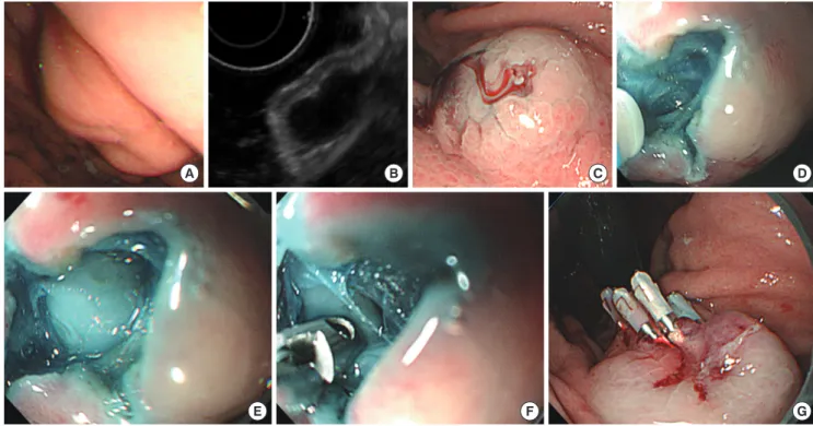
Fig. 1. Forceps biopsy technique after small submucosal dissection. (A) Endoscopic view of a gastric subepithelial tumor (SET). (B) Endoscopic ultrasonography image of a gas- tric SET. (C) Submucosal saline injection. (D) Cross-shaped dissection of the mucosa and submucosa with a dual knife. (E) Confirming the exposure of the underlying tumor. (F) Biopsy forceps were introduced through the hole, taking multiple tissue samples from inside. (G) Prophylactic or hemostatic clipping.
A B C D
E F G
adverse events.
EUS-FNA was also performed by three experienced endos- copists. EUS-FNA was performed by using a linear echoendo- scope (GF-UCT 260; Olympus) with a 19 or 22 gauge (EUSN3 EchoTip; Wilson-Cook Medical Inc., Winston-Salem, NC, USA) according to standard techniques, under real-time US guid- ance and color/pulsed Doppler control. Three to 5 (mean 3.3) passes were performed for each lesion. The patients who un- derwent EUS-FAN were hospitalized and carefully monitored for 24 hours after the EUS-FNA.
The software program SPSS Version 18 (SPSS, Inc., Chicago, IL, USA) was used for statistical analyses. Student’s t-test was used to compare numerical variables between the two groups and the χ2 or the Fisher’s exact test was used to compare cate- gorical variables.
Ethics statement
The study protocol was approved by the institutional review board of Kangbuk Samsung Hospital (IRB No. KBC13042). All patients who agreed to participate in the study signed a written informed consent form. This study has been registered with the Clinical Research Information Service (CRIS), Republic of Ko- rea (Clinical trial registration No. KCT0000730).
RESULTS
SESD biopsies, as described, was attempted in 42 patients (mean
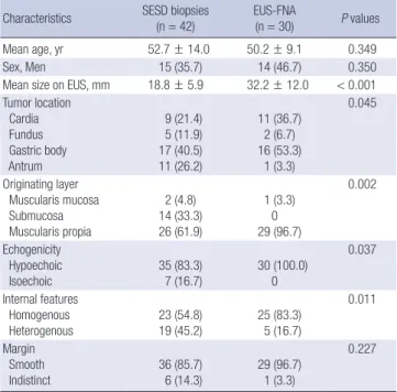
age 53 years) and EUS-FNA was performed in 30 patients (mean age 50 years). Table 1 summarizes the demographic character- istics of patients and endoscopic characteristics of the gastric SETs. The average lesion size measured with EUS in SESD biop- sies group was smaller than that in EUS-FNA group (mean 18.8
± 5.9 mm vs. 32.2 ± 12.0 mm, P < 0.001). The SET locations in SESD biopsies group included: cardia (n = 9, 21.4%), fundus (n = 5, 11.9%), gastric body (n = 17, 40.5%), and antrum (n = 11, 26.2%). The SET locations in EUS-FNA group included: cardia (n = 11, 36.7%), fundus (n = 2, 6.7%), gastric body (n = 16, 53.3%), and antrum (n = 1, 3.3%). In SESD biopsies group, 2 patients (4.8%) had lesions that arose from the muscularis mucosa, 14 (33.3%) from the submucosa, and 26 (61.9%) from the muscu- laris propria, whereas in EUS-FNA group, only one patient (3.3%) had lesion that arose from the muscularis mucosa and all the other (96.7%) from the muscularis propria (P = 0.002). Hypo- echoic and homogenous lesions were more frequent in EUS- FNA group than in SESD biopsies group.
The diagnostic yield of SESD biopsies was comparable to that of EUS-FNA (35 of 42, 83.3% vs. 24 of 30, 80.0%, P = 0.717) (Ta- ble 2). In addition, the diagnostic yield of SESD biopsies was comparable to that of EUS-FNA among lesions originated from muscularis propria layer (21 of 26, 80.8% vs. 24 of 29, 82.8%, P = 1.000). In SESD biopsies group, the diagnostic lesions in- cluded 15 leiomyomas, 7 GISTs, 10 heterotopic pancreases, 2 li- pomas, and one other lesion. The other lesion was an 18-mm isoechoic, heterogeneous mass that originated from the sub- mucosal layer. Pathological analysis of this lesion revealed tiny pieces of fibrovascular tissue with mucinous material sugges- tive of mucin containing benign lesions or mucinous adenocar- cinoma. Given the possibility of mucinous adenocarcinoma in this case, ESD was performed. ESD confirmed the pathological diagnosis of mucinous adenocarcinoma with invasion into the submucosa (SM 3). The deep resection margin of the ESD spec- Table 1. Demographic characteristics of patients and endoscopic characteristics of
subepithelial lesions
Characteristics SESD biopsies
(n = 42) EUS-FNA
(n = 30) P values
Mean age, yr 52.7 ± 14.0 50.2 ± 9.1 0.349
Sex, Men 15 (35.7) 14 (46.7) 0.350
Mean size on EUS, mm 18.8 ± 5.9 32.2 ± 12.0 < 0.001 Tumor location
Cardia Fundus Gastric body Antrum
9 (21.4) 5 (11.9) 17 (40.5) 11 (26.2)
11 (36.7) 2 (6.7) 16 (53.3)
1 (3.3)
0.045
Originating layer Muscularis mucosa Submucosa Muscularis propia
2 (4.8) 14 (33.3) 26 (61.9)
1 (3.3) 0 29 (96.7)
0.002
Echogenicity Hypoechoic Isoechoic
35 (83.3) 7 (16.7)
30 (100.0) 0
0.037
Internal features Homogenous
Heterogenous 23 (54.8)
19 (45.2) 25 (83.3) 5 (16.7)
0.011
Margin Smooth
Indistinct 36 (85.7)
6 (14.3) 29 (96.7) 1 (3.3)
0.227
Data are presented as mean ± SD or number (%).
SESD biopsies = forceps biopsies after small endoscopic submucosal dissection, EUS- FNA = endoscopic ultrasonography-guided fine-needle aspiration.
Table 2. Outcomes of SESD biopsies group versus EUS-FUA group
Outcomes SESD biop-
sies (n = 42) EUS-FNA (n = 30) P values
Diagnosis achieved 35 (83.3) 24 (80.0) 0.717
Diagnosis achieved in lesions originated
from muscularis propria layer 21/26 (80.8) 24/29 (82.8) 1.000 Final diagnosis
Leiomyoma GIST
Neuroendocrine tumor Heterotopic pancreas Lipoma
Other
15 (42.9) 7 (20.0)
0 10 (28.6)
2 (5.7) 1 (2.9)
11 (42.3) 14 (53.8) 1 (3.8)
0 0 0
0.007
Number of forceps biopsies or needle passes 3.9 ± 1.3 3.3 ± 0.7 0.021 Procedure time, min 10.0 ± 4.1 37.3 ± 10.6 < 0.001 Data are presented as mean ± SD or number (%).
SESD biopsies = forceps biopsies after small endoscopic submucosal dissection, EUS- FNA = endoscopic ultrasonography-guided fine-needle aspiration, GIST = gastrointes- tinal stromal tumors.
imen also had carcinomatous tissue. Therefore, the patient un- derwent a gastrectomy. In EUS-FNA group, the diagnostic le- sions included 11 leiomyomas, 14 GISTs, and 1 neuroendocrine tumor.
The mean numbers of forceps biopsies in SESD biopsies group was 3.9 and needle passes in EUS-FNA group were 3.3 (P = 0.021). The mean procedure time of SESD biopsies was shorter than that of EUS-FNA (10 vs. 37 minutes, P < 0.001). In SESD biopsies group, all incisions were closed with clips. In some cases, there was a small amount of bleeding from the cut surface immediately after incision of the overlying mucosa and submucosa or from the tumor surface after biopsy. The bleed- ing was controlled by clipping. There were no procedure-relat- ed adverse events including perforation or massive bleeding in both groups.
The results of seven GIST cases in SESD biopsies group are shown in Table 3. The forceps biopsy specimens obtained from these cases were not large enough to evaluate the mitotic index in 50 consecutive high power fields (HPFs) (range 5-42 HPFs).
Of the seven patients with GIST, four underwent a surgical re- section. According to Fletcher’s classification, three of these four patients were classified to be at very low risk for a malig- nancy and only one patient was at intermediate risk (20).
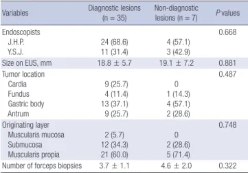
Among SESD biopsies group, there was no significant differ- ence in the diagnostic yield between the endoscopists. In addi- tion, there were no significant differences between the diagnos- tic and nondiagnostic cases with regard to SET size, location, originating layer, and the number of forceps biopsies obtained (Table 4).
DISCUSSION
It is important to differentiate the various types of SETs because these lesions have different prognoses and require different management or therapeutic options. EUS findings alone have limited specificity for the diverse subtypes of SETs and fail to distinguish between benign and malignant lesions. Our study
demonstrated that the diagnostic yield of SESD biopsies was comparable to that of EUS-FNA (83% vs. 80%).
Currently, there is no consensus regarding the optimal man- agement strategy for small (< 3 cm in diameter) asymptomatic SETs. Therefore, endosonographic clinical practice patterns in surveillance and management are highly variable. A recent up- date in the National Comprehensive Cancer Network Guide- lines on the management of GISTs recommended that inciden- tally encountered small GISTs (< 2 cm in diameter) be followed endoscopically until they grow or become symptomatic (21).
Another study reported that GIST lesions (diagnosed by EUS) that are < 3 cm in size with low malignant potential can be ob- served with EUS rather than cytohistologically diagnosed or re- moved (19). However, current literature suggests that all GIST lesions have malignant potential, even those just 1 cm in size (22). Therefore, optimal pathologic examination of tumor tissue is mandatory, especially for lesions that are hypoechoic, located in the muscularis propria of the stomach, or are > 2 cm.
Several diagnostic methods have been proposed for the tis- sue diagnosis of GI SETs. EUS-FNA is currently considered the standard method for GI SETs samples; however, it has limited value for the cytologic diagnosis of nonmesenchymal lesions (with nondiagnostic samples up to 100%) (3). In addition, EUS- FNA has a high failure rate in immunostaining with regard to mesenchymal tumors; this limitation decreases its diagnostic yield from 70%-74% to 34%-53% after immunohistochemical analysis (3,4). Unfortunately, immunohistochemical analysis is not always feasible with EUS-FNA samples because there is in- sufficient material obtained by aspiration. EUS-TCB emerged as a method to solve the limitations of EUS-FNA. Theoretically, this procedure provides core-tissue specimens that would in- crease the diagnostic yield with thicker samples. Despite this prediction, the diagnostic yield of TCB in GI SETs is not superi- Table 3. Results of the seven patients with GIST in SESD biopsies group
Patient Tumor location
No. of forceps biopsies
Size on EUS, mm
Mitoses per HPF Biopsy after
submucosal dissection
Surgical resection
1 Body 5 30 2/42 9/50 (intermediate risk)
2 Body 3 13 0/22 1/50 (very low risk)
3 Fundus 3 14 0/21 0/50 (very low risk)
4 Fundus 3 15 0/17 1/50 (very low risk)
5 Antrum 3 18 0/7 NA
6 Fundus 5 15 0/5 NA
7 Body 6 20 1/19 NA
SESD biopsies = forceps biopsies after small endoscopic submucosal dissection, EUS=
endoscopic ultrasonography, GIST = gastrointestinal stromal tumors, HPF = high pow- er field, NA = not available.
Table 4. Comparison between diagnostic and non-diagnostic lesions in SESD biop- sies group
Variables Diagnostic lesions (n = 35)
Non-diagnostic
lesions (n = 7) P values Endoscopists
J.H.P.
Y.S.J. 24 (68.6)
11 (31.4) 4 (57.1) 3 (42.9)
0.668
Size on EUS, mm 18.8 ± 5.7 19.1 ± 7.2 0.881
Tumor location Cardia Fundus Gastric body Antrum
9 (25.7) 4 (11.4) 13 (37.1) 9 (25.7)
0 1 (14.3) 4 (57.1) 2 (28.6)
0.487
Originating layer Muscularis mucosa Submucosa Muscularis propia
2 (5.7) 12 (34.3) 21 (60.0)
0 2 (28.6) 5 (71.4)
0.748
Number of forceps biopsies 3.7 ± 1.1 4.6 ± 2.0 0.322 Data are presented as mean ± SD or number (%).
SESD biopsies = forceps biopsies after small endoscopic submucosal dissection, EUS = endoscopic ultrasonography.
or to that of EUS-FNA (47%-63%) (4,5,23). There is a high rate of technical failure with TCB because the device is very stiff, which hinders the needle from obtaining tissue (24). In addition, there are safety concerns regarding TCB. A previous study reported that among 52 TCB procedures performed for gastric SETs, there were two cases of sepsis (5).
This study presents several reasons why SESD biopsies may be preferable to EUS-FNA or EUS-TCB. First, SESD biopsies provide a high diagnostic yield. Although its diagnostic yield was compared to the retrospective data of EUS-FNA cases, our results show that its diagnostic yield is not inferior to that of EUS-FNA. Second, SESD biopsies also provide sufficient tissue for accurate diagnosis in a cost-effective manner, while avoid- ing unnecessary follow-up examinations or repeated explor- atory surgeries. According to our data, the pathological diagno- ses of 35 included 7 GISTs, 15 leiomyomata, and 10 heterotopic pancreases. Since GISTs have malignant potential, they neces- sitate lifelong follow-up or resection. Patients who were diag- nosed with benign lesions such as a leiomyoma or heterotopic pancreas do not require annual EUS examinations or surgical resection. Third, another advantage to SESD biopsies is that it can be easily performed regardless of the lesion’s anatomic lo- cation. In contrast, EUS-FNA and EUS-TCB have high failure rates with regard to tissue acquisition when the SET lesion is in the cardia or fundus because the stiff device has difficulty ac- cessing these areas. The diagnostic yield of SESD biopsies is high, even when SETs are in the cardia or fundus (13 of 14, 93%).
Fourth, SESD biopsies can be easily performed regardless of the lesion’s size. Our results showed that the average lesion size in SESD biopsies group was smaller than that in EUS-FNA group (19 vs. 32 mm). EUS-FNA has difficulty in needle passing and aspiration of small sized SETs. Fifth, SESD biopsy is a safe pro- cedure. There were no procedure-related adverse events in this study including perforation, bleeding, or sepsis. Finally, the procedure time of SESD biopsies was shorter than that of EUS- FNA (10 vs. 39 minutes) and thus it can save time.
Similarly to EUS-FNA and EUS-TCB, the main limitation of the SESD biopsies is that it does not provide sufficient tissue (with at least 50 HPFs) to evaluate the malignant potential of GISTs. If more forceps biopsies are performed, it is possible to obtain 50 consecutive HPFs in order to evaluate the mitotic in- dex. However, biopsy-obtained tissue is unlikely to represent the mitotic activity of the entire tumor. The mitotic index mea- sured over 50 HPFs may actually be inaccurate because of the heterogeneous distribution of mitotic activity. In addition, the mitotic index should be measured from the most mitotically active area.
Another group recently developed US-guided single-incision with needle knife (SINK) and deep forceps biopsy for the histo- logic diagnosis of upper GI SETs (25). They found that the diag- nostic yield of the SINK biopsy was 92.8% (13 of 14). Of eight
GIST cases, the SINK specimens were sufficient for immuno- histochemical analysis in seven cases and for measurement of the mitotic index in five cases. The diagnostic yield of our meth- od was not superior to that of the SINK biopsy (83.3% vs. 92.8%).
However, our study included a larger number of patients than did the SINK biopsy study (42 vs. 14 cases). In addition, our study was prospective, while the SINK study was retrospective. Our methods involved a cross-shaped dissection of mucosa and submucosa, exposure of the underlying tumor, and then tissue extraction through the forceps biopsy. In contrast, the SINK bi- opsy involves a linear incision without confirming the exposure of the underlying tumor. Although SINK specimens allowed the evaluation of the mitotic index in some of GIST cases, this index may not represent the entire tumor, as described previously.
More similar to our study, Lee et al. (26) performed endoscopic biopsy of nine gastric SETs using the ESD technique. Their pro- cedure involved making a 5-mm-diameter hole with a flex knife and then a 15-mm-diameter round incision. Next, they used an IT2 knife to make a submucosal dissection and performed mul- tiple endoscopic biopsies. This technique is somewhat incon- venient because it employs both a flex knife and an IT2 knife, as opposed to our method, which only required a dual knife. Their study population was also very small.
Recently, several diagnostic methods using the unroofing technique have been proposed to obtain a sufficient amount of GI SET tissue. These techniques include partial resection (27), retract-ligate-unroof-biopsy with loop (17), and suck-ligate-un- roof-biopsy by using loop (18). These methods have high diag- nostic yields (94%, 81%, and 100%, respectively) and provide sufficient tumor tissue for immunohistochemical analysis and mitotic index calculation. However, incomplete endoscopic re- section of a GIST tumor can allow for peritoneal dissemination of disease (11). Furthermore, the retract-ligate-unroof-biopsy and suck-ligate-unroof-biopsy with loop techniques cannot provide an en bloc specimen of the entire tumor or negative margin assessment; therefore, any neoplastic lesion with ma- lignant potential needs close serial follow-up by expensive EUS.
An unexpected finding in our study is that leiomyoma was the most common gastric SET (although non-diagnostic lesions were considered). GIST is known to be the most frequent SET in the stomach, whereas leiomyoma is the most frequent SET in the esophagus (28). In our study, among SESD biopsies group, most SETs (8 of 9) in the cardia were leiomyomas and only one case was heterotopic pancreas. Leiomyoma seems to be fre- quently found in the upper stomach near the esophagogastric junction. If a SET > 20 mm is incidentally found during routine endoscopy in the cardia of the stomach, there is high probabili- ty that it is a leiomyoma. After immunohistochemical confir- mation, this lesion would not require surgery.
This study has several limitations. First, the sample size was small and SESD biopsies were performed at a single center by
only two endoscopists. It is possible that the operator-related factors influenced our results and that the diagnostic yields may be lower than those in an average practice. Future investigation at multiple centers with more endoscopists is needed to clarify the efficacy of SESD biopsies. Another limitation to this study is that the diagnostic yield of SESD biopsies was compared to the retrospective data of EUS-FNA cases. Finally, SESD biopsies specimens were not large enough to evaluate mitotic index in 50 consecutive HPFs.
This study focused on the acquisition of tissue specimens with SESD biopsies for diagnostic purposes. SESD biopsies was found to be an effective, easy, and safe technique for the histo- logic diagnosis of gastric SETs and its diagnostic yield is compa- rable to that of EUS-FNA. It may be a reliable alternative to con- ventional EUS-FNA and TCB. However, further prospective com- parative studies with larger sample sizes are needed to confirm this method’s efficacy.
DISCLOSURE
The authors have no potential conflicts of interest to disclose.
AUTHOR CONTRIBUTION
Study concept and design: Jung YS, Park JH. Acquisition of data:
Jung YS, Lee H, Kim HJ, Park JH. Interpretation of data: Jung YS, Kim K. Writing the first draft: Jung YS. Critical revision of the manuscript: Jung YS, Lee H, Kim K, Sohn JH, Kim HJ, Park JH.
Final approval: all authors.
ORCID
Yoon Suk Jung http://orcid.org/0000-0002-1963-7170 Hyuk Lee http://orcid.org/0000-0003-4271-7205 Kyungeun Kim http://orcid.org/0000-0001-7938-4673 Jin Hee Sohn http://orcid.org/0000-0002-4735-3853 Hong Joo Kim http://orcid.org/0000-0003-4121-6329 Jung Ho Park http://orcid.org/0000-0002-8367-4371 REFERENCES
1. Akahoshi K, Sumida Y, Matsui N, Oya M, Akinaga R, Kubokawa M, Moto- mura Y, Honda K, Watanabe M, Nagaie T. Preoperative diagnosis of gas- trointestinal stromal tumor by endoscopic ultrasound-guided fine needle aspiration. World J Gastroenterol 2007; 13: 2077-82.
2. Williams DB, Sahai AV, Aabakken L, Penman ID, van Velse A, Webb J, Wil- son M, Hoffman BJ, Hawes RH. Endoscopic ultrasound guided fine nee- dle aspiration biopsy: a large single centre experience. Gut 1999; 44: 720-6.
3. Philipper M, Hollerbach S, Gabbert HE, Heikaus S, Böcking A, Pomjanski N, Neuhaus H, Frieling T, Schumacher B. Prospective comparison of en- doscopic ultrasound-guided fine-needle aspiration and surgical histolo-
gy in upper gastrointestinal submucosal tumors. Endoscopy 2010; 42:
300-5.
4. Fernández-Esparrach G, Sendino O, Solé M, Pellisé M, Colomo L, Pardo A, Martínez-Pallí G, Argüello L, Bordas JM, Llach J, et al. Endoscopic ul- trasound-guided fine-needle aspiration and trucut biopsy in the diagno- sis of gastric stromal tumors: a randomized crossover study. Endoscopy 2010; 42: 292-9.
5. Polkowski M, Gerke W, Jarosz D, Nasierowska-Guttmejer A, Rutkowski P, Nowecki ZI, Ruka W, Regula J, Butruk E. Diagnostic yield and safety of endoscopic ultrasound-guided trucut [corrected] biopsy in patients with gastric submucosal tumors: a prospective study. Endoscopy 2009; 41: 329- 34.
6. Cantor MJ, Davila RE, Faigel DO. Yield of tissue sampling for subepithelial lesions evaluated by EUS: a comparison between forceps biopsies and endoscopic submucosal resection. Gastrointest Endosc 2006; 64: 29-34.
7. Ji JS, Lee BI, Choi KY, Kim BW, Choi H, Huh M, Chung WC, Chae HS, Chung IS. Diagnostic yield of tissue sampling using a bite-on-bite technique for incidental subepithelial lesions. Korean J Intern Med 2009; 24: 101-5.
8. Białek A, Wiechowska-Kozłowska A, Pertkiewicz J, Polkowski M, Milkie- wicz P, Karpińska K, Ławniczak M, Starzyńska T. Endoscopic submucosal dissection for treatment of gastric subepithelial tumors (with video). Gas- trointest Endosc 2012; 75: 276-86.
9. Li QL, Yao LQ, Zhou PH, Xu MD, Chen SY, Zhong YS, Zhang YQ, Chen WF, Ma LL, Qin WZ. Submucosal tumors of the esophagogastric junction originating from the muscularis propria layer: a large study of endoscopic submucosal dissection (with video). Gastrointest Endosc 2012; 75: 1153-8.
10. Hwang JC, Kim JH, Kim JH, Shin SJ, Cheong JY, Lee KM, Yoo BM, Lee KJ, Cho SW. Endoscopic resection for the treatment of gastric subepithelial tumors originated from the muscularis propria layer. Hepatogastroenter- ology 2009; 56: 1281-6.
11. Waterman AL, Grobmyer SR, Cance WG, Hochwald SN. Is endoscopic resection of gastric gastrointestinal stromal tumors safe? Am Surg 2008;
74: 1186-9.
12. von Renteln D, Riecken B, Walz B, Muehleisen H, Caca K. Endoscopic GIST resection using FlushKnife ESD and subsequent perforation clo- sure by means of endoscopic full-thickness suturing. Endoscopy 2008; 40 Suppl 2: E224-5.
13. Park YS, Park SW, Kim TI, Song SY, Choi EH, Chung JB, Kang JK. Endo- scopic enucleation of upper-GI submucosal tumors by using an insulat- ed-tip electrosurgical knife. Gastrointest Endosc 2004; 59: 409-15.
14. Huang WH, Feng CL, Lai HC, Yu CJ, Chou JW, Peng CY, Yang MD, Chiang IP. Endoscopic ligation and resection for the treatment of small EUS-sus- pected gastric GI stromal tumors. Gastrointest Endosc 2010; 71: 1076-81.
15. Lee SH, Park JH, Park DH, Chung IK, Kim HS, Park SH, Kim SJ, Cho HD.
Endoloop ligation of large pedunculated submucosal tumors (with vid- eos). Gastrointest Endosc 2008; 67: 556-60.
16. Sun S, Ge N, Wang C, Wang M, Lü Q. Endoscopic band ligation of small gastric stromal tumors and follow-up by endoscopic ultrasonography.
Surg Endosc 2007; 21: 574-8.
17. Binmoeller KF, Shah JN, Bhat YM, Kane SD. Retract-ligate-unroof-biopsy:
a novel approach to the diagnosis and therapy of large nonpedunculated stromal tumors (with video). Gastrointest Endosc 2013; 77: 803-8.
18. Binmoeller KF, Shah JN, Bhat YM, Kane SD. Suck-ligate-unroof-biopsy by using a detachable 20-mm loop for the diagnosis and therapy of small subepithelial tumors (with video). Gastrointest Endosc 2014; 79: 750-5.
19. Palazzo L, Landi B, Cellier C, Cuillerier E, Roseau G, Barbier JP. Endosono- graphic features predictive of benign and malignant gastrointestinal stro- mal cell tumours. Gut 2000; 46: 88-92.
20. Fletcher CD, Berman JJ, Corless C, Gorstein F, Lasota J, Longley BJ, Miet- tinen M, O’Leary TJ, Remotti H, Rubin BP, et al. Diagnosis of gastrointesti- nal stromal tumors: A consensus approach. Hum Pathol 2002; 33: 459-65.
21. Demetri GD, Benjamin RS, Blanke CD, Blay JY, Casali P, Choi H, Corless CL, Debiec-Rychter M, DeMatteo RP, Ettinger DS, et al. NCCN Task Force report: management of patients with gastrointestinal stromal tumor (GIST)- -update of the NCCN clinical practice guidelines. J Natl Compr Canc Netw 2007; 5 Suppl 2: S1-29.
22. Huang HY, Li CF, Huang WW, Hu TH, Lin CN, Uen YH, Hsiung CY, Lu D.
A modification of NIH consensus criteria to better distinguish the highly lethal subset of primary localized gastrointestinal stromal tumors: a sub- division of the original high-risk group on the basis of outcome. Surgery 2007; 141: 748-56.
23. Hoda KM, Rodriguez SA, Faigel DO. EUS-guided sampling of suspected GI stromal tumors. Gastrointest Endosc 2009; 69: 1218-23.
24. Ginès A, Wiersema MJ, Clain JE, Pochron NL, Rajan E, Levy MJ. Prospec-
tive study of a Trucut needle for performing EUS-guided biopsy with EUS- guided FNA rescue. Gastrointest Endosc 2005; 62: 597-601.
25. de la Serna-Higuera C, Pérez-Miranda M, Díez-Redondo P, Gil-Simón P, Herranz T, Pérez-Martín E, Ochoa C, Caro-Patón A. EUS-guided single- incision needle-knife biopsy: description and results of a new method for tissue sampling of subepithelial GI tumors (with video). Gastrointest En- dosc 2011; 74: 672-6.
26. Lee HL, Kwon OW, Lee KN, Jun DW, Eun CS, Lee OY, Jeon YC, Han DS, Yoon BC, Choi HS, et al. Endoscopic histologic diagnosis of gastric GI sub- mucosal tumors via the endoscopic submucosal dissection technique.
Gastrointest Endosc 2011; 74: 693-5.
27. Lee CK, Chung IK, Lee SH, Lee SH, Lee TH, Park SH, Kim HS, Kim SJ, Cho HD. Endoscopic partial resection with the unroofing technique for reli- able tissue diagnosis of upper GI subepithelial tumors originating from the muscularis propria on EUS (with video). Gastrointest Endosc 2010;
71: 188-94.
28. Nishida T, Kawai N, Yamaguchi S, Nishida Y. Submucosal tumors: com- prehensive guide for the diagnosis and therapy of gastrointestinal sub- mucosal tumors. Dig Endosc 2013; 25: 479-89.