J Korean Soc Radiol 2015;73(6):403-407 http://dx.doi.org/10.3348/jksr.2015.73.6.403
INTRODUCTION
A hemangioma of the bone is a benign tumor that may affect any bone and can occur at any age; however, this tumor com- monly affects the vertebral bodies and the skull. Involvement of a long bone is infrequent and most hemangiomas that occur in a long tubular bone are located in the metadiaphysis or diaphy- sis (1, 2). An intraosseous hemangioma rarely involves in the epiphysis of the long bone and only a few cases of an epiphyseal hemangioma have been previously reported (3-6).
We present a case of a symptomatic hemangioma that affect- ed the epiphysis of the humeral head and describe the associated radiographic, computed tomography (CT), and magnetic reso-
nance imaging (MRI) findings. As the imaging findings were similar to findings for the vertebral body hemangiomas on CT and MRI, the diagnosis was considered preoperatively.
CASE REPORT
A 20-year-old man presented with dull pain for three months in the right shoulder. The patient had no traumatic history or shoulder dislocation, and he had not been suffering from known arthritis. Radiographs of the right shoulder joint (Fig. 1) showed no gross abnormality in the humeral head. On CT images (Fig.
2), an irregular and lobulating osteolytic lesion was visible with a peripheral sclerotic rim and focal cortical defects. Magnetic
Epiphyseal Hemangioma of the Humeral Head: Imaging Findings and Literature Review
상완골두에 발생한 골단 혈관종: 영상 소견 및 선행 문헌 검토
Woo Jin Yang, MD
1, Wook Jin, MD
1*, Young Soo Chun, MD
2, Gou Young Kim, MD
3, So Young Park, MD
1, Ji Seon Park, MD
4, Kyung Nam Ryu, MD
4Departments of 1Radiology, 2Orthopaedic Surgery, 3Pathology, Kyung Hee University Hospital at Gangdong, College of Medicine, Kyung Hee University, Seoul, Korea
4Department of Radiology, Kyung Hee University Hospital, College of Medicine, Kyung Hee University, Seoul, Korea
We describe a case of an epiphyseal hemangioma in the humeral head of a 20-year- old man. On plain radiographs, the lesion showed no gross abnormality. The com- puted tomography images demonstrated the presence of an irregular and lobulat- ing osteolytic lesion with a peripheral sclerotic rim and focal cortical defects. The magnetic resonance images showed an ill-defined low signal intensity on T1-weight- ed images and mixed low and high signal intensities on T2-weighted images. Addi- tionally, ill-defined marrow enhancement with inner low signal lines was noted in this lesion. The patient was treated with curettage and a bone chip graft. The present case is instructive in the differential diagnosis of epiphyseal bone tumors; furthermore, the possibility of an intraosseous hemangioma should also be considered.
Index terms
Intraosseous Hemangioma Epiphysis
Humerus X-Ray CT MRI
Received May 21, 2015 Revised June 28, 2015 Accepted July 6, 2015
*Corresponding author: Wook Jin, MD
Department of Radiology, Kyung Hee University Hospital at Gangdong, 892 Dongnam-ro, Gangdong- gu, Seoul 05278, Korea.
Tel. 82-2-440-6933 Fax. 82-2-440-6932 E-mail: [email protected]
This is an Open Access article distributed under the terms of the Creative Commons Attribution Non-Commercial License (http://creativecommons.org/licenses/by-nc/3.0) which permits unrestricted non-commercial use, distri- bution, and reproduction in any medium, provided the original work is properly cited.
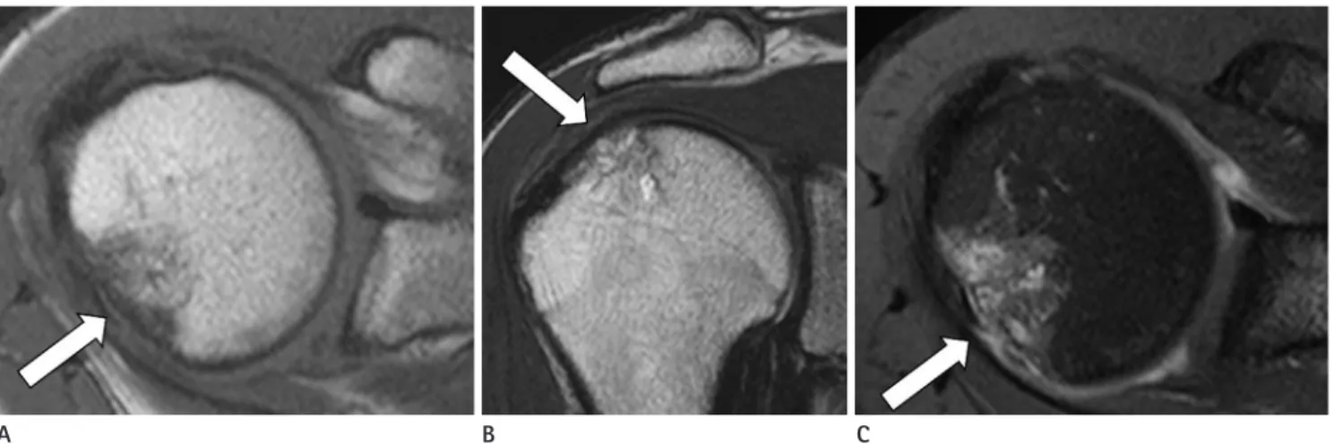
resonance images (Fig. 3) showed irregular low signal intensity on T1-weighted images, mixed low and high signal intensities on T2-weighted images, and inhomogeneous enhancement. In addition, inner and peripheral low signal lines in this lesion were observed on all MR sequences. We thought that these low
signal intensity lines were probably due to thickened bony tra- beculae. Based on the above multi-modality imaging findings, we diagnosed the intraosseous lesion of the humeral head as an intraosseous hemangioma.
The patient underwent open surgery, including a careful cu- rettage and a bone chip graft. During surgery, multiple dark brownish fragments were obtained and a histopathological ex- amination of these fragments showed the presence of multiple dilated vascular channels and the irregular architecture of the thickened bony trabeculae (Fig. 4A). Immunostaining for the expression of factor VIII revealed a variety of different sizes and shapes of vascular channels, lined by endothelial cells (Fig. 4B).
Based on these findings, an intraosseous hemangioma was diag- nosed.
DISCUSSION
A hemangioma may affect any bone, but commonly affects the vertebral bodies and the skull. Intraosseous hemangiomas can be easily diagnosed when lesions are located at these typical sites (7). When a lesion is located in the vertebral body, it has a coarse, vertical trabecular pattern as seen on radiographs and CT.
Typically, a CT scan demonstrates a characteristic appearance–
the so-called polka-dot pattern or a honeycomb pattern (1, 2).
Fig. 1. An anteroposterior radiograph of the right shoulder shows no gross abnormalities in the humeral head.
Fig. 2. CT scans show an irregular and lobulating osteolytic lesion with a peripheral sclerotic rim and thickened internal trabeculae (arrow). On coronal reformatted images, a focal cortical defect (arrow) is suggested.
Generally, a vertebral hemangioma shows high signal intensity on both T1- and T2-weighted images (8). The loss of hemato- poietic cells in the gaps of hemangiomas and apparent increase in the fat component can produce an increased signal intensity on T1-weighted images in hemangiomas of the vertebral bod- ies (7). However, vertebral hemangiomas can demonstrate vari- able signal intensity on T1-weighted images, based on the de- gree of adipose tissue present (9).
Intraosseous hemangiomas rarely affect the long tubular bones and hemangiomas of the long tubular bones usually oc-
cur in the metadiaphyseal or diaphyseal portion of the bones of the lower extremities and ribs (1, 2, 7). Radiographically, the in- traosseous hemangioma is seen as an osteolytic area with tra- beculation, creating a soap bubble or honeycomb appearance as a result of the expanding, proliferating, engorging vessels and thickened, remodeled bone trabeculae (7). MR findings of an intraosseous hemangioma have not been well documented, ex- cept for vertebral body or skull hemangiomas (10). Generally, medullary hemangiomas of long bones show a low signal on T1-weighted images and a high signal on T2-weighted images.
Fig. 3. Magnetic resonance images show irregular low signal intensity on T1-weighted images (A, arrow; T1WI; axial plane; TR/TE, 600/20 ms), mixed low and high signal intensities on T2-weighted images (B, arrow; T2WI; coronal plane; TR/TE, 4400/80 ms) and inhomogeneous enhance- ment on gadolinium-enhanced fat-suppressed T1WI (C, arrow; CE-FS T1WI; axial plane; TR/TE, 670/20 ms). In addition, inner and peripheral low signal lines in this lesion are demonstrated on all MR sequences.
CE-FS = contrast-enhanced fat-suppressed, TE = echo time, TR = repetition time
A B C
Fig. 4. A photomicrograph (A) of an intraosseous hemangioma reveals numerous vascular channels (arrows) of various sizes and shapes. The ir- regular architecture of the thickened bony trabeculae (T) is noted (hematoxylin and eosin staining, original magnification, × 40). Immunostaining for factor VIII (B) shows vascular channels of a variety of shapes and sizes (arrows), lined by endothelial cells (polymer method; original magnifi- cation, × 200).
A B
The high signal on T2-weighted images can be explained by the fluid content of the tumor vessels (7). In addition, Resnick (2) described the multiple channel-like appearance of the high sig- nal intensity on T2-weighted images. The appearance was simi- lar to that of hemangiomas in the soft tissues in a case of tibial hemangioma. The enhancement pattern of intraosseous heman- giomas was reported to be variable, with whole enhancement, peripheral enhancement, or lattice-like enhancement observed (4, 9, 10).
In reviewing the literature, several hemangiomas involving the epiphyses of the long tubular bones have been reported (Ta- ble 1) (3-6). Among these cases, three hemangiomas were seen as well-defined osteolytic lesions on radiographs. In the present case, there was no gross bony abnormality in the humeral head.
Two of the three cases where the CT findings were reported sh- owed partial cortical breakthrough on CT images. In the present case, CT imaging showed a focal cortical defect, similar to the findings of the above two cases. On MRI, one case showed a multilocular and hemorrhagic lesion with fluid-fluid level, and the other cases showed hypointensity on T1-weighted images, hyperintensity with internal hypointense septa on T2-weighted images, and lattice-like enhancement. In the present case, the lesion showed inhomogeneous enhancement and intervening low signal lines on MRI. In agreement with the histopathologi- cal findings, the portion of inhomogeneous enhancement might
be related to the multiple dilated vascular channels while the por- tion of intervening low signal lines might be related to the thick- ened trabeculae.
In conclusion, we report an unusual case of a hemangioma involving the epiphysis of the humeral head that was seen with inhomogeneous enhancement and intervening low signal lines.
These findings were similar to findings of hemangiomas in the vertebral bodies.
REFERENCES
1. Unni KK. Benign vascular tumors. In Unni KK, Inwards CY.
Dahlin’s Bone Tumors: General Aspects and Data on 10,165 Cases. Philadelphia: Lippincott Williams & Wilkins, 2010:
262-271
2. Resnick D. Tumors of vascular differentiation. In Resnick D.
Diagnosis of Bone and Joint Disorders. Philadelphia: Saun- ders, 1995:3821-3846
3. Boumdin H, Rachid K, Mahi M, Chaouir S, Benameur M.
[Hemangioma of the humerus: value of imaging]. J Radiol 2002;83(9 Pt 1):1088-1089
4. Yamamoto T, Kurosaka M, Mizuno K. Juxta-articular hem- angioma of long bone. Skeletal Radiol 2000;29:535-537 5. Mirra JM. Vascular tumors. In Mirra JM, Picci P, Gold RH.
Bone Tumors: Clinical, Radiologic, and Pathologic Correla- Table 1. Cases of Intraosseous Hemangiomas Involving in the Epiphysis of the Long Tubular Bones
Authors Sex/Age Location CR CT MR Others
Boumdin et al. (3)
M/22 Humeral head A well-defined osteolytic lesion with marginal sclerosis
An osteolytic lesion with coarse trabecular pattern & partial cortical breakthrough
A multilocular and hemorrhagic lesion with fluid-fluid level
Cavernous hemangioma
Yamamoto et al. (4)
M/70 Proximal tibia A well-defined osteolytic lesion with marginal sclerosis
A well-defined osteolytic lesion with partial cortical breakthrough
Hypointense lesion on T1WI
Hyperintense areas with internal, hypointense septa on T2WI
Lattice-like enhancement
Cavernous hemangioma
Mirra (5) (-) Tibial epiphysis (-) (-) (-) Associated with
soft tissue hemangioma Pandey and
Pandey (6)
F/7 Greater tuberosity of the humerus
A localized osteolytic lesion
(-) (-) (-)
(-) = absent or not available, CR = conventional radiograph, WI = weighted image
tions. Philadelphia: Lea & Febiger, 1989:1338-1478 6. Pandey S, Pandey AK. Osseous haemangiomas. Arch Orthop
Trauma Surg 1981;99:23-28
7. Dorfman HD, Czerniak B. Vascular lesions. In Dorfman HD, Czerniak B. Bone Tumors. St. Louis: Mosby, 1998:729-814 8. Ross JS, Masaryk TJ, Modic MT, Carter JR, Mapstone T, Den-
gel FH. Vertebral hemangiomas: MR imaging. Radiology 1987;165:165-169
9. Murphey MD, Fairbairn KJ, Parman LM, Baxter KG, Parsa MB, Smith WS. From the archives of the AFIP. Musculoskel- etal angiomatous lesions: radiologic-pathologic correla- tion. Radiographics 1995;15:893-917
10. Matsumoto K, Ishizawa M, Okabe H, Taniguchi I. Hemangi- oma of bone arising in the ulna: imaging findings with em- phasis on MR. Skeletal Radiol 2000;29:231-234
상완골두에 발생한 골단 혈관종: 영상 소견 및 선행 문헌 검토
양우진
1· 진 욱
1* · 전영수
2· 김교영
3· 박소영
1· 박지선
4· 류경남
4저자들은 상완골두에 골단 혈관종(epiphyseal hemangioma)이 발생한 20세 남자 환자의 증례를 보고하고자 한다. 단순 방사선사진에서 병변은 이상 소견을 보이지 않았다. 전산화단층촬영에서 병변은 경계가 불규칙한 소엽모양의 종괴로 보였 으며 주변의 경화성 테두리(sclerotic rim)와 국소 피질 결손을 동반하였다. 자기공명영상에서 병변은 경계가 불명확하였 으며 T1 강조영상에서 저신호강도를 보이고 T2 강조영상에서 혼합신호강도를 보였다. 또한 병변의 내부에 경계가 불명확 한 골수의 조영증강과 선형의 저신호강도가 보였다. 환자는 소파술과 파편골 이식술을 받았다. 본 증례는 골단부의 골 종 양 감별에서 골내 혈관종(intraosseous hemangioma)의 가능성도 고려해야 함을 시사하는 교훈적인 증례이기에 이를 보고 한다.
경희대학교 의과대학 강동경희대학교병원 1영상의학과, 2정형외과, 3병리과, 4경희대학교 의과대학 경희대학교병원 영상의학과