Diagnosis of Hypersensitivity Pneumonitis: 2020 Clinical Practice Guideline
2020년 개정 진료 치침에 따른 과민성폐렴의 진단
Soojung Park, MD
1, Yu-Whan Oh, MD
1, Eun-Young Kang, MD
2, Hwan Seok Yong, MD
2, Cherry Kim, MD
3,
Ki Yeol Lee, MD
3, Sung Ho Hwang, MD
1*
1Department of Radiology, Korea University Anam Hospital, College of Medicine, Korea University, Seoul, Korea
2Department of Radiology, Korea University Guro Hospital, College of Medicine, Korea University, Seoul, Korea
3Department of Radiology, Korea University Ansan Hospital, College of Medicine, Korea University, Ansan, Korea
Hypersensitivity pneumonitis (HP) is an interstitial lung disease (ILD) characterized by an in- haled inciting antigen that leads to the inflammation of the lung parenchyma and small airway with immunologic reactions. Over the last decades, the most effective therapeutic option for HP has been limited to antigen avoidance. The differential diagnosis of HP from other ILDs is the beginning of treatment as well as diagnosis. However, the presence of several overlapping clinical and radiologic features makes differentiating HP from other ILDs particularly challeng- ing. In 2020, a multidisciplinary committee of experts from the American Thoracic Society, Jap- anese Respiratory Society, and Asociación Latinoamericana del Tórax suggested a new clinical practice guideline classifying HP into nonfibrotic and fibrotic phenotypes on the basis of chest high-resolution CT (HRCT) findings. Therefore, we introduced a new diagnostic algorithm based on chest HRCT in the clinical practice guideline for the diagnosis of HP.
Index terms Hypersensitivity Pneumonitis; Interstitial Lung Disease; Diagnosis;
Tomography, X-Ray Computed; Practice Guideline
서론
과민성폐렴(hypersensitivity pneumonitis)은 흡입된 항원에 의한 면역반응으로 인해 폐 간질에서 육아종과 염증이 발생하는 질환이다(1). 흔히 직업적 또는 주변 환경으로부터 노출 되는 다양한 유기물들이 항원이 되어 병리학적으로 외인성 알레르기성 폐포염(extrinsic al- lergic alveolitis)을 유발한다(2).
Received April 30, 2021 Revised June 26, 2021 Accepted June 29, 2021
*Corresponding author Sung Ho Hwang, MD Department of Radiology, Korea University Anam Hospital, 73 Goryeodae-ro, Seongbuk-gu, Seoul 02841, Korea.
Tel 82-2-920-6289 Fax 82-2-929-3796
E-mail sungho.hwng@gmail.com This is an Open Access article distributed under the terms of the Creative Commons Attribu- tion Non-Commercial License (https://creativecommons.org/
licenses/by-nc/4.0) which permits unrestricted non-commercial use, distribution, and reproduc- tion in any medium, provided the original work is properly cited.
ORCID iDs Soojung Park https://
orcid.org/0000-0001-9478-2610 Yu-Whan Oh
https://
orcid.org/0000-0003-2646-0497 Eun-Young Kang
https://
orcid.org/0000-0002-4848-509X Hwan Seok Yong
https://
orcid.org/0000-0003-0247-8932 Cherry Kim
https://
orcid.org/0000-0002-3361-5496 Ki Yeol Lee
https://
orcid.org/0000-0002-0323-1280 Sung Ho Hwang
https://
orcid.org/0000-0003-1850-0751
과민성폐렴은 초기에 경미한 염증으로 시작되나 항원에 대한 노출이 반복될수록 폐섬유화 (lung fibrosis)를 동반한 영구적인 폐 손상까지 일으킬 수 있다(3, 4). 때문에 과민성폐렴은 병의 진행 과정에서 다양한 형태의 폐 손상을 보일 수 있고 때로는 급성과 만성 폐 손상의 특징들이 동 시에 나타나기도 한다(5). 과거에는 임상적 특징에 따라 과민성폐렴을 급성, 아급성과 만성으로 분 류해왔지만 임상양상 중 많은 부분이 겹칠 수 있어 임상양상을 통한 분류는 명확한 진단이 어려울 때가 많다(6, 7). 비가역적 폐섬유화와 환자의 예후와의 유의한 상관관계를 근거로 과민성폐렴을 폐섬유화의 유무를 통해 구분하자는 주장이 관련 전문가들 사이에 많은 지지를 얻고 있다(8-10).
하지만 종종 폐 섬유화를 동반하는 과민성폐렴은 간질성폐질환(interstitial lung disease; 이하 ILD) 중 특히 특발성 폐섬유증(idiopathic pulmonary fibrosis)과 감별이 어려울 수 있다(11-13).
2020년 미국흉부학회(American Thoracic Society; 이하 ATS), 일본호흡기학회 (Japanese Re- spiratory Society; 이하 JRS), 라틴아메리카흉부학회(Asociación Latinoamericana del Tórax;
이하 ALAT)는 과민성폐렴 진단의 어려움을 해결하고자 특별위원회를 구성하여 과민성폐렴 진단 을 위한 진료지침을 개발하였다(5). 이번 진료지침에서 흥미로운 점은 과민성폐렴의 진단 초반에 고해상도 전산화단층촬영(high-resolution CT; 이하 HRCT)을 적극 활용하며 이후 단계적 접근 과정 이에 따른 진단의 신뢰도를 제시한 것이다. 이에 본 종설은 새롭게 소개된 과민성폐렴 진단 과정에서 HRCT의 활용 방법과 중요 HRCT 소견들을 중심으로 과민성폐렴 진단의 전반을 살펴보 도록 하겠다.
원인과 기전
과민성폐렴은 다양한 원인항원과 개개인별 면역 반응의 차이로 인해 임상양상 역시 다양하다.
현재까지 200여 종 이상의 과민성폐렴 원인항원이 규명되었고, 진균 포자, 부패균, 동식물의 유기 물질, 화학물질과 독성 가스 등이 원인항원으로 작용할 수 있다(2). 이런 원인항원은 주로 직업, 주 거지 및 취미활동 중 노출될 수 있으며 항원의 농도, 노출 횟수 및 기간과 과민성폐렴의 발병률은 비례한다(14).
과민성폐렴의 병태생리는 복합적인 면역반응 기전으로 해석될 수 있다. 초기 과민성폐렴은 형 질세포(plasma cell)의 제3형 과민반응으로 시작되다가 항원에 지속적으로 노출되면 큰포식세포 (macrophage)의 제4형 과민반응을 통해 육아종과 림프구 침윤이 이루어진다. 이 과정이 만성화 되면 섬유모세포(fibroblast)가 자극되어 아교질(collagen) 분비가 촉진되어 세포외기질이 증가되 면서 폐섬유화가 만들어진다. 전통적으로 과민성폐렴의 임상 발현은 급성, 아급성, 만성으로 나누 고 있으나 발발 시기에 따른 임상증상의 양상이 겹칠 경우 정확한 분류가 어려울 수 있다(15).
진단 검사법
일반적으로 과민성폐렴 진단은 환자 주변 환경과 병력에 대한 조사를 통해 과민성폐렴에 대한
의심을 하면서 시작된다고 할 수 있다(16). 과민성폐렴에 대한 최종 확진을 위해선 다음의 세 가지
가 필요하다. 첫째, 노출항원의 확인, 둘째, HRCT에서의 과민성폐렴 소견, 그리고 마지막으로 기 관지 폐포 세척액(bronchoalveolar lavage; 이하 BAL)에서의 림프구증가(lymphocytosis) 또는 조직병리학적 증거들이 과민성폐렴 진단을 위한 충족 조건들이다(5). 노출항원 확인을 위해선 환 자의 과거력 조사와 함께 혈청항체검사(serum immunoglobulin G test)가 활용되기도 한다. 하 지만 혈청항체검사는 높은 민감도를 가진다는 장점이 있으나 위음성의 가능성이 높다는 한계점 역시 있다(17). 과민성폐렴 환자의 BAL 검사에서는 림프구가 정상의 4배까지도 증가할 수 있고 그 중에서도 CD8+ 림프구의 증가가 두드러지면 사코이드증(sarcoidosis)과 특발성 폐섬유증 등의 감별진단에 도움이 되기도 한다(17). 때로는 불명확한 검사 결과들로 인해 과민성폐렴과 다른 간 질성폐질환(ILD)과의 감별이 어려워 폐생검(lung biopsy)이 시행되는 경우도 있다(18). 과민성폐 렴의 조직병리 소견은 상피모양세포가 성글게 모여 있는 비괴사성 육아종 소견(“poorly formed nonnecrotizing granulomas”)이 특징적이며 이를 통해 다른 간질성폐질환(ILD)과의 감별이 가 능하다(5). 하지만 침습적인 폐생검은 가장 마지막에 고려되는 검사이며 다학제 토의를 거쳐 전문 가들 및 환자의 동의가 선행된 후 폐생검이 진행되어야 할 것이다.
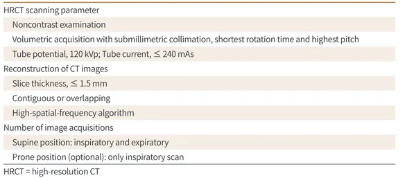
HRCT 검사 방법
과민성폐렴 환자에서도 간질성폐질환(ILD) 진단을 위해 추천되는 HRCT 검사 방법을 사용한다 (Table 1). HRCT 검사는 다중절편 나선식 CT (multi detector-row spiral CT)를 이용해 동일 촬 영시간 동안 흉부 전체를 스캔하는 방식(volumetric scanning)이 권고된다. 더 나아가 HRCT 검 사는 기계적 촬영 조건 및 영상 재구성 조건에서 균형과 최적화가 중요하다(19). 일반적으로 HRCT 촬영 시 X선 튜브(X-ray tube)는 120 kVp의 관전압과 240 mAs 이하의 관전류로 설정하면 서 환자에 노출되는 방사선량은 약 1~3 mSv 범위를 유지해야 한다(5, 19). 따라서 방사선 노출을 줄이기 위해 환자의 체구에 따라 HRCT의 관전압과 관전류에 대한 조정이 필요하다. HRCT 검사 는 환자가 테이블에 똑바로 누운 상태에서 최대 흡기 시와 호기 시에 두 차례 촬영되기도 한다. 흡
Table 1. Chest HRCT in the Diagnosis of Hypersensitivity Pneumonitis HRCT scanning parameter
Noncontrast examination
Volumetric acquisition with submillimetric collimation, shortest rotation time and highest pitch Tube potential, 120 kVp; Tube current, ≤ 240 mAs
Reconstruction of CT images Slice thickness, ≤ 1.5 mm Contiguous or overlapping High-spatial-frequency algorithm Number of image acquisitions
Supine position: inspiratory and expiratory Prone position (optional): only inspiratory scan HRCT = high-resolution CT
기 HRCT가 가장 기본적인 검사이지만 소기도질환을 시사하는 공기가둠(air trapping)은 호기 스 캔에서 볼 수 있는 소견이다. 일부 전문가들은 공기가둠 유무 확인을 통해 진단 신뢰도를 향상시 킬 수 있다는 점 때문에 호기 HRCT 촬영을 추천한다(5). 경우에 따라서는 엎드린 자세로 최대 흡 기 HRCT 촬영을 추가할 수도 있지만 이 역시 필수검사는 아니다. HRCT 촬영 시 피치(pitch)를 최대로 하면 CT 겐트리와 환자 테이블이 빠르게 움직이면서 환자의 움직임으로 인한 인공물을 예 방할 수 있다. HRCT 촬영 후 영상정보는 얇은 콜리메이션(collimation)과 고공간주파수 알고리 즘(high spatial frequency algorithm)을 이용해 탁월한 해상도의 영상으로 재구성된다. 이때 세 기관지를 포함한 폐내 작은 구조에 대한 영상화를 위해선 1.5 mm 이하의 절편두께로 연속되거나 또는 중첩된 단면들로 HRCT 영상을 재구성할 것을 추천한다.
과민성폐렴 의심 환자에서 진행하는 HRCT는 기본적으로 혈관 내 조영제 주입을 필요로 하지 않는 비조영증강 영상검사이다. 하지만 급성 호흡곤란을 보이는 환자에서는 급성 폐색전증(acute pulmonary embolism) 감별을 위해 HRCT 촬영에 이어서 CT 혈관조영술이 추가될 수 있다.
HRCT 소견
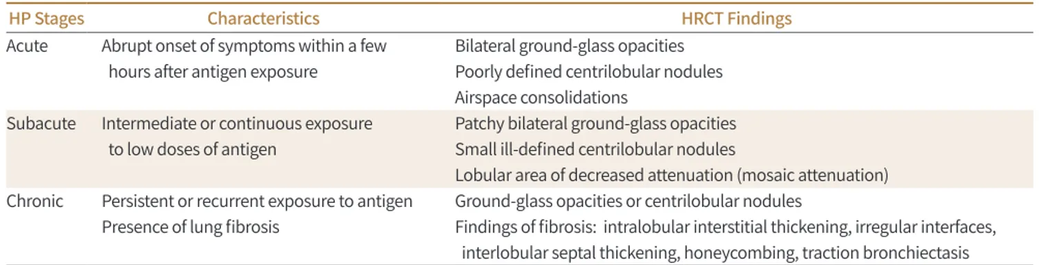
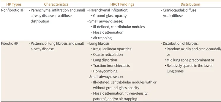
과민성폐렴의 HRCT 소견은 폐실질과 소기도(small airway)의 염증세포 침윤과 폐섬유화 등의 병리학적 특징을 반영한다. 일반적으로 과민성폐렴에서 소기도에 대한 염증세포의 침윤은 불분명 한 경계의 중심소엽성 결절(ill-defined centrilobular nodule), 간유리음영(ground-glass opaci- ty), 모자이크 음영(mosaic attenuation), 및 공기가둠 등으로 나타날 수 있다(20). 한편 과민성폐 렴에서 염증세포의 폐실질 침윤은 HRCT 상 미만성의(diffuse) 간유리음영과 흔하지 않지만 경화 (consolidation)와 낭종(cyst)을 동반하기도 한다(21). 이런 폐실질의 침윤은 폐섬유화로 진행하면 서 거친 그물 형태(reticulation), 폐실질의 뒤틀림(architectural distortion)과 벌집모양(honey- combing) 등으로 나타난다. 이런 HRCT 소견들은 과민성폐렴의 병기(급성, 아급성 그리고 만성) 또는 폐섬유화 동반 여부를 기준으로 과민성폐렴을 구분하는데 활용될 수 있다(Tables 2, 3).
비섬유화 과민성폐렴은 염증 초기 폐실질의 가역적 손상을, 섬유화 과민성폐렴은 만성 염증으 로 인한 비가역적인 폐섬유화 상태를 의미한다. 질환의 손상 정도 특히 폐섬유화와 관련된 HRCT
Table 2. HRCT Features of HP Depending on the Stage of the Disease
HP Stages Characteristics HRCT Findings
Acute Abrupt onset of symptoms within a few hours after antigen exposure
Bilateral ground-glass opacities Poorly defined centrilobular nodules Airspace consolidations
Subacute Intermediate or continuous exposure to low doses of antigen
Patchy bilateral ground-glass opacities Small ill-defined centrilobular nodules
Lobular area of decreased attenuation (mosaic attenuation) Chronic Persistent or recurrent exposure to antigen
Presence of lung fibrosis
Ground-glass opacities or centrilobular nodules
Findings of fibrosis: intralobular interstitial thickening, irregular interfaces, interlobular septal thickening, honeycombing, traction bronchiectasis HP = hypersensitivity pneumonitis, HRCT = high-resolution CT
소견의 유무를 통해 과민성폐렴을 구분하는 것은 환자의 예후 예측에 있어 유용하다(22, 23). 일반 적으로 비섬유화 과민성폐렴은 HRCT에서 중심소엽성 결절과 간유리음영을 주로 보인다(Fig. 1).
이때 폐실질의 침윤과 함께 소기도가 좁아졌다면 흡기 HRCT에서 불분명한 경계의 중심소엽성 결절과 모자이크 음영을, 호기 HRCT에서는 공기가둠이 나타난다(5). 이러한 비섬유화 과민성폐 렴의 병변들은 양측 폐에서 대칭적으로 미만성 분포를 보이는데 폐 중하부에서 흔하다(5). 한편 섬유화 과민성폐렴에서 폐실질의 섬유화와 기관지 폐쇄가 공존하는 모습을 보인다(24). 폐실질과 소기도의 염증은 섬유화가 진행되면서 HRCT 상 폐실질 내 불규칙 선형 음영(irregular linear density), 거친 그물 형태, 폐실질의 뒤틀림과 견인성 기관지 확장(traction bronchiectasis) 등으 로 나타난다(Fig. 2). 이런 섬유화 과민성폐렴에서도 간유리음영을 동반할 수 있으며 심각한 수준
Table 3. Typical HRCT Features of HP Depending on the Presence or Absence of Lung FibrosisHP Types Characteristics HRCT Findings Distribution
Nonfibrotic HP - Parenchymal infiltration and small airway disease in a diffuse distribution
- Parenchymal infiltration:
• Ground-glass opacity - Small airway disease:
• Ill-defined, centrilobular nodules
• Mosaic attenuation
• Air trapping
- Craniocaudal: diffuse - Axial: diffuse
Fibrotic HP - Patterns of lung fibrosis and small airway disease
- Lung fibrosis:
• Irregular linear opacities
• Coarse reticulation
• Lung distortion
• Traction bronchiectasis
• Honeycombing - Small airway disease:
• Ill-defined, centrilobular nodules with or without ground-glass opacity
• Mosaic attenuation, “three-density pattern”, and/or air trapping
- Distribution of fibrosis:
• Random axially and craniocaudally or
• Mid lung zone predominant or
• Relatively spared in the lower lung zones
HP = hypersensitivity pneumonitis, HRCT = high-resolution CT
Fig. 1. HRCT images of nonfibrotic hypersensitivity pneumonitis.
A. HRCT at the level of the right upper lobe bronchus shows extensive bilateral ground-glass opacities, cen- trilobular nodules, and small consolidations, which may represent the acute stage.
B. HRCT at the level of the lower lung zones shows bilateral poorly-defined centrilobular nodules with a dif- fuse distribution in the subacute stage.
HRCT = high-resolution CT
A B
의 섬유화 과민성폐렴은 벌집모양을 보일 수 있다. 벌집모양을 동반한 과민성폐렴은 특발성 폐섬 유증과의 감별진단에 있어 주의가 요구된다. 이때 과민성폐렴은 다수의 모자이크 음영을 보이며 폐 기저부(basal lung)의 섬유화가 흔하지 않다는 점들이 감별에 도움이 된다(25).
과민성폐렴의 HRCT 소견에서 흔히 언급되는 모자이크 음영은 서로 다른 CT 음영들이 폐 안에 서 혼재되어 있는 모습을 의미한다. 과민성폐렴의 HRCT에서 세 가지 서로 다른 음영들: 1) 낮은 CT 음영의 소기도의 폐쇄, 2) 높은 CT 음영의 폐실질의 침윤, 그리고 3) 정상 폐실질의 CT 음영이 함께 보일 수 있는데, 이를 삼상 폐밀도 경향(three-different density pattern) 또는 헤드치즈사 인(headcheese sign)이라 부른다(5, 20). 이는 과민성폐렴에서 두드러지는 HRCT 소견이긴 하나 세기관지염에 동반된 폐렴과 폐부종(pulmonary edema)에서도 나타날 수 있기에 다소 비특이 적이다(26).
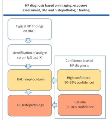
진단과정과 신뢰도
과민성폐렴은 다음 3가지 진단기준인 1) 항원의 식별, 2) HRCT 영상 소견, 그리고 3) BAL 림프 구증가증/전형적인 조직병리학 결과들을 종합적으로 평가하여 진단된다. 과민성폐렴 진단은 기 본적으로 여러 검사 결과들에 대한 종합적인 해석에 기반하기에 호흡기내과, 흉부외과, 영상의학 과 그리고 병리학과 전문가들이 함께하는 다학제 토의 과정이 필요하다. 한편 위에 언급한 진단 검사 방법들은 검사의 용이성 및 침습성을 고려해 최적의 검사 진행 순서와 조합을 구성해야 한 다. 단계별 진단과정에서 확인된 검사 결과와 증거 수준에 따라 과민성폐렴 진단의 신뢰도는 달라 진다. 이번에 발표된 진료지침에 따르면, 다학제 토의를 통해 도출할 수 있는 과민성폐렴 진단의 신뢰도는 1) 확실한 진단(definite diagnosis; 신뢰도, > 90%), 2) 높은 신뢰도(high confidence; 신 뢰도, 80~89%), 3) 보통의 신뢰도(moderate confidence; 신뢰도, 70~79%), 그리고 4) 낮은 신뢰 도(low confidence; 신뢰도, 51~69%) 4등급으로 나눌 수 있다(5). 예를 들면, 과민성폐렴의 전형
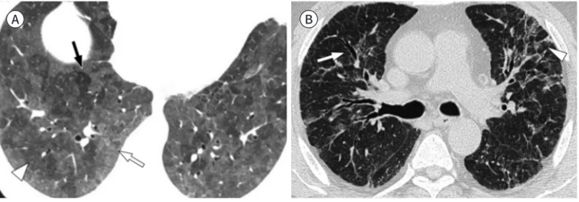
Fig. 2. HRCT images of fibrotic hypersensitivity pneumonitis.
A. HRCT at the level of the lower lobe shows the three-density pattern, comprising ground-glass opacity (white arrow), normal lung parenchyma (arrowhead), and lucent areas (black arrow). The lucent areas re- flect the mosaic attenuation associated with bronchiolar obstruction.
B. HRCT at the level of the right upper lobe bronchus shows evidence of fibrosis with irregular reticular opacities, traction bronchiectasis (arrow), and architectural distortion (arrowhead).
HRCT = high-resolution CT
A B
적인 HRCT 소견을 보이는 환자에서 항원식별과 BAL의 림프구증가 조건이 충족되었다면 이때 과민성폐렴에 대한 진단은 80% 이상의 신뢰도를 가지게 된다(Fig. 3). 이 경우 폐조직검사를 생략 할 수 있는데, 이는 과민성폐렴을 포함해 간질성폐질환(ILD)에서의 진단 목표가 최소 침습적 방법 을 이용해 신뢰도 높은 진단에 도달하는 것이기 때문이다.
결론
2020년에 미국흉부학회(ATS), 일본 호흡기학회(JRS), 라틴아메리카흉부학회(ALAT) 소속 전문 가들은 심층 토의를 통해 과민성폐렴 진단에 대한 임상진료지침을 개발하여 공식 발표하였다. 이 번 지침은 여타의 ILD 감별과 마찬가지로 과민성폐렴 진단 역시 다학제 전문가들 간의 협의를 통 한 단계적 접근을 추천하고 있다. 이 단계적 진단과정에서 HRCT는 침습적 검사 시행 여부 결정에 있어 중요한 역할을 한다. 또한 과민성폐렴 진단 후 HRCT에서 보이는 폐병변의 형태적 특징을 통 해 과민성폐렴을 1) 비섬유화 과민성폐렴과 2) 섬유화 과민성폐렴으로 구분할 것을 이번 임상진료 지침은 추천하고 있다. 이에 과민성폐렴의 진단에서 HRCT 역할과 관련 영상 소견에 대한 이해가 보다 중요해졌다.
Author Contributions
Conceptualization, all authors; data curation, P.S.; investigation, all authors; methodology H.S.H.;
project administration, K.E.Y.; resources, P.S., H.S.H.; software, H.S.H.; supervision, H.S.H.; visualiza- tion, P.S., H.S.H.; writing—original draft, P.S., O.Y.W., H.S.H.; and writing—review & editing, P.S., O.Y.W., H.S.H.
Fig. 3. Diagnostic algorithm of HP with multiple diagnostic tools.
BAL = bronchoalveolar lavage, HP = hypersensitivity pneumonitis, HRCT = high-resolution CT, IgG = immunoglob- ulin G
HP diagnosis based on imaging, exposure assessment, BAL and histopathologic finding
Typical HP findings on HRCT
Identification of antigen serum IgG test (+)
Confidence level of HP diagnosis BAL lymphocytosis
HP histopathology
High confidence (80–89% confidence)
Definite (≥ 90% confidence)
Conflicts of Interest
Eun-Young Kang was appointed as the Guest Editor in the July issue of Journal of the Korean Society of Radiology; however, she was not involved in the peer reviewer selection, evaluation, or decision process of this article. Otherwise, no other potential conflicts of interest relevant to this article were reported.
Funding None
Acknowledgments
This research was technically supported by the Korea University Anam Imaging Data Bank.
REFERENCES
1. Lacasse Y, Selman M, Costabel U, Dalphin JC, Ando M, Morell F, et al. Clinical diagnosis of hypersensitivity pneumonitis. Am J Respir Crit Care Med 2003;168:952-958
2. Vasakova M, Selman M, Morell F, Sterclova M, Molina-Molina M, Raghu G. Hypersensitivity pneumonitis: cur- rent concepts of pathogenesis and potential targets for treatment. Am J Respir Crit Care Med 2019;200:301- 308
3. Vogelmeier C, Krombach F, Münzing S, König G, Mazur G, Beinert T, et al. Activation of blood neutrophils in acute episodes of farmer’s lung. Am Rev Respir Dis 1993;148:396-400
4. Pardo A, Barrios R, Gaxiola M, Segura-Valdez L, Carrillo G, Estrada A, et al. Increase of lung neutrophils in hy- persensitivity pneumonitis is associated with lung fibrosis. Am J Respir Crit Care Med 2000;161:1698-1704 5. Raghu G, Remy-Jardin M, Ryerson CJ, Myers JL, Kreuter M, Vasakova M, et al. Diagnosis of hypersensitivity
pneumonitis in adults. An official ATS/JRS/ALAT clinical practice guideline. Am J Respir Crit Care Med 2020;
202:e36-e69
6. Richerson HB, Bernstein IL, Fink JN, Hunninghake GW, Novey HS, Reed CE, et al. Guidelines for the clinical evaluation of hypersensitivity pneumonitis. Report of the subcommittee on hypersensitivity pneumonitis. J Allergy Clin Immunol 1989;84:839-844
7. Hansell DM, Moskovic E. High-resolution computed tomography in extrinsic allergic alveolitis. Clin Radiol 1991;43:8-12
8. Walsh SL, Sverzellati N, Devaraj A, Wells AU, Hansell DM. Chronic hypersensitivity pneumonitis: high resolu- tion computed tomography patterns and pulmonary function indices as prognostic determinants. Eur Ra- diol 2012;22:1672-1679
9. Mooney JJ, Elicker BM, Urbania TH, Agarwal MR, Ryerson CJ, Nguyen MLT, et al. Radiographic fibrosis score predicts survival in hypersensitivity pneumonitis. Chest 2013;144:586-592
10. Johannson KA, Elicker BM, Vittinghoff E, Assayag D, de Boer K, Golden JA, et al. A diagnostic model for chronic hypersensitivity pneumonitis. Thorax 2016;71:951-954
11. Silva CI, Müller NL, Lynch DA, Curran-Everett D, Brown KK, Lee KS, et al. Chronic hypersensitivity pneumoni- tis: differentiation from idiopathic pulmonary fibrosis and nonspecific interstitial pneumonia by using thin- section CT. Radiology 2008;246:288-297
12. Jinta T, Miyazaki Y, Kishi M, Akashi T, Takemura T, Inase N, et al. The pathogenesis of chronic hypersensitivity pneumonitis in common with idiopathic pulmonary fibrosis: expression of apoptotic markers. Am J Clin Pathol 2010;134:613-620
13. Salisbury ML, Gu T, Murray S, Gross BH, Chughtai A, Sayyouh M, et al. Hypersensitivity pneumonitis: radio- logic phenotypes are associated with distinct survival time and pulmonary function trajectory. Chest 2019;
155:699-711
14. Fernández Pérez ER, Swigris JJ, Forssén AV, Tourin O, Solomon JJ, Huie TJ, et al. Identifying an inciting anti- gen is associated with improved survival in patients with chronic hypersensitivity pneumonitis. Chest 2013;
144:1644-1651
15. Lacasse Y, Selman M, Costabel U, Dalphin JC, Morell F, Erkinjuntti-Pekkanen R, et al. Classification of hyper- sensitivity pneumonitis: a hypothesis. Int Arch Allergy Immunol 2009;149:161-166
16. Vasakova M, Morell F, Walsh S, Leslie K, Raghu G. Hypersensitivity pneumonitis: perspectives in diagnosis
and management. Am J Respir Crit Care Med 2017;196:680-689
17. Park MS. Diagnosis and treatment of hypersensitivity pneumonitis. J Korean Med Assoc 2009;52:49-58 18. Walsh SLF, Lederer DJ, Ryerson CJ, Kolb M, Maher TM, Nusser R, et al. Diagnostic likelihood thresholds that
define a working diagnosis of idiopathic pulmonary fibrosis. Am J Respir Crit Care Med 2019;200:1146-1153 19. Raghu G, Remy-Jardin M, Myers JL, Richeldi L, Ryerson CJ, Lederer DJ, et al. Diagnosis of idiopathic pulmo-
nary fibrosis. An official ATS/ERS/JRS/ALAT clinical practice guideline. Am J Respir Crit Care Med 2018;198:
e44-e68
20. Webb WR, Müller NL, Naidich DP. Hypersensitivity pneumonitis and eosinophilic lung diseases. Webb WR, Müller NL, Naidich DP, eds. High-resolution CT of the lung. 5th ed. Philadelphia: Lippincott Williams &
Wilkins, 2014:376-396
21. Franquet T, Müller NL. Disorders of the small airways: high-resolution computed tomographic features. Se- min Respir Crit Care Med 2003;24:437-444
22. Gimenez A, Storrer K, Kuranishi L, Soares MR, Ferreira RG, Pereira CAC. Change in FVC and survival in chronic fibrotic hypersensitivity pneumonitis. Thorax 2018;73:391-392
23. Sahin H, Brown KK, Curran-Everett D, Hale V, Cool CD, Vourlekis JS, et al. Chronic hypersensitivity pneumo- nitis: CT features comparison with pathologic evidence of fibrosis and survival. Radiology 2007;244:591-598 24. Chung JH, Montner SM, Adegunsoye A, Oldham JM, Husain AN, Vij R, et al. CT findings associated with sur-
vival in chronic hypersensitivity pneumonitis. Eur Radiol 2017;27:5127-5135
25. Barnett J, Molyneaux PL, Rawal B, Abdullah R, Hare SS, Vancheeswaran R, et al. Variable utility of mosaic at- tenuation to distinguish fibrotic hypersensitivity pneumonitis from idiopathic pulmonary fibrosis. Eur Respir J 2019;54:1900531
26. Chung MH, Edinburgh KJ, Webb EM, McCowin M, Webb WR. Mixed infiltrative and obstructive disease on high-resolution CT: differential diagnosis and functional correlates in a consecutive series. J Thorac Imaging 2001;16:69-75
2020년 개정 진료 치침에 따른 과민성폐렴의 진단
박수정
1· 오유환
1· 강은영
2· 용환석
2· 김채리
3· 이기열
3· 황성호
1*
과민성폐렴(hypersensitivity pneumonitis)은 기도를 통해 흡입된 항원물질이 세기관지와 폐포에 면역매개 염증병변을 일으켜 발생하는 간질성폐질환(interstitial lung disease)이다.
다양한 유기물질이 발생원인으로 작용할 수 있기에 환자의 흉부영상검사와 임상증상을 통해 과민성폐렴을 의심하고 직업 또는 주변 환경을 통해 노출되는 항원을 파악하는 것이 과민성 폐렴 진단의 핵심이다. 하지만 다양한 임상증상과 진행 형태로 인해 간질성폐질환 환자의 진 단에서 과민성폐렴을 정확히 감별할 수 있느냐는 풀기 쉽지 않은 주제이다. 이에 2020년 미 국흉부학회, 일본호흡기학회 그리고 라틴아메리카흉부학회는 과민성폐렴 진단에 대한 새로 운 임상진료지침을 발표하였다. 이번 임상진료지침은 과민성폐렴 진단에 있어서 흉부 고해 상도 전산화단층촬영(high-resolution CT; 이하 HRCT)의 역할을 강조하며 과민성폐렴에 대한 새로운 분류 기준도 제시하고 있다. 본 종설은 흉부 HRCT 내용을 포함해 새롭게 소개 된 과민성폐렴 진단에 대한 전반을 살펴보고자 한다.
1고려대학교 의과대학 안암병원 영상의학과,
2고려대학교 의과대학 구로병원 영상의학과,
3고려대학교 의과대학 안산병원 영상의학과