Ann Clin Microbiol Vol. 21, No. 1, March, 2018 https://doi.org/10.5145/ACM.2018.21.1.8
pISSN 2288-0585⋅eISSN 2288-6850
Surveillance Culture of Carbapenemase-Producing Enterobacteriaceae in a Tertiary-Care Hospital
Eunyoung Lee, Yangsoon Lee
Department of Laboratory Medicine, Hanyang University College of Medicine, Seoul, Korea
Background: Carbapenem-resistant Enterobacteriaceae (CRE) are increasingly being reported throughout the world, which is a significant problem for patient treat- ment and infection control. Carbapenem-resistance in Enterobacteriaceae is mainly due to carbapenem- hydrolyzing β-lactamase, which tends to spread through genetic mobile elements. Therefore, the de- tection of carbapenemase-producing Enterobacteria- ceae (CPE) carriers is particularly important for the prevention and epidemiological monitoring of these infections. In this study, we performed surveillance cultures for CPE in patients admitted to the hospital and evaluated the prevalence of CPE.
Methods: Stool cultures were obtained from a total of 228 patients at our tertiary-care hospital between March and May 2017. Stool specimens were in- oculated on ChromID CARBA agar (bioMérieux, France) and incubated for 18-24 hours. Suspicious colonies with pink or bluish-green color were screened for CPE by the modified Hodge test (MHT)
and carbapenemase inhibition test (CIT). We per- formed PCR to detect five carbapenemase genes, blaKPC, blaIMP, blaVIM, blaNDM, and blaOXA-48.
Results: Among 228 isolates, seven were suspicious for CPE: four Klebsiella pneumoniae, one Escherichia coli, one Enterobacter aerogenes, and one Serratia marcescens. Two K. pneumoniae isolates showed positive reactions in both the modified Hodge test and inhibition test with phenylboronic acid. By PCR, blaKPC was identified in these two K. pneumoniae isolates.
Conclusion: Our results showed a very low preva- lence (2/228, 0.9%) of CPE in our tertiary-care hospi- tal based on surveillance culture in a recent three month period. (Ann Clin Microbiol 2018;21:8-11) Key Words: Carbapenemase-producing Enterobacteria-
ceae, Healthcare-associated infection, In- fection control
8
Received 29 September, 2017, Revised 28 October, 2017, Accepted 1 November, 2017
Correspondence: Yangsoon Lee, Department of Laboratory Medicine, Hanyang University Seoul Hospital, Hanyang University College of Medicine, 222-1 Wangsimni-ro, Seongdong-gu, Seoul 04763, Korea. (Tel) 82-2-2290-9655, (Fax) 82-2-2290-9193, (E-mail) [email protected]
ⓒ The Korean Society of Clinical Microbiology.
This is an Open Access article distributed under the terms of the Creative Commons Attribution Non-Commercial License (http://creativecommons.org/licenses/by-nc/4.0) which permits unrestricted non-commercial use, distribution, and reproduction in any medium, provided the original work is properly cited.
INTRODUCTION
Carbapenem-resistant Enterobacteriaceae (CRE) are increas- ingly being reported in throughout the world, which is a sig- nificant problem for patient treatment and infection control [1,2]. CRE are also resistant to β-lactam, fluoroquinolone, ami- noglycoside and trimethoprim/sulfamethoxazole, and the severe infections caused by CRE are very likely to cause high mortal- ity and morbidity [2]. The risk factors for CRE include intensive care unit (ICU) hospitalization, tracheotomy, mechanical ven- tilation and antibiotic usage [3,4]. Carbapenem-resistance in Enterobacteriaceae is mainly due to carbapenem-hydrolyzing β- lactamase, which tends to spread through genetic mobile ele- ments [5]. Therefore, the detection of carbapenemase-producing
Enterobacteriaceae (CPE) carriers is particularly important for the prevention and epidemiological monitoring of these infections.
Ambler classification class A β-carbapenemases such as Klebsiella pneumoniae carbapenemase (KPC); class B metallo- β-lactamases such as imipenemase (IMP), Verona integron- encoded metallo-β-lactamase (VIM), and New Delhi metallo- β-lactamase (NDM); and class D such as oxacillinase (OXA-48) are important carbapenemases [6,7]. The Korea Centers for Disease Control and Prevention (KCDC) reported 174 CPE isolates in hospitals with over 300 beds in Korea in 2014 [8]. In the KCDC report, the most common isolate was K.
pneumoniae (59.8%), while OXA-232 was the most common carbapenemase gene (27.6%) based on the results of active sur- veillance of a hospital outbreak. The second most frequently de-
Eunyoung Lee and Yangsoon Lee : Surveillance Culture for CPE
9
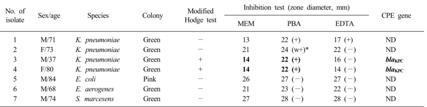
Table 1. Isolates suspicious for carbapenemase-producing Enterobacteriaceae on chromogenic agar No. of
isolate Sex/age Species Colony Modified
Hodge test
Inhibition test (zone diameter, mm)
CPE gene
MEM PBA EDTA
1 M/71 K. pneumoniae Green − 13 22 (+) 17 (+) ND
2 F/73 K. pneumoniae Green − 21 24 (w+)* 22 (−) ND
3 M/37 K. pneumoniae Green + 14 22 (+) 16 (−) blaKPC
4 F/80 K. pneumoniae Green + 14 22 (+) 14 (−) blaKPC
5 M/84 E. coli Pink − 26 27 (−) 27 (−) ND
6 M/68 E. aerogenes Green − 21 23 (−) 22 (−) ND
7 M/74 S. marcesens Green − 27 28 (−) 28 (−) ND
*Indicates a positive result when the size difference was 4 mm or more and weakly positive at 3 mm.
Abbreviations: MEM, meropenem; PBA, phenylboronic acid; EDTA, ethylenediaminetetraacetic acid; CPE, carbapenemase-producing Enterobacteriaceae; M, male; ND, not detected; F, female.
tected gene was the KPC type (23.6%).
Although there have been several hospitals reporting CPE, there have not been cases of CPE acquired from a hospi- tal-associated environment or outbreak at our institute until now.
However, we might have missed the presence of colonized CPE in stool specimens because we did not perform surveillance cul- tures for CPE. Therefore, in this study, we performed surveil- lance cultures for CPE in patients admitted to our institute and evaluated the prevalence of CPE.
MATERIALS AND METHODS
Stool cultures were obtained from a total of 228 patients at a tertiary-care hospital from March to May 2017. There were 119 male and 109 female patients with mean age of 67 years (range 20-96). We carried out CPE culture using the remaining stool specimens from patients, which were requested for routine bacterial culture, Clostridium difficile culture, or vancomycin-re- sistant Enterococcus culture at the clinical microbiology laboratory. During this period, the isolates were consecutively recovered as one isolate per patient. Stool specimens were in- oculated on ChromID CARBA agar (bioMérieux, Marcy l’Etoile, France) and incubated for 18-24 hours. Each isolate was identified by MicroScan Walkaway (Beckman Coulter, Brea, CA, USA) or Bruker Biotyper (Bruker Daltonics, Bremen, Germany) matrix-assisted laser desorption ionization-time of flight (MALDI-TOF) mass spectrometry systems. Suspicious colonies with pink or bluish-green color on ChromID CARBA agar (bioMérieux) were screened for CPE by the modified Hodge test (MHT) and carbapenemase inhibition test (CIT). CIT were performed using phenylboronic acid (PBA, Sigma, Korea)
and ethylenediaminetetraacetic acid (EDTA, Sigma, Korea) to detect class A and class B carbapenemases, respectively. In ad- dition, we performed PCR to detect the five carbapenemase genes blaKPC, blaIMP, blaVIM, blaNDM, and blaOXA-48 as previously described [9].
RESULTS AND DISCUSSION
Among 228 isolates, seven were suspicious for CPE: four K.
pneumoniae, one Escherichia coli, one Enterobacter aerogenes and one Serratia marcesens (Table 1). The E. coli colonies had a pink color, while the other colonies were green. Two K. pneu- moniae isolates (nos. 3 and 4) showed positive reactions for both the MHT and CIT with PBA. As a result, blaKPC was iden- tified in these two K. pneumoniae isolates. One K. pneumoniae isolate (no. 1) showed a negative MHT reaction but positive CIT with both PBA and EDTA. However, none of the five car- bapenemase genes were detected by PCR. This isolate may hy- perproduce AmpC β-lactamase with porin loss and reveal car- bapenem resistance as previous studies [9,10]. E. coli, E. aero- genes and S. marcesens isolates were negative in the MHT and CIT, and PCR did not detect any carbapenemase genes in these isolates.
Two KPC-producing K. pneumoniae (nos. 3 and 4) isolates were obtained from stool specimens from an 82-year-old female with septic shock and a 37-year-old male with cerebral in- farction, respectively. These patients required prolonged hospi- talization in ICU for 28 days and 25 days, respectively. The two KPC-producing isolates were resistant to ampicillin/sulbactam, cefotaxime, cefepime, imipenem, meropenem, ertapenem, levo- floxacin, and trimethrprim/sulfamethoxazole, but were suscep-
10
Ann Clin Microbiol 2018;21(1):8-11tible to colistin and tigecycline.
Jeong et al. [11] suggested that the prevalence and predom- inant genotypes of CPE in Korea showed hospital-specific dif- ferences such as epidemic presence, sporadic presence, and absence. Therefore, they suggested that CPE dissemination is at an early stage in Korea. There are two limitations in this study:
the short period of hospital surveillance and the limited number of patients referred to the laboratory for stool culture. The limi- tation of this study was that the surveillance culture for evalua- tion the prevalence was not performed for all patients admitted in hospital or intensive-care unit.
In this study, our institute may be a sporadic presence hospi- tal in this time. Our results indicated a very low prevalence (2/228, 0.9%) of CPE in a tertiary-care hospital based on sur- veillance culture. However, continuous monitoring and infection control for CPE should be performed to prevent transmission of this superbug.
ACKNOWLEDGMENTS
This work was supported by the research fund of Hanyang University (HY-2016-2782).
REFERENCES
1. Gupta N, Limbago BM, Patel JB, Kallen AJ. Carbapenem-resistant Enterobacteriaceae: epidemiology and prevention. Clin Infect Dis
2011;53:60-7.
2. Nordmann P, Naas T, Poirel L. Global spread of Carbapenemase- producing Enterobacteriaceae. Emerg Infect Dis 2011;17:1791-8.
3. Falagas ME, Rafailidis PI, Kofteridis D, Virtzili S, Chelvatzoglou FC, Papaioannou V, et al. Risk factors of carbapenem-resistant Klebsiella pneumoniae infections: a matched case control study. J Antimicrob Chemother 2007;60:1124-30.
4. Vardakas KZ, Matthaiou DK, Falagas ME, Antypa E, Koteli A, Antoniadou E. Characteristics, risk factors and outcomes of carbapenem-resistant Klebsiella pneumoniae infections in the intensive care unit. J Infect 2015;70:592-9.
5. Pesesky MW, Hussain T, Wallace M, Wang B, Andleeb S, Burnham CA, et al. KPC and NDM-1 genes in related Entero- bacteriaceae strains and plasmids from Pakistan and the United States. Emerg Infect Dis 2015;21:1034-7.
6. Yigit H, Queenan AM, Anderson GJ, Domenech-Sanchez A, Biddle JW, Steward CD, et al. Novel carbapenem-hydrolyzing beta-lactamase, KPC-1, from a carbapenem-resistant strain of Klebsiella pneumoniae. Antimicrob Agents Chemother 2001;45:
1151-61.
7. Jeong SH, Lee KM, Lee J, Bae IK, Kim JS, Kim HS, et al. Clonal and horizontal spread of the blaOXA-232 gene among Entero- bacteriaceae in a Korean hospital. Diagn Microbiol Infect Dis 2015;82:70-2.
8. Park JW, Lee EJ, Lee DH. Status of carbapenemase producing Enterobacteriaceae in Korea, 2014. Public Health Weekly Report 2014;9:9-13.
9. Lee JS and Song WG, eds. Guidelines for diagnosis of CPE. 1st ed. Osong: Korea Centers for Disease Control and Prevention;
2014:20-37.
10. Thomson KS. Extended-spectrum-beta-lactamase, AmpC, and carbapenemase issues. J Clin Microbiol 2010;48:1019-25.
11. Jeong SH, Kim HS, Kim JS, Shin DH, Kim HS, Park MJ, et al.
Prevalence and molecular characteristics of carbapenemase- producing Enterobacteriaceae from five hospitals in Korea. Ann Lab Med 2016;36:529-35.
Eunyoung Lee and Yangsoon Lee : Surveillance Culture for CPE
11
=국문초록=
카바페넴분해효소 생성 장내세균에 대한 감시배양
한양대학교 의과대학 진단검사의학교실 이은영, 이양순
배경: 카바페넴 내성 장내세균(Carbapenem-resistant Enterobacteriaceae, CRE)은 전세계적으로 점차 증가하고 있으며, 환자 의 치료 및 감염관리에 매우 중요하다. CRE의 카바페넴 내성은 주로 이동성을 가진 카바페넴분해 효소 때문인 것으로 알려져 있다. 따라서, 카바페넴분해효소를 분비하는 장내세균(carbapenemase-producing Enterobacteriaceae, CPE)의 보균자 검출은 원내 감염의 예방 및 감시에 중요하다. 본 연구에서는 국내 1개 3차병원에서 CPE 감시배양을 시행하여 발생률을 조사하였다.
방법: 2017년 3월부터 5월까지 1개 3차병원에 내원한 총 228명 환자의 대변배양을 시행하였다. 대변은 ChromID CARBA agar (bioMérieux, France)에 접종하고, 18-24시간 배양하였다. CPE가 의심되는 집락에 대해서 modified Hodge test (MHT) 와 carbapenemase inhibition test (CIT)을 시행하였고, blaKPC, blaIMP, blaVIM, blaNDM 및 blaOXA-48 유전자에 대해서 PCR 및 염기서열을 분석하였다.
결과: CPE가 의심되는 균주는 228균주 중에서 7주로, Klebsiella pneumoniae 4주, Escherichia coli 1주, one Enterobacter aeroginosa 1주, Serratia marcescens 1주였다. K. pneumoniae 2주가 MHT와 CIT에 양성이었고, KPC 유전자가 검출되었다.
본 기관의 최근 3개월간 CPE 발생률은 0.9% (2/228)임을 알 수 있었다.
결론: CPE 감시 배양을 통해서 낮은 발생률(2/228, 0.9%)을 확인하였으며, 지속적인 감시배양이 필요할 것으로 사료된다.
[Ann Clin Microbiol 2018;21:8-11]
교신저자 : 이양순, 04763, 서울시 성동구 왕십리로 222-1 한양대학교 의과대학 진단검사의학교실 Tel: 02-2290-9655, Fax: 02-2290-9193 E-mail: [email protected]