INTRODUCTION
Atopic dermatitis (AD) is one of the most common chronic in- flammatory skin diseases, characterized by xerosis, eczematous skin lesions, pruritus, immunodysregulation, epidermal barrier dysfunction and IgE-mediated sensitization to food and envi- ronmental allergens.1 Our understanding of the disease patho- genesis of AD has increased, and it is clear that no single defect accounts for the array of clinical features associated with AD.
Lifetime prevalence of AD varies from approximately 8% to 18%
worldwide.2 AD is on the rise in many developing countries, af- fecting over 10% of the population, and has reached a plateau of
~20% in Western countries.3,4 The striking increase in AD inci- dence observed in recent decades has been attributed to the re- settlement of populations from rural to urban areas. According to the hygiene hypothesis,5 a lack of early childhood exposure to a variety of microbes results in reduced immune tolerance.
Of the various pathogenic factors suggested for AD, impaired epidermal permeability barrier function is considered to be im- portant. Many studies have suggested that a defective epider- mal barrier combined with an abnormal immune response might contribute to the pathophysiology of AD, supporting the outside-inside hypothesis.6,7 In addition, the clinical manifesta- tions of AD predate the development of asthma and allergic rhi- nitis, suggesting that AD is an entry point for subsequent aller-
Epidermal Permeability Barrier Defects and Barrier Repair Therapy in Atopic Dermatitis
Hae-Jin Lee,
1Seung-Hun Lee
2*
1Medical Corps of Sangmudae Army Service Support Group, Republic of Korea Army Training and Doctrine Command, Jangsung, Korea
2 Department of Dermatology, Gangnam Severance Hospital, Cutaneous Biology Research Institute, Yonsei University College of Medicine, Seoul, Korea
gic disease in a process called the atopic march. The concept of atopic march was hypothesized to describe the progression of atopic disorders from AD in infants to asthma and allergic rhini- tis in children. Kubo et al.1 suggested a theoretical model of bar- rier disruption followed by percutaneous sensitization, which may apply to the pathogenesis of the atopic march. Therefore, the importance of barrier disruption in AD has gained increas- ing attention. In this review, we discuss the recent progress in understanding the functions of the epidermal permeability bar- rier, its immunologic role in human and animal subjects, and barrier repair therapies in AD.
Epidermal permeability barrier dysfunctions in AD
The skin, as an interface between the organism and the exter- nal environment, plays a major role in protecting and support- ing the life it encloses. The outermost layer of the skin, the stra- tum corneum (SC), is the primary mediator of this epidermal permeability barrier function.8 Atopic dry skin displays impaired Atopic dermatitis (AD) is a multifactorial inflammatory skin disease perpetuated by gene-environmental interactions and which is characterized by genetic barrier defects and allergic inflammation. Recent studies demonstrate an important role for the epidermal permeability barrier in AD that is closely related to chronic immune activation in the skin during systemic allergic reactions. Moreover, acquired stressors (e.g., Staphylococcus aureus infection) to the skin barrier may also initiate inflammation in AD. Many studies involving patients with AD revealed that defective skin barriers com- bined with abnormal immune responses might contribute to the pathophysiology of AD, supporting the outside-inside hypothesis. In this review, we discuss the recent advances in human and animal models, focusing on the defects of the epidermal permeability barrier, its immunologic role and barrier repair therapy in AD.
Key Words: Atopic dermatitis; barrier repair therapy; skin barrier
This is an Open Access article distributed under the terms of the Creative Commons Attribution Non-Commercial License (http://creativecommons.org/licenses/by-nc/3.0/) which permits unrestricted non-commercial use, distribution, and reproduction in any medium, provided the original work is properly cited.
Correspondence to: Seung Hun Lee, MD, PhD, Department of Dermatology, Gangnam Severance Hospital, Yonsei University College of Medicine, 211 Eonju-ro, Gangnam-gu, Seoul 135-720, Korea.
Tel: +82-2-2019-3360; Fax: +82-2-3463-6136; E-mail: ydshderm@yuhs.ac Received: December 8, 2013; Accepted: January 6, 2014
•There are no financial or other issues that might lead to conflict of interest.
Allergy Asthma Immunol Res. 2014 July;6(4):276-287.
http://dx.doi.org/10.4168/aair.2014.6.4.276 pISSN 2092-7355 • eISSN 2092-7363
barrier function, indicated by an increased transepidermal wa- ter loss (TEWL) and decreased water-binding capacity due main- ly to altered levels of inter/intracellular components in the SC.
Filaggrin and its gene
Filaggrin (FLG) is named after the filament aggregating pro- tein, and the FLG gene is located on chromosome 1q21 within the epidermal differentiation complex.9 FLG protein is local- ized in the stratum granulosum (SG). Profilaggrin, a 400-kDa polyprotein, is the main component of keratohyaline granules.
In the process of keratinocyte differentiation, profilaggrin is de- phosphorylated and cleaved into 10-12 FLG molecules. In vitro, FLG monomers aggregate and align keratin bundles, which contribute to the mechanical strength and integrity of the SC.6 To promote corneocyte compaction, FLG monomers are de- graded into their constituent amino acids, including glutamine, arginine and histidine. These amino acids are then hydrolyzed further into acidic polycarboxylic acid osmolytes, which main- tain SC hydration (the so-called natural moisturizing factors, NMFs), by caspase 14, peptidylarginine deiminases or bleomy- cin hydrolase to maintain hydration of the upper SC and to re- duce skin surface pH.5,10 Maintaining acidic pH is key for many protective functions, including permeability barrier homeosta- sis, SC integrity and cohesion, antimicrobial defense, and it is important for the activation of enzymes involved in ceramide metabolism and modulation of the serine protease cascade re- quired for coordinated epidermal differentiation and cornified cell envelope formation.6,11 Moreover, FLGs are crucial proteins for terminal differentiation of epidermal keratinocytes, which form a skin barrier with the SC intercellular lipids. Pyrrolidone carboxylic acid, a major component of NMFs in skin, is primar- ily produced from FLG proteins in the SC and plays a vital role in maintaining hydration of the SC.8 If these proteins are de- creased or absent due to FLG mutations, the FLG-associated SC barrier is disrupted. Such SC barrier disruption results in de- creased formation and secretion of the lamellar body (LB), cor- nified envelope, and corneodesmosome, elevated pH and de- creased tight junction (TJ) proteins, leading to increased epi- sodes of percutaneous allergen exposure (Fig. 1).1 Kezic et al.12 confirmed that individuals with FLG-null mutations have signif- icantly reduced levels of NMFs in the SC of their forearms and palms. Moreover, significantly lower NMF levels were observed in individuals with a history of AD who were carriers of FLG mu- tations compared with those who were non-carriers. The au- thors demonstrated higher TEWL in the carriers of FLG muta- tions compared with non-carriers. FLG has been suggested to contribute to the formation of acid mantle (described below) within the SC through the production of urocanic acid (UCA) via the filaggrin-histidine-UCA cascade.13 Consequently, FLG- deficiency in AD lesions leads to defects in the formation of the cornified envelope, a decreased ability to maintain SC hydra- tion, and a parallel elevation in pH. The increase in pH enhanc-
es the KLK5 and KLK7 activities, which are optimal at neutral pH, resulting in over-degradation of corneodesmosomes and decreased SC integrity and cohesion. Patients with FLG muta- tions might have a higher risk of allergic sensitization compared with those with wild-type FLG.14 FLG mutations are significant- ly correlated with increased risk of developing atopic diseases, including AD, atopic asthma, allergic rhinitis and nickel and food allergies, even though FLG is not found in the bronchial epithelium.15
Nevertheless, FLG null mutations may not be sufficient to in- duce the findings typical of AD. The median prevalence of FLG mutations among Europeans and Asians is 7.7% and 3.0%, re- spectively.16 In European studies, the prevalence of FLG muta- tions in AD subjects is 3% in Italy, 15.2%-22.9% in Germany, 40.2%-42% in the UK and 45.2%-55.8% in Ireland, and this pat- tern suggests a higher tendency for FLG mutation prevalence in AD patients who reside in countries of higher latitudes than in those of lower latitudes.16 Patients with FLG mutations can de- velop ichthyosis vulgaris without manifestations of AD, and ap- proximately 50%-90% of AD patients have no FLG defects.16,17 In fact, mice with the FLG null mutation exhibited no spontane- ous AD-like skin lesions.18 However, flaky tail mice homozygous for the 5303delA mutation in the FLG and Tmem79/Matt genes showed FLG and NMF deficiencies, presumably representing the characteristics of AD after exposure to allergens.19 A recent study revealed that the matted gene Matt is a predisposing gene for AD in mice, and a common Matt (Pl use consistent designa- tions when describing flaky tail mice in this section and later in the manuscript) single nucleotide polymorphism is associated with AD in human subjects.19 In addition, Sasaki et al.20 showed that the Tmem79 (ma/ma) mutation is responsible for the spon- taneous dermatitis phenotype in matted mice, probably due to the impairment of the lamellar granule secretory system and al- tered SC barrier function, which has also been identified in Caucasians with FLG mutations in a dose-dependent manner.21 Fig. 1. Barrier dysfunction associated with filaggrin deficiency leads to lipid bi- layer disorganization, delayed bilayer maturation, as well as decreased SC cohe- sion, paracellular permeability barrier, and photoprotection, which all may play important roles in the pathogenesis of atopic dermatitis (AD). NMF, natural mois- turizing factors; SC, stratum corneum; TEWL, transepidermal water loss.
A recent study by Kim et al. showed that IL-25 inhibits the ex- pression of FLG and acts synergistically with Th2 cytokines to inhibit FLG expression.22 We assume that the barrier breakage in AD is due not only to the FLG gene alone, but that FLG com- bined with other genes might cause or exacerbate AD skin le- sions.
TJ barrier
TJs are involved in the control of paracellular migration of in- flammatory cells. Increasing evidence shows that skin TJs, which localize in the SG, contribute to epidermal barrier formation,23 however, the role of TJs in AD is still unknown. Among TJ pro- teins, claudin (Cldn)-1, a TJ-specific integral membrane pro- tein, and Cldn-4 play important roles in the barrier function of the skin. In a recent study, Cldn-1 knockout mice demonstrat- ed normal SC structure but abnormal SC formation and SC barrier defects. These Cldn-1 knockout mice died shortly after birth and had increased TEWL.24 In humans, a lack of Cldn-1 causes NISCH syndrome, which is characterized by ichthyosis, scalp hypotrichosis, scarring alopecia, neonatal sclerosing cholangitis and leukocyte vacuolization. Kubo et al.25 reported that dendrites of activated Langerhans cells express Cldn-1 and ZO-1. Therefore, functional bicellular and tricellular TJs, which can take up external allergens easily when the SC barrier is dis- rupted, are present between Langerhans cells and keratino- cytes.1 In a recent study, patients with AD demonstrated a de- fect in TJ barrier function and composition.26 Furthermore, Yuki et al.27 reported that impaired TJ barriers affect polar lipids and profilaggrin processing by disturbing the pH of the SC.
Based on these reports, we assume that decreased TJ compo- nents in lesional AD promote epicutaneous penetration of al- lergens, including the house dust mite antigen, which leads to the increased possibility of developing systemic allergy or atop- ic march. Recently, Kuo et al.28 showed that the activation of toll-like receptor (TLR)-2, which shows reduced expressed in AD, enhances TJ function in mice and human keratinocytes, suggesting the use of TLR-2 enhancers as a potential therapeu- tic strategy in patients with AD. Further investigation of the crosstalk between TJs and the SC barrier is needed to clarify the role of TJs in the pathogenesis of AD.
PH, proteases and protease activated receptor (PAR) 2
The skin surface pH, which ranges from 4.5 to 5.5 in humans, is slightly acidic compared with the normal physiologic pH.8 The importance of pH in vivo was first reported in experiments in which the permeability barrier function was disrupted acute- ly, producing a parallel increase in pH.29 The pH of the SC influ- ences key epidermal functions, including permeability barrier homeostasis, desquamation of corneocytes, initiation of inflam- mation, processing of secreted LB polar lipids and antimicrobi- al defense (Fig. 2A). The UCA breakdown products, free fatty ac- ids and sodium hydrogen antiporter (NHE1) are the three main
factors responsible for maintaining acidic pH in the skin.9 In AD, the baseline skin pH is more alkaline than the average baseline pH of healthy skin.30 The perturbation of lipid metabolism and its molecular organization as well as elevated skin pH induce the growth of bacteria such as Staphylococcus aureus (S. aureus).
Trans-UCA (t-UCA) is an endogenous acidifier of the SC. Hence, decreased production of FLG products could result in an initial increase in skin surface pH that is sufficient to activate multiple serine proteases in SC, which exhibit neutral-to-alkaline pH optima.31 If prolonged, the pH-induced increase in serine pro- tease activity could precipitate downstream structural and functional alterations, as mentioned below (Fig. 2B). Moreover, increased pH leads to higher activities of Kallikrein (KLK) 5 and 7 (SC chymotryptic enzyme), which have optimal activity at neutral pH, resulting in the over-degradation of corneodesmo- somes and a decrease in SC integrity and cohesion.13 Moreover, elevated skin pH inhibits the activity of lipid-processing en- zymes, including β-glucocerebrosidase (β-GlcCer’ase), acid sphingomyelinase (aSMase) and secretory phospholipase A2 (sPLA2), which are essential in the production of ceramide and free fatty acids, thereby causing impaired lipid processing and defects in the epidermal permeability barrier.
SC acidity is important in antimicrobial barrier function in that it inhibits the growth of pathogens. The growth of S. aureus, which readily colonizes in the lesional skin of AD, is normally inhibited at low skin pH and grows best at pH 7.5. In AD, the in- creased pH in lesional skin leads to bacterial growth, resulting in allergic inflammation and aggravation of AD. Hatano et al.11 showed that the acidification of SC alone substantially prevents the development of barrier abnormalities and downstream im- mune abnormalities during the elicitation phase in the murine oxazolone-induced AD model. This study suggests that the maintenance of a normal or hyperacidic pH could be a possible novel treatment for reversing or preventing AD in humans.
PAR is a G protein-coupled receptor, characterized by a unique mechanism of self-activation following specific proteolytic cleavage of its extracellular domain.13 Four PAR members have been identified: PAR-1, -3, and -4 are activated by thrombin and are involved in homeostasis and thrombosis, whereas PAR-2 is activated by trypsin-like serine proteases, but not by thrombin.
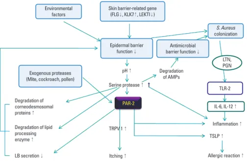
PAR-2 is widely distributed throughout the mammalian body13 and is expressed by almost all skin cell types, especially kerati- nocytes. Previous studies demonstrated that PAR-2 is expressed in the suprabasal epidermal layers of both humans and mice, and that this expression is most prominent in the granular lay- er, implying that PAR-2 expression might depend on the state of epidermal differentiation.32 PAR-2 localizes in the suprabasal layers of the mouse and human epidermis with high levels in the SG, supporting PAR-2 as the primary sensor of barrier-initi- ated serine protease activity.33 The lesional skin of AD expresses high levels of PAR-2. Furthermore, PAR-2 activation is likely to be involved in pruritus of AD.13 Indeed, high expression rates of
PAR-2 and trypsin have been observed in the lesional skin of patients with AD, and PAR-2 agonist peptides induce pruritus in patients with AD.34 PAR-2 in the skin is involved in pruritus via the transient receptor potential vanilloid receptor (TRPV1) inflammation, and thymic stromal lymphopoietin (TSLP) acti- vation, implying that PAR-2 antagonists represent potentially useful therapeutics for treating AD (Fig. 3).35 The association between PAR-2 and TSLP is discussed below.
Many studies have provided evidence that genes associated with proteases/protease inhibitors become deregulated in pa- tients with AD, shifting the balance between proteases and pro- tease inhibitors toward increased protease activity. A link has been demonstrated between AD and a 4-base pair (AACC) in- sertion into the 3’-untranslated region of the KLK7 gene, which increases the half-life of KLK7 mRNA and the enzymatic activi- ty of KLK7.13 Lymphoepithelial Kazal-type-related inhibitor (LEKTI) is an inhibitor of multiple serine proteases in skin, and it regulates the enzymatic activities of serine proteases, includ- ing KLK5, -6, -7, -13, and -14, thereby controlling epidermal barrier function.36 A premature stop codon mutation in the SPINK5 gene, which encodes LEKTI, is associated with Nether-
ton syndrome (NS), a rare ichthyosiform dermatosis character- ized by congenital ichthyosiform erythroderma, severe atopic manifestations and hair shaft abnormalities. SPINK5-deficient mice demonstrate increased proteolytic activities of KLK5 and KLK7 in the epidermis and abnormal degradation of desmo- glein 1, resulting in abnormal corneodesmosome cleavage and premature desquamation. These data suggest that LEKTI is a key regulator of KLK5 and KLK7 activity, and defective SC adhesion caused by epidermal protease hyperactivity is the primary pathogenic event in NS.13 In addition, several studies reported that the polymorphisms in the SPINK5 gene are associated with AD.37,38 Moreover, cystatin A, a cysteine protease inhibitor, inhib- its the endogenous cathepsins B, -H, and -L and the exogenous proteases from house dust mites of dermatophagoides pteron- yssinus (Der p) 1 and dermatophagoides farinae (Der f) 1.13,39 The cystatin A-encoding gene (CSTA) polymorphism (+344c variant) is associated with AD.39 This CSTA variant causes re- duced levels of cystatin A in the skin surface and in sweat, allow- ing exogenous proteases to break down the integrity of the SC, which enhances the penetration of allergens and aggravates AD.
These genetic variants in protease and protease inhibitor genes Fig. 2. The factors involved in acidic pH maintenance and their role in normal epidermis, and the consequences of altered pH in patients with AD. (A) Exogenous free fatty acids are derived from sweat glands or catalyzed from sebaceous gland-derived triglyc- erol moieties via microorganism- secreted lipases. Endogenous free fatty acids are derived from phospholipids by phospholipase A2 (PLA2), both of which are se- creted by lamellar bodies (LBs) at the SC–stratum granulosum junc- tion. Additionally, the Na+/H+ an- tiporter is involved in maintaining the skin acid mantle. Therefore, the skin acid mantle regulates SC integrity and cohesion, antimicro- bial function, processing of LB polar lipids, structural organiza- tion of lamellar membrane, and β-glucocerebrosidase/sphingo- myelinase function. (B) AD or oth- er stressors alter the function of the skin acid mantle. The altered acid mantle increases serine pro- tease activity and decreases the function of corneodesmosomes, resulting in decreased production of ceramides and LB secretion, altered SC cohesion and inflam- mation activation.
A
B
coexist in some patients with AD, resulting in a more severe de- fect in the skin barrier.13
The proteolytic activity from various contact allergens and aeroallergens are considered important factors in the initiation and aggravation of AD. Proteolytic activity plays a role in the pathogenesis of allergic diseases, including allergic rhinitis, asthma and AD, by inducing Th2 allergic inflammation and di- rectly affecting the structure and function of the epidermal bar- rier, which facilitates further penetration of allergens through the defective skin barrier.40 House dust mite and cockroach al- lergens, important environmental factors in the pathogenesis of AD, have been shown to have proteolytic activity.41 House dust mite allergens contain several cysteine and serine proteas- es. The serine proteases from the mite allergens, Der p 3 and Der p 9, activate PAR-2 on keratinocytes to produce cytokines and contribute to the pathogenesis of AD, whereas the cysteine protease activity in Der p 1 stimulates inflammation via a PAR- 2-independent mechanism. S. aureus produce extracellular V8 protease, which causes defective epidermal permeability barri- er function in the skin of hairless mice by directly degrading DSG1.42 These reports imply that proteolytically active allergens could break down the skin barrier via PAR-2-mediated inhibi- tion of LB secretion or PAR-2-non-mediated mechanisms, in- cluding the degradation of corneodesmosomal proteins and lipid processing enzymes. This degradation further triggers al- lergen penetration through the disrupted epithelial barrier to aggravate Th2-mediated inflammation and possibly causes the non-IgE-associated form of AD (atopiform AD) to switch to the typical IgE-associated form of AD.13 Various roles of serine pro- teases and PAR-2 are described in Fig. 3.
Peroxisome proliferator-activated receptors (PPARs)
PPARs are ligand-activated transcription factors involved in
the genetic regulation of lipid metabolism and energy homeo- stasis. PPARs belong to a subfamily of nuclear hormone recep- tors comprising three different isoforms of PPARs: PPARα, PPARβ/δ and PPARγ.43 After ligand binding, PPARs can regulate gene expression by binding to peroxisome proliferator response elements in target genes, after heterodimerizing with the reti- noid X receptors. Activators of PPARs and liver X receptors dis- play potent but largely positive effects on the epidermal struc- ture and function in normal and diseased skin, including the upregulation of FLG, anti-inflammatory activity, and reverse epidermal hyperplasia and differentiation.44 In AD, PPARs re- duce several inflammatory mediators in the skin and regulate epidermal barrier homeostasis by stimulating epidermal differ- entiation and lipid production. In fact, PPAR ligands inhibit T helper cell responses by inhibiting IL-2 production in T cell clones.45 Since patients with AD exhibit primary abnormalities in the epidermal barrier function, the PPAR/LXR activators might possess potential utility in AD by stimulating epidermal differentiation and lipid production.44 Lower expression of PPARα is observed in AD lesional skin, suggesting that decreased PPARα signaling might be involved in the relationship between permeability barrier abrogation and allergic inflammation in AD.46 Experiments using PPARα knockout mice revealed a modest decrease in the epidermal expression of differentiation markers (profilaggrin and loricrin), a thinner SG than that in wild type mice, decreased keratohyaline granules, normal per- meability barrier function and decreased β-GlcCer’ase activi- ty.47 Indeed, the combined therapy of topical PPARα activators/
ligands and steroids showed not only potent anti-inflammatory benefits in the murine AD model, but also almost no rebound flares, which is one of the considerable side effects of steroid therapy.48 Furthermore, PPARα prevents the side effects of ste- roid-induced structural and functional abnormalities and im-
Degradation of AMPs
LTN, PGN
TLR-2 IL-6, IL-12 ↑ Inflammation ↑
Allergic reaction ↑ TSLP ↑ PAR-2
Degradation of corneodesmosomal proteins ↑ Degradation of lipid processing enzyme ↑
TRPV 1 ↑ pH ↑ Serine protease ↑
Itching ↑ LB secretion ↓
Environmental factors
S. Aureus colonization Skin barrier-related gene
(FLG↓, KLK7↑, LEKTI↓)
Exogenous proteases (Mite, cockroach, pollen)
Epidermal barrier function ↓
Antimicrobial barrier function ↓
Fig. 3. The roles of serine protease and PAR- 2 in AD. AMP, antimicrobial peptide; FLG, fil- aggrin; KLK, kallikrein; LB, lamellar body; LE- KTI, lymphoepithelial Kazal-type-related in- hibitor; LTN, lipoglycan; PAR-2, protease ac- tivated receptor-2; PGN, peptidoglycan; TLR, toll-like receptor; TSLP, thymic stromal lym- phopoietin.
proves paracellular permeability.48 Recent analysis of AD skin lesions showed increased PPARγ expression in keratinocytes and in infiltrating T cells and monocytes.49 PPARγ plays a criti- cal role in the regulation of genes involved in cellular prolifera- tion, in specific components of the Th2 inflammatory pathway and in maintenance of the skin barrier.45 Systemic treatment with a PPARγ agonist, rather than with topical treatment, decreased the amount of surface area affected, the severity of lesions and the number of flares in patients with severe AD.50 Therefore, PPARs and their corresponding ligands are potential therapeutic targets for treating AD. Further studies are needed to verify the underlying mechanism of PPARs in AD pathogenesis.
Ceramides and lipid processing
The major lipid species of the SC are ceramides, fatty acids and cholesterol. The majority of SC lipids are derived from the LB and synthesized in the keratinocytes of the upper stratum spinosum and SG.8 In human AD, a selective reduction in ce- ramide content, especially ceramide 1, is observed. Indeed, Na- kajima et al.51 reported that a ceramide deficiency in the epider- mis leads to psoriasis-like lesions in the serine palmitoyltrans- ferase knockout mouse model. The abnormal neutral skin pH in patients with AD inhibits the activity of lipid-processing en- zymes, including β-GlcCer’ase, aSMase and sPLA2, thereby causing impaired lipid processing and a defective lipid barrier, as mentioned above. Other minor mechanisms that might ac- count for the frequently colonized microbial pathogens in AD involve acidic ceramidase activity, which could decrease ce- ramide content.52 Moreover, altered ceramide metabolism in AD results in decreased levels of sphingosine, which is pro- duced from ceramide by ceramidase in the SC. Since sphingo- sine is known to have a powerful antimicrobial activity on S. au- reus at physiologic levels, downregulation of sphingosine is one explanation for the susceptibility of patients with AD to S. aure- us colonization.37,38
Antimicrobial peptides
Antimicrobial peptides (AMPs) are important molecules in- volved in the evolutionarily conserved innate immune defense system of the skin against bacteria, viruses, fungi, parasites and tumor cells.53 More than 1,700 AMPs have been identified.54 In normal human skin, keratinocytes are the main source of AMPs, and AMP synthesis occurs primarily in the SG.53 In addition to their antimicrobial activity, AMPs in the skin have variable functions, including modulation of host inflammatory respons- es and promotion of wound healing.55 Moreover, AMPs have been shown to be associated with permeability barrier function in a knockout mouse model for cathelin-related antimicrobial peptide (CRAMP), a murine homologue of LL-37. These knock- out mice showed a significant delay in permeability barrier re- covery after tape stripping as well as structural abnormalities in LB content.56 In addition, a recent study suggested that human
β-defensin-2 (HBD2) can be used as a marker for disease sever- ity and barrier properties in AD.53
AD skin has significantly lower AMP production than does pso- riatic skin, while both disorders are associated with inflamma- tion and defective barrier function.57 Nomura et al.58 proposed that this defect in AD is caused by AMP suppression via elevated levels of T-helper 2 cytokines, IL-4 and IL-13. Lower AMP levels diminished the antimicrobial barrier function in AD subjects, re- sulting in increased susceptibility to S. aureus and other microbi- al superinfections, such as eczema herpeticum.53,59 In fact, LL-37 exhibited antiviral activity against herpes simplex virus (HSV), and mice deficient in the LL-37 homologue showed higher levels of HSV-2 replication in their skin. Moreover, the skin of AD sub- jects with eczema herpeticum exhibited significantly lower levels of LL-37 than that of AD patients. Therefore, reduced levels of LL- 37 may render AD subjects susceptible to eczema herpeticum.59,60 Many reports have demonstrated that AMPs are induced in the skin in AD. Reduced gene expression of hBD-3 was not ob- served in the epidermis of patients with AD compared with psoriasis,60 and increased expression levels of hBD-3 and other AMPs, such as hBD-2, psoriasin, elafin and calprotectin, were found in AD skin compared with healthy skin.61 However, an- other study did not detect differences in hBD-2 and LL-37 ex- pression levels between the nonlesional skin of AD patients and non-AD patients, indicating that expression of hBD-2 and LL-37 is not decreased at baseline in the epidermis of patients with AD.62 Ballardini et al.63 and Gambichler et al.64 detected higher levels of AMPs in lesional skin than in nonlesional skin in AD. Based on these contradictory reports, we assume that the increased AMP amounts are lower at the onset of inflam- mation in the lesional skin in AD patients compared with that in non-AD patients. Further studies are needed to clarify the causality of AMP expression on lesional and non-lesional epi- dermis in AD.
Ultraviolet irradiation and AD
Ultraviolet (UV) C radiation is absorbed by the ozone layer;
however, the skin is constantly exposed to UVA and UVB irradi- ation, and this UV radiation affects FLG degradation. Using his- tidine as a substrate, histidase produces t-UCA, a major UVB- absorbing epidermal chromophore. t-UCA is photoisomerized to cis-UCA with UVB exposure, and cis-UCA may produce oxi- dative DNA damage and initiate the translation of genes associ- ated with apoptosis and immunosuppression.16,65 Although UV irradiation of the skin induces disruption of the epidermal bar- rier, phototherapy is generally used in patients with AD to re- lieve acute flares (UVA1) and chronic lesions (narrow band UVB).66 The mechanism of action in AD has not been elucidat- ed; however, the proper UV dose has positive effects on the epi- dermal permeability barrier function by immunomodulating apoptosis of inflammatory cells, reducing the number of cuta- neous nerve fibers, inhibiting the Langerhans cells and altering
cytokine production.67 In addition, a suberythemal dose of UVB on the skin exerts beneficial effects on the SC barrier function by activating the cutaneous vitamin D system.68,69 In addition, UV initiates endoplasmic reticulum (ER) stress responses in- cluding downstream activation of the transcription factor, NF- κB.70 In the epidermis, ER stress increases human cathelicidin antimicrobial peptide (CAMP) expression, which stimulates in- nate immunity.71 A recent study suggested that UV radiation enhances cortisol activity in a waveband-dependent manner, and cortisol production protects and/or restores epidermal barrier homeostasis.72 In fact, most patients affected by AD im- prove during the sunny summer season, and artificial UV radi- ation, including psoralen-UVA and narrow-band UVB, is fre- quently utilized in the treatment of AD. Further comparative clinical studies are needed to clarify the proper dose and wave- length of UV therapy in AD.
Link between the epidermal permeability barrier function and the systemic immune reaction in ad: thymic stromal
lymphopoietin (TSLP)
TSLP is an interleukin-7-like cytokine produced by epithelial cells that triggers dendritic cells to induce Th2 inflammatory re- sponses.73 TSLP enhances the effector stage of the allergic re- sponse and amplifies secretion of Th2 cytokines in synergy with IL-1 and TNF-α.74 Furthermore, TSLP is induced by exogenous stimuli, such as defects in barrier differentiation, infections, TLR ligation and trauma, as well as by topical application of active vitamin D3 and low-calcemic analogue treatment.75 TSLP is overexpressed in keratinocytes in AD skin lesions and triggers a pathway underlying AD progression and atopic march. In addi- tion to early protection from skin barrier defects in AD, the se- lective inhibition of TSLP might be a key intervention strategy to block the progression of atopic march. Previous studies have reported increased levels of TSLP in tissues biopsied from acute and chronic AD skin lesions and from the serum of patients with AD.76 Overexpression of TSLP in epidermal keratinocytes is a systemic driver of bronchial hyperresponsiveness, and TSLP in asthmatic airway epithelial cells is correlated with reduced lung function.77 The PAR-2–TSLP relationship, in particular, has been emphasized in many studies.35 Indeed, PAR-2 might be a parent signal that induces the overexpression of TSLP in epithelial cells.78 A recent study revealed high TSLP expression when keratino- cytes of flaky tail mice were exposed to a PAR-2 agonist, but low TSLP expression in the presence of a PAR-2 antagonist, suggest- ing that PAR-2 is at least partly responsible for the induction of TSLP production.79 These results suggest that TSLP could be a link between skin barrier function and the systemic immune reaction in AD.
Animal models in AD
Many research groups have established animal models of AD.
In this section, we discuss several AD animal models that have
been used most frequently and which seem to correlate with the outside–inside hypothesis in AD. Flaky tail mice have been used to investigate the role of FLG in AD. The spontaneous flaky tail (ft) mouse arose on the background of an existing recessive hair phenotype, matted (ma). Flaky tail mice have a 1-bp frame- shift mutation in the FLG gene. Flgft mice have spontaneous dermatitis with increased IgE levels, endogenous protease (KLK5, 7, and 14) activity, TSLP expression, and basophil accumula- tion in the skin, showing many characteristics of AD.79 They ap- pear normal at birth, and the flaky phenotype becomes visible 3 days after birth with the presence of dry scaly skin and tail constrictions. Under specific pathogen-free conditions, the ma- jority of Flgft mice developed clinical and histological eczema- tous skin lesions similar to human AD with outside-inside skin barrier dysfunction, as evaluated by newly devised methods.80 Moreover, the topical application of dermatophagoides pteron- yssinus in Flgft mice induces skin lesions that are clinically and histologically similar to those seen in AD.79 The relative contri- bution of Flg and ma to the compound phenotype has yet to be defined fully. As mentioned above, the matted mouse gene is a predisposing gene for AD, and the Tmem79 (ma/ma) mutation is responsible for the spontaneous dermatitis phenotype in mat- ted mice, which can also be found in Caucasians with FLG mu- tations in a dose-dependent manner.21 Based on these reports, the flaky tail mouse is perhaps the most represented animal model of AD corresponding with FLG mutation in human AD.
Oxazolone (Ox) is commonly used to provoke allergic contact dermatitis, and it evokes a Th1-dominated response to the hap- ten.81 However, multiple challenges of Ox to the skin of hairless mice over an extended period cause the skin inflammation to shift from a Th1-dominated delayed type hypersensitivity re- sponse to a chronic Th2-dominated inflammatory response similar to human AD.11,44,48,81,82 Nine to 10 Ox challenges to hair- less mice produced a chronic Th2-like skin inflammation, which was characterized by dermal infiltration of Th2 lymphocytes (expressing CRTH2), mast cells and eosinophils, increased ex- pression of IL-4 in the dermis and highly elevated IgE levels.
Repeated Ox challenges led to increased epidermal hyperplasia and decreased expression of keratinocyte differentiation mark- ers. Skin barrier abnormalities were associated with increased TEWL. Additionally, impaired LB secretion along with decreased SC ceramide content and hydration resulted in reduced lamel- lar membranes, similar to those observed in patients with AD.
Furthermore, as in human AD, increased epidermal serine pro- tease activity in the SC and decreased expression of 2 LB-de- rived antimicrobial peptides (CRAMP and mBD3) paralleled the decrease of their human homologues in AD skin lesions. Al- though the repeated hapten sensitization model is not geneti- cally driven, this model may be applicable to the acquired form of AD with respect to extrinsic allergens. Because of its repro- ducibility, predictability, low cost, and relative rapidity, the re- peated Ox sensitization model could be useful for evaluating
pathomechanisms and potential treatments for AD.
Nc/Nga mice, an inbred mouse strain reported by Matsuda et al., provided the first model of murine AD.83 Nc/Nga mice have mutations on chromosome 9 linked to increased IgE production and Th2 responses. Skin lesions develop spontaneously in Nc/
Nga mice after exposure to various exogenous aeroallergens, closely mimicking human AD. In addition, Nc/Nga mice show skin barrier abnormalities with increased TEWL and abnormal skin conductivity under conventional conditions and impaired ceramide metabolism.84 However, Yagi et al.85 suggested that AD-like skin changes in Nc/Nga mice are IgE/Th2-independent because STAT6-deficient Nc/Nga mice exhibit skin changes, comparable to those of STAT6-positive Nc/Nga littermates, and undetectable IgE serum levels. Therefore, the Th2-mediated immune response is not necessary for the development of AD- like skin disease in Nc/Nga mice.86
Several studies have employed the occlusive patch of ovalbu- min (OVA) to generate AD and allergic march models. Although asthma-like lung lesions developed in these models, AD-like skin lesions, including eczematous lesions, and disrupted SC barrier function did not fully develop. Zhu et al.87 used IL-13 transgenic mice treated with OVA. Demehri et al.88 used RBP- jCKO mice treated with OVA, and Hershko et al.89 used Ox chal- lenge with OVA treatment. Although all of these models ex- pressed AD-like skin lesions combined with asthma-like lung lesions, the methods relied on intraperitoneal injections of OVA for sensitization, not epicutaneous sensitization via damaged skin barriers. We suggest that the allergic march model corre- lating with the outside-inside hypothesis might be generated using topically applied allergens (such as house dust mites) onto AD-like skin lesions of Flgft mice, Ox-induced AD model mice and Nc/Nga mice with disrupted barriers. In addition, in- halation of the allergens might reveal physiologic mechanisms of allergen penetration, systemic circulation, and allergen chal- lenges.
Barrier repair therapy in AD
Although anti-inflammatory treatments might reduce the dis- ease severity by suppressing the immune reaction, they do not fully correct the primary underlying barrier abnormalities that drive disease pathogenesis in AD. The ideal emollients for bar- rier repair therapy should normalize the epidermal barrier function by reducing TEWL, improving SC hydration and pro- tecting the skin from allergen penetration. AD treatment must aim to restore the barrier and its function, as well as suppress inflammation and control the pruritus. In addition, it is impor- tant to educate patients by highlighting the chronic nature of AD, the importance of maintenance therapy and the need for prompt suppression of flare-ups.90 General considerations and approaches to treating AD by restoring the barrier system in- clude education (about soaps, hydration and lowering stress levels), hydration (using emollients to decrease the use of ste-
roids), decreasing S. aureus carriage, breaking the itch-scratch cycle use of antihistamines, which are also helpful for barrier function,91 reduction of pH in the SC (hyperacidification),11 and use of epidermal barrier-enhancing agents, including serine protease inhibitors, topical PAR2 antagonists, PPAR and LXR activators, AMP enhancing agents and TJ component replace- ment therapies, some of which need further clinical research to clarify the treatment effect on AD.
Moisturizers have a pivotal role in improving and maintaining the skin-barrier function and in reducing skin susceptibility to irritants.92 Properties of physiologic lipid-based products differ from nonphysiologic agents. Compared with other emollients that form a more superficial occlusive barrier (e.g., petrolatum), ceramide-dominant physiologic lipid-based products are thought to participate in physiologic lipid replacement therapy by per- meating the SC, incorporating into keratinocyte synthesis, be- coming processed in the LBs and being secreted back into the SC.93 The ceramide-dominant, triple-physiologic lipid barrier repair therapy for AD (Cer:cholesterol:free fatty acids at a 3:1:1 molar ratio) addresses the problems of both a global reduction in lipids and the further decline in Cer in AD.52,94 Studies on AD and barrier repair treatment showed that adequate lipid re- placement therapy reduces inflammation and restores epider- mal barrier function comparable to that by topical fluticasone cream.95,96 Due to the decreased amount of ceramide in AD, the approach of ceramide supplementation was introduced using pseudoceramide, which showed a close similarity to that of SC intercellular lipids. The physiologic lipid vehicle mixture con- taining pseudoceramide showed positive results in an AD model.97,98 Hatano et al.48 reported that steroid therapy com- bined with PPARα ligand therapy is not only effective, but it also prevents the development of steroid-induced side effects, thereby reducing the amount of steroid required in a murine AD model. These studies imply that barrier repair therapy re- duces the usage and side effects of steroid, which enhances the efficacy of AD treatment. These studies suggest the theoretical possibility that the combined treatment of calcineurin inhibi- tors and barrier repair therapy might be a useful modality for Table 1. Advantages of emollient therapy
1. Emollient has a barrier corrective function
- Normalizes the barrier (decreases cytokine cascade) and pH (decreases serine protease activity)
- Prevents allergen/hapten ingress
- Possesses anti-inflammatory and antimicrobial (AMP↑) properties 2. Emollient therapy could decrease the amount of steroid usage
3. Emollient therapy could decrease and/or inhibit the side effects of steroid therapy
4. Emollient therapy could be used as a maintenance treatment in AD - Might be a potentially useful treatment when combined with proac-
tive therapy such as calcineurin inhibitors and/or topical fluticasone propionate
AD treatment (Table 1). Further clinical studies involving the combined treatment of steroid and/or other proactive thera- pies and barrier repair therapies are needed. Urea, used in vari- ous topical emollients, has been shown to restore barrier func- tion (transglutaminase-1, involucrin, loricrin, FLG and epider- mal lipid synthetic enzymes) and AMP expression (LL-37 and HBD2) by regulating epidermal gene expression in a murine AD model.99 This study suggests the potential use of urea in therapeutic applications for AD treatment. Histamine could ag- gravate AD via its effects on epidermal keratinocyte differentia- tion. In a recent study using an animal model of AD, Gschwandt- ner et al.100 reported that histamine inhibits epidermal keratino- cyte differentiation and skin barrier formation in organotypic skin models. The ability of topical histamine type 1 and 2 recep- tor (H1/2r) antagonists to target epidermal H1/2r could translate into increased efficacy in the treatment of inflammatory derma- toses, likely due to decreased inflammation and enhanced barri- er function by stimulating the synthesis and secretion of epider- mal lipids.91 Therefore, H1r and H2r antagonists could be used as a topical barrier repair therapy to treat AD.
The anti-inflammatory features of barrier repair therapy could be explained by several mechanisms. Normalizing the barrier function induces decreased cytokine cascade, prevention of al- lergen and/or hapten ingress and increased antimicrobial de- fense. Furthermore, certain free fatty acids have anti-inflamma- tory properties. In addition, normalizing the pH using barrier repair therapy leads to downregulation of serine protease activ- ity, which causes reduced Th2 inflammation, IL-1 activation and PAR2-mediated pruritus. Regarding the pathomechanisms described above, barrier repair therapy for AD should include a prolonged pH reduction in the SC (hyperacidification) and the application of serine protease inhibitors, topical PAR2 antago- nists, PPAR and LXR activators, AMP-enhancing agents or re- placement of TJ components. Further studies are needed to de- velop novel and effective barrier repair therapies for AD on the basis of AD pathogenesis.
CONCLUSIONS
The epidermal permeability barrier plays a crucial role in main- taining skin homeostasis. Perturbations in barrier function have significant effects on overall skin quality. The barrier anomalies facilitate sustained antigen ingress through the defective barri- er, which can bring about a Th2-dominant response. It enhanc- es the TEWL, resulting in dry skin and leading to the release of preformed proinflammatory cytokines and to a cascade of in- flammatory events. These events negatively affect the epider- mal permeability barrier function, which causes a vicious cycle in the skin, perhaps leading to systemic inflammation. The con- verging pathogenic features described above provide a basis for the development of specific strategies to restore the barrier function in AD.
The defective epidermal barrier in AD could allow the so-called atopic march, characterized by the epicutaneous delivery of antigens that induce asthma and allergic rhinitis. Although FLG is not expressed in either bronchial or other non-keratinizing mucosal epithelia, FLG deficiency is associated with mucosal atopy independent of AD. Based on this observation, barrier re- pair therapy might block the development of atopic march.
REFERENCES
1. Kubo A, Nagao K, Amagai M. Epidermal barrier dysfunction and cutaneous sensitization in atopic diseases. J Clin Invest 2012;122:
440-7.
2. Shaw TE, Currie GP, Koudelka CW, Simpson EL. Eczema preva- lence in the United States: data from the 2003 National Survey of Children’s Health. J Invest Dermatol 2011;131:67-73.
3. Yura A, Shimizu T. Trends in the prevalence of atopic dermatitis in school children: longitudinal study in Osaka Prefecture, Japan, from 1985 to 1997. Br J Dermatol 2001;145:966-73.
4. Olesen AB, Bang K, Juul S, Thestrup-Pedersen K. Stable incidence of atopic dermatitis among children in Denmark during the 1990s.
Acta Derm Venereol 2005;85:244-7.
5. Elias PM. Therapeutic implications of a barrier-based pathogene- sis of atopic dermatitis. Ann Dermatol 2010;22:245-54.
6. McAleer MA, Irvine AD. The multifunctional role of filaggrin in al- lergic skin disease. J Allergy Clin Immunol 2013;131:280-91.
7. Kuo IH, Yoshida T, De Benedetto A, Beck LA. The cutaneous in- nate immune response in patients with atopic dermatitis. J Allergy Clin Immunol 2013;131:266-78.
8. Lee SH, Jeong SK, Ahn SK. An update of the defensive barrier function of skin. Yonsei Med J 2006;47:293-306.
9. Levin J, Friedlander SF, Del Rosso JQ. Atopic dermatitis and the stratum corneum: part 1: the role of filaggrin in the stratum corne- um barrier and atopic skin. J Clin Aesthet Dermatol 2013;6:16-22.
10. Sandilands A, Sutherland C, Irvine AD, McLean WH. Filaggrin in the frontline: role in skin barrier function and disease. J Cell Sci 2009;122:1285-94.
11. Hatano Y, Man MQ, Uchida Y, Crumrine D, Scharschmidt TC, Kim EG, Mauro TM, Feingold KR, Elias PM, Holleran WM. Maintenance of an acidic stratum corneum prevents emergence of murine atop- ic dermatitis. J Invest Dermatol 2009;129:1824-35.
12. Kezic S, Kemperman PM, Koster ES, de Jongh CM, Thio HB, Camp- bell LE, Irvine AD, McLean WH, Puppels GJ, Caspers PJ. Loss-of- function mutations in the filaggrin gene lead to reduced level of natural moisturizing factor in the stratum corneum. J Invest Der- matol 2008;128:2117-9.
13. Lee SE, Jeong SK, Lee SH. Protease and protease-activated recep- tor-2 signaling in the pathogenesis of atopic dermatitis. Yonsei Med J 2010;51:808-22.
14. Mócsai G, Gáspár K, Nagy G, Irinyi B, Kapitány A, Bíró T, Gyimesi E, Tóth B, Maródi L, Szegedi A. Severe skin inflammation and filag- grin mutation similarly alter skin barrier in atopic dermatitis pa- tients. Br J Dermatol. Forthcoming 2013.
15. Ring J, Möhrenschlager M, Weidinger S. Molecular genetics of atopic eczema. Chem Immunol Allergy 2012;96:24-9.
16. Thyssen JP, Godoy-Gijon E, Elias PM. Ichthyosis vulgaris: the filag- grin mutation disease. Br J Dermatol 2013;168:1155-66.
17. Fallon PG, Sasaki T, Sandilands A, Campbell LE, Saunders SP,
Mangan NE, Callanan JJ, Kawasaki H, Shiohama A, Kubo A, Sund- berg JP, Presland RB, Fleckman P, Shimizu N, Kudoh J, Irvine AD, Amagai M, McLean WH. A homozygous frameshift mutation in the mouse Flg gene facilitates enhanced percutaneous allergen priming. Nat Genet 2009;41:602-8.
18. Kawasaki H, Nagao K, Kubo A, Hata T, Shimizu A, Mizuno H, Ya- mada T, Amagai M. Altered stratum corneum barrier and enhanced percutaneous immune responses in filaggrin-null mice. J Allergy Clin Immunol 2012;129:1538-46.e6.
19. Saunders SP, Goh CS, Brown SJ, Palmer CN, Porter RM, Cole C, Campbell LE, Gierlinski M, Barton GJ, Schneider G, Balmain A, Prescott AR, Weidinger S, Baurecht H, Kabesch M, Gieger C, Lee YA, Tavendale R, Mukhopadhyay S, Turner SW, Madhok VB, Sulli- van FM, Relton C, Burn J, Meggitt S, Smith CH, Allen MA, Barker JN, Reynolds NJ, Cordell HJ, Irvine AD, McLean WH, Sandilands A, Fallon PG. Tmem79/Matt is the matted mouse gene and is a pre- disposing gene for atopic dermatitis in human subjects. J Allergy Clin Immunol 2013;132:1121-9.
20. Sasaki T, Shiohama A, Kubo A, Kawasaki H, Ishida-Yamamoto A, Yamada T, Hachiya T, Shimizu A, Okano H, Kudoh J, Amagai M. A homozygous nonsense mutation in the gene for Tmem79, a com- ponent for the lamellar granule secretory system, produces spon- taneous eczema in an experimental model of atopic dermatitis. J Allergy Clin Immunol 2013;132:1111-20.e4.
21. Gruber R, Elias PM, Crumrine D, Lin TK, Brandner JM, Hachem JP, Presland RB, Fleckman P, Janecke AR, Sandilands A, McLean WH, Fritsch PO, Mildner M, Tschachler E, Schmuth M. Filaggrin genotype in ichthyosis vulgaris predicts abnormalities in epider- mal structure and function. Am J Pathol 2011;178:2252-63.
22. Kim BE, Bin L, Ye YM, Ramamoorthy P, Leung DY. IL-25 enhances HSV-1 replication by inhibiting filaggrin expression, and acts syn- ergistically with Th2 cytokines to enhance HSV-1 replication. J In- vest Dermatol 2013;133:2678-85.
23. Baek JH, Lee SE, Choi KJ, Choi EH, Lee SH. Acute modulations in stratum corneum permeability barrier function affect claudin ex- pression and epidermal tight junction function via changes of epi- dermal calcium gradient. Yonsei Med J 2013;54:523-8.
24. Sugawara T, Iwamoto N, Akashi M, Kojima T, Hisatsune J, Sugai M, Furuse M. Tight junction dysfunction in the stratum granulosum leads to aberrant stratum corneum barrier function in claudin- 1-deficient mice. J Dermatol Sci 2013;70:12-8.
25. Kubo A, Nagao K, Yokouchi M, Sasaki H, Amagai M. External anti- gen uptake by Langerhans cells with reorganization of epidermal tight junction barriers. J Exp Med 2009;206:2937-46.
26. De Benedetto A, Rafaels NM, McGirt LY, Ivanov AI, Georas SN, Cheadle C, Berger AE, Zhang K, Vidyasagar S, Yoshida T, Bogunie- wicz M, Hata T, Schneider LC, Hanifin JM, Gallo RL, Novak N, Wei- dinger S, Beaty TH, Leung DY, Barnes KC, Beck LA. Tight junction defects in patients with atopic dermatitis. J Allergy Clin Immunol 2011;127:773-86.e1-7.
27. Yuki T, Komiya A, Kusaka A, Kuze T, Sugiyama Y, Inoue S. Impaired tight junctions obstruct stratum corneum formation by altering polar lipid and profilaggrin processing. J Dermatol Sci 2013;69:
148-58.
28. Kuo IH, Carpenter-Mendini A, Yoshida T, McGirt LY, Ivanov AI, Barnes KC, Gallo RL, Borkowski AW, Yamasaki K, Leung DY, Geo- ras SN, De Benedetto A, Beck LA. Activation of epidermal toll-like receptor 2 enhances tight junction function: implications for atopic dermatitis and skin barrier repair. J Invest Dermatol 2013;133:988-98.
29. Mauro T, Holleran WM, Grayson S, Gao WN, Man MQ, Kriehuber E, Behne M, Feingold KR, Elias PM. Barrier recovery is impeded at neutral pH, independent of ionic effects: implications for extracel- lular lipid processing. Arch Dermatol Res 1998;290:215-22.
30. Rippke F, Schreiner V, Doering T, Maibach HI. Stratum corneum pH in atopic dermatitis: impact on skin barrier function and colo- nization with Staphylococcus Aureus. Am J Clin Dermatol 2004;5:
217-23.
31. Brattsand M, Stefansson K, Lundh C, Haasum Y, Egelrud T. A pro- teolytic cascade of kallikreins in the stratum corneum. J Invest Dermatol 2005;124:198-203.
32. Hachem JP, Houben E, Crumrine D, Man MQ, Schurer N, Roelandt T, Choi EH, Uchida Y, Brown BE, Feingold KR, Elias PM. Serine protease signaling of epidermal permeability barrier homeostasis.
J Invest Dermatol 2006;126:2074-86.
33. Rattenholl A, Steinhoff M. Proteinase-activated receptor-2 in the skin: receptor expression, activation and function during health and disease. Drug News Perspect 2008;21:369-81.
34. Steinhoff M, Neisius U, Ikoma A, Fartasch M, Heyer G, Skov PS, Luger TA, Schmelz M. Proteinase-activated receptor-2 mediates itch: a novel pathway for pruritus in human skin. J Neurosci 2003;23:
6176-80.
35. Briot A, Lacroix M, Robin A, Steinhoff M, Deraison C, Hovnanian A. Par2 inactivation inhibits early production of TSLP, but not cu- taneous inflammation, in Netherton syndrome adult mouse mod- el. J Invest Dermatol 2010;130:2736-42.
36. Ovaere P, Lippens S, Vandenabeele P, Declercq W. The emerging roles of serine protease cascades in the epidermis. Trends Biochem Sci 2009;34:453-63.
37. Zhao LP, Di Z, Zhang L, Wang L, Ma L, Lv Y, Hong Y, Wei H, Chen HD, Gao XH. Association of SPINK5 gene polymorphisms with atopic dermatitis in Northeast China. J Eur Acad Dermatol Vene- reol 2012;26:572-7.
38. Kato A, Fukai K, Oiso N, Hosomi N, Murakami T, Ishii M. Associa- tion of SPINK5 gene polymorphisms with atopic dermatitis in the Japanese population. Br J Dermatol 2003;148:665-9.
39. Vasilopoulos Y, Cork MJ, Teare D, Marinou I, Ward SJ, Duff GW, Tazi-Ahnini R. A nonsynonymous substitution of cystatin A, a cys- teine protease inhibitor of house dust mite protease, leads to de- creased mRNA stability and shows a significant association with atopic dermatitis. Allergy 2007;62:514-9.
40. Roelandt T, Heughebaert C, Hachem JP. Proteolytically active al- lergens cause barrier breakdown. J Invest Dermatol 2008;128:
1878-80.
41. Jeong SK, Kim HJ, Youm JK, Ahn SK, Choi EH, Sohn MH, Kim KE, Hong JH, Shin DM, Lee SH. Mite and cockroach allergens activate protease-activated receptor 2 and delay epidermal permeability barrier recovery. J Invest Dermatol 2008;128:1930-9.
42. Hirasawa Y, Takai T, Nakamura T, Mitsuishi K, Gunawan H, Suto H, Ogawa T, Wang XL, Ikeda S, Okumura K, Ogawa H. Staphylococcus aureus extracellular protease causes epidermal barrier dysfunc- tion. J Invest Dermatol 2010;130:614-7.
43. Sertznig P, Reichrath J. Peroxisome proliferator-activated receptors (PPARs) in dermatology: Challenge and promise. Dermatoendo- crinol 2011;3:130-5.
44. Hatano Y, Man MQ, Uchida Y, Crumrine D, Mauro TM, Feingold KR, Elias PM, Holleran WM. Murine atopic dermatitis responds to peroxisome proliferator-activated receptors alpha and beta/delta (but not gamma) and liver X receptor activators. J Allergy Clin Im-
munol 2010;125:160-9.e1-5.
45. Wohlfert EA, Nichols FC, Nevius E, Clark RB. Peroxisome prolifer- ator-activated receptor gamma (PPARgamma) and immunoregu- lation: enhancement of regulatory T cells through PPARgamma- dependent and -independent mechanisms. J Immunol 2007;178:
4129-35.
46. Adachi Y, Hatano Y, Sakai T, Fujiwara S. Expressions of peroxisome proliferator-activated receptors (PPARs) are directly influenced by permeability barrier abrogation and inflammatory cytokines and depressed PPARalpha modulates expressions of chemokines and epidermal differentiation-related molecules in keratinocytes. Exp Dermatol 2013;22:606-8.
47. Staumont-Sallé D, Abboud G, Brénuchon C, Kanda A, Roumier T, Lavogiez C, Fleury S, Rémy P, Papin JP, Bertrand-Michel J, Tercé F, Staels B, Delaporte E, Capron M, Dombrowicz D. Peroxisome pro- liferator-activated receptor alpha regulates skin inflammation and humoral response in atopic dermatitis. J Allergy Clin Immunol 2008;
121:962-8.e6.
48. Hatano Y, Elias PM, Crumrine D, Feingold KR, Katagiri K, Fujiwara S. Efficacy of combined peroxisome proliferator-activated receptor- alpha ligand and glucocorticoid therapy in a murine model of atopic dermatitis. J Invest Dermatol 2011;131:1845-52.
49. Dahten A, Koch C, Ernst D, Schnöller C, Hartmann S, Worm M.
Systemic PPARgamma ligation inhibits allergic immune response in the skin. J Invest Dermatol 2008;128:2211-8.
50. Behshad R, Cooper KD, Korman NJ. A retrospective case series re- view of the peroxisome proliferator-activated receptor ligand rosi- glitazone in the treatment of atopic dermatitis. Arch Dermatol 2008;
144:84-8.
51. Nakajima K, Terao M, Takaishi M, Kataoka S, Goto-Inoue N, Setou M, Horie K, Sakamoto F, Ito M, Azukizawa H, Kitaba S, Murota H, Itami S, Katayama I, Takeda J, Sano S. Barrier abnormality due to ceramide deficiency leads to psoriasiform inflammation in a mouse model. J Invest Dermatol 2013;133:2555-65.
52. Elias PM; with the editorial assistance of Joan Wakefield. Lipid ab- normalities and lipid-based repair strategies in atopic dermatitis.
Biochim Biophys Acta 2014;1841:323-30.
53. Afshar M, Gallo RL. Innate immune defense system of the skin. Vet Dermatol 2013;24:32-8.e8-9.
54. Wang G, Epand RF, Mishra B, Lushnikova T, Thomas VC, Bayles KW, Epand RM. Decoding the functional roles of cationic side chains of the major antimicrobial region of human cathelicidin LL-37. Anti- microb Agents Chemother 2012;56:845-56.
55. Lai Y, Gallo RL. AMPed up immunity: how antimicrobial peptides have multiple roles in immune defense. Trends Immunol 2009;30:
131-41.
56. Aberg KM, Man MQ, Gallo RL, Ganz T, Crumrine D, Brown BE, Choi EH, Kim DK, Schröder JM, Feingold KR, Elias PM. Co-regula- tion and interdependence of the mammalian epidermal permea- bility and antimicrobial barriers. J Invest Dermatol 2008;128:917-25.
57. Ong PY, Ohtake T, Brandt C, Strickland I, Boguniewicz M, Ganz T, Gallo RL, Leung DY. Endogenous antimicrobial peptides and skin infections in atopic dermatitis. N Engl J Med 2002;347:1151-60.
58. Nomura I, Goleva E, Howell MD, Hamid QA, Ong PY, Hall CF, Darst MA, Gao B, Boguniewicz M, Travers JB, Leung DY. Cytokine milieu of atopic dermatitis, as compared to psoriasis, skin prevents in- duction of innate immune response genes. J Immunol 2003;171:
3262-9.
59. Howell MD, Wollenberg A, Gallo RL, Flaig M, Streib JE, Wong C,
Pavicic T, Boguniewicz M, Leung DY. Cathelicidin deficiency pre- disposes to eczema herpeticum. J Allergy Clin Immunol 2006;117:
836-41.
60. Kopfnagel V, Harder J, Werfel T. Expression of antimicrobial pep- tides in atopic dermatitis and possible immunoregulatory func- tions. Curr Opin Allergy Clin Immunol 2013;13:531-6.
61. de Jongh GJ, Zeeuwen PL, Kucharekova M, Pfundt R, van der Valk PG, Blokx W, Dogan A, Hiemstra PS, van de Kerkhof PC, Schalkwi- jk J. High expression levels of keratinocyte antimicrobial proteins in psoriasis compared with atopic dermatitis. J Invest Dermatol 2005;125:1163-73.
62. Goo J, Ji JH, Jeon H, Kim MJ, Jeon SY, Cho MY, Lee SH, Choi EH.
Expression of antimicrobial peptides such as LL-37 and hBD-2 in nonlesional skin of atopic individuals. Pediatr Dermatol 2010;27:
341-8.
63. Ballardini N, Johansson C, Lilja G, Lindh M, Linde Y, Scheynius A, Agerberth B. Enhanced expression of the antimicrobial peptide LL-37 in lesional skin of adults with atopic eczema. Br J Dermatol 2009;161:40-7.
64. Gambichler T, Skrygan M, Tomi NS, Othlinghaus N, Brockmeyer NH, Altmeyer P, Kreuter A. Differential mRNA expression of anti- microbial peptides and proteins in atopic dermatitis as compared to psoriasis vulgaris and healthy skin. Int Arch Allergy Immunol 2008;147:17-24.
65. Gibbs NK, Norval M. Urocanic acid in the skin: a mixed blessing? J Invest Dermatol 2011;131:14-7.
66. Garritsen FM, Brouwer MW, Limpens J, Spuls PI. Photo(chemo) therapy in the management of atopic dermatitis: an updated sys- tematic review with the use of GRADE and implications for practice and research. Br J Dermatol. Forthcoming 2013.
67. Gambichler T, Kreuter A, Tomi NS, Othlinghaus N, Altmeyer P, Skrygan M. Gene expression of cytokines in atopic eczema before and after ultraviolet A1 phototherapy. Br J Dermatol 2008;158:
1117-20.
68. Hong SP, Kim MJ, Jung MY, Jeon H, Goo J, Ahn SK, Lee SH, Elias PM, Choi EH. Biopositive effects of low-dose UVB on epidermis:
coordinate upregulation of antimicrobial peptides and permeabil- ity barrier reinforcement. J Invest Dermatol 2008;128:2880-7.
69. Vähävihu K, Ylianttila L, Salmelin R, Lamberg-Allardt C, Viljakain- en H, Tuohimaa P, Reunala T, Snellman E. Heliotherapy improves vitamin D balance and atopic dermatitis. Br J Dermatol 2008;158:
1323-8.
70. Zhang K, Kaufman RJ. From endoplasmic-reticulum stress to the inflammatory response. Nature 2008;454:455-62.
71. Park K, Elias PM, Oda Y, Mackenzie D, Mauro T, Holleran WM, Uchida Y. Regulation of cathelicidin antimicrobial peptide expres- sion by an endoplasmic reticulum (ER) stress signaling, vitamin D receptor-independent pathway. J Biol Chem 2011;286:34121-30.
72. Skobowiat C, Sayre RM, Dowdy JC, Slominski AT. Ultraviolet radia- tion regulates cortisol activity in a waveband-dependent manner in human skin ex vivo. Br J Dermatol 2013;168:595-601.
73. Zhang Y, Zhou X, Zhou B. DC-derived TSLP promotes Th2 polar- ization in LPS-primed allergic airway inflammation. Eur J Immu- nol 2012;42:1735-43.
74. Allakhverdi Z, Comeau MR, Jessup HK, Yoon BR, Brewer A, Chart- ier S, Paquette N, Ziegler SF, Sarfati M, Delespesse G. Thymic stro- mal lymphopoietin is released by human epithelial cells in response to microbes, trauma, or inflammation and potently activates mast cells. J Exp Med 2007;204:253-8.