Usefulness of
18 F-FDG PET/CT and Multiphase CT in the Differential Diagnosis of Hepatocellular
Carcinoma and Combined Hepatocellular Carcinoma- Cholangiocarcinoma
간세포암종과 혼합성 간세포암종-담관암종에서 다위상 전산단층촬영술 소견과 18F-FDG PET/CT에서 섭취율 차이에 대한 분석
Jae Chun Park, MD1 , Jung Gu Park, MD1* , Gyoo-Sik Jung, MD1 , Hee Kang, MD1 , Sungmin Jun, MD2
Departments of 1Radiology, 2Nuclear Medicine, Kosin University Gospel Hospital, Kosin University College of Medicine, Busan, Korea
Purpose The purpose of this study was to evaluate the usefulness of multiphasic CT and 18F-flu- orodeoxyglucose (FDG) PET/CT for the differentiation of combined hepatocellular carcinoma- cholangiocarcinoma (cHCC-CCA) from hepatocellular carcinoma (HCC).
Materials and Methods From January 2007 to April 2016, 93 patients with pathologically con- firmed HCC (n = 84) or cHCC-CCA (n = 9) underwent CT and PET/CT imaging. Contrast enhance- ment patterns were divided into three types based on the attenuation of the surrounding liver parenchyma: type I (early arterial enhancement with delayed washout), type II (early arterial enhancement without delayed washout), and type III (early hypovascular, infiltrative appear- ance, or peripheral rim enhancement).
Results cHCC-CCAs (89%) had a higher PET/CT positive rate than did HCCs (61%), but the PET/
CT positive rate did not differ significantly (p = 0.095). Among the 19 cases of the type II enhance- ment pattern, 3 (21%) of 14 HCCs and 4 (80%) of 5 cHCC-CCAs were PET/CT positive. cHCC-CCAs had a significantly higher PET/CT positive rate (p = 0.020) in the type II enhancement pattern.
Received August 20, 2019 Revised November 12, 2019 Accepted January 28, 2020
*Corresponding author Jung Gu Park, MD Department of Radiology, Kosin University Gospel Hospital, Kosin University
College of Medicine, 262 Gamcheon-ro, Seo-gu, Busan 49267, Korea.
Tel 82-51-990-6341 Fax 82-51-255-2764 E-mail [email protected] This is an Open Access article distributed under the terms of the Creative Commons Attribu- tion Non-Commercial License (https://creativecommons.org/
licenses/by-nc/4.0) which permits unrestricted non-commercial use, distribution, and reproduc- tion in any medium, provided the original work is properly cited.
ORCID iDs Jae Chun Park https://
orcid.org/0000-0003-1870-1510 Jung Gu Park
https://
orcid.org/0000-0002-1229-5745 Gyoo-Sik Jung
https://
orcid.org/0000-0002-6863-1517 Hee Kang
https://
orcid.org/0000-0001-8065-5477 Sungmin Jun
https://
orcid.org/0000-0003-0838-9236
Conclusion The PET/CT positive rate of cHCC-CCA was significantly higher than that of HCC in lesions with a type II enhancement pattern. The 18F-FDG PET/CT can be useful for the differentiation of cHCC- CCA from HCC in lesions with a type II enhancement pattern on multiphasic CT.
Index terms Carcinoma, Hepatocellular; Positron Emission Tomography Computed Tomography;
Tomography, X-Ray Computed
INTRODUCTION
Primary liver cancers are divided into three histologic categories: hepatocellular carcinoma (HCC), cholangiocarcinoma (CCA), and combined hepatocellular carcinoma-cholangiocarci- noma (cHCC-CCA). cHCC-CCA, also known as mixed hepatobiliary carcinoma, is defined as a primary liver cancer consisting of unequivocal elements of both HCC and CCA that are close- ly admixed, with a reported incidence ranging from 1.0% to 6.5% of all primary liver cancers (1, 2). In 2010, the WHO classified cHCC-CCA into two types: the classical type and a subtype with stem-cell features. Classical type cHCC-CCA is characterized by the presence of typical HCC-like and CCA-like areas within the same tumor. cHCC-CCA with stem cell features is pre- dominantly composed of tumor cells with hepatic stem/progenitor cell differentiation (1). How- ever, later studies showed that the 2010 WHO classification of cHCC-CCA does not fully account for the histologic heterogeneity of cHCC-CCA. Thus, the community of international patholo- gists, radiologists, and clinicians has recently proposed a new consensus classification and ter- minology for primary liver cancer with both hepatocyte and cholangiocytic differentiation (3).
Although the clinical and prognostic implications of this classification system remain contro- versial, accurate diagnosis of cHCC-CCA is important because it has a different prognosis com- pared with other primary liver cancers (4-6).
According to the current guidelines of the American Association for the Study of Liver Dis- eases, the European Association for the Study of the Liver, and Japanese Society of Hepatology for the management of HCCs, HCCs in cirrhotic livers can be diagnosed through typical im- aging features on multiphasic liver CT or on MRI without histologic confirmation. The guide- lines define a typical HCC vascular pattern, which is the presence of homogenous hyperen- hancement (wash-in) in the arterial phase followed by wash-out in the venous or late phase (7, 8). However, some cases of cHCC-CCA show marked enhancement on the arterial-phase of multiphasic liver CT that resembles typical findings of HCC (9, 10). Based on a previous study using multiphasic CT findings, in cHCC-CCA, the gross appearance of HCC-component-pre- dominant lesions and CCA-component-predominant lesions resembled those of HCC and CCA, respectively (11, 12).
Positron emission tomography/computed tomography (PET/CT) using 18F-fluorodeoxyglu- cose (FDG) is a useful diagnostic tool for tumor characterization (13). However, several reports suggest that the sensitivity of 18F-FDG PET for detection of HCC is insufficient (50–65%) (14, 15). On the other hand, the sensitivity of FDG PET/CT for detection of CCA is higher than that for HCC (84%–94%) (16, 17). However, whether 18F-FDG PET/CT can be used for differential diagnosis of HCC and cHCC-CCA is uncertain. The purpose of this study was to evaluate mul-
tiphasic CT and 18F-FDG PET/CT findings for their usefulness in differentiating cHCC-CCA from HCC.
MATERIALS AND METHODS PATIENTS
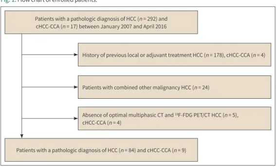
We retrospectively searched the database and records of the Department of Pathology dat- ing from January 2007 to April 2016 using the search terms HCC and cHCC-CCA. The diagno- sis of HCC and cHCC-CCA was based on the 2010 WHO classification. We identified 292 pa- tients with a diagnosis of HCC and 17 patients with a diagnosis of cHCC-CCA, as confirmed by a pathologist (B.H, with 29 years of experience in pathology). The study population was select- ed from these patients using the following inclusion criteria: preoperative optimal multipha- sic liver CT studies, including unenhanced, hepatic arterial phase, portal venous phase, and equilibrium phase, and availability of 18F-FDG PET/CT. Exclusion criteria for the study were as follows: (a) a history of previous adjuvant treatment such as transarterial chemoemboliza- tion, radiofrequency ablation (RFA), and surgery and (b) other combined malignancy. The fi- nal study population comprised 84 patients diagnosed with HCC and 9 patients diagnosed with cHCC-CCA (Fig. 1). Diagnosis in 33 patients was histologically confirmed by examination of a surgical specimen and in 60 patients, it was histologically confirmed by examination of gun-biopsy specimen. A retrospective review of imaging studies, clinical data, and demograph- ic information for the patients was also conducted. A total of 93 patients (mean age, 57 years; age range, 36–78 years), consisting of 80 men and 13 women, met our inclusion criteria and formed our study population (Table 1). This study was approved by our Institutional Review Board (IRB No. 2018-07-016-003).
Patients with a pathologic diagnosis of HCC (n = 292) and cHCC-CCA (n = 17) between January 2007 and April 2016
Patients with a pathologic diagnosis of HCC (n = 84) and cHCC-CCA (n = 9)
History of previous local or adjuvant treatment HCC (n = 178), cHCC-CCA (n = 4)
Patients with combined other malignancy HCC (n = 24)
Absence of optimal multiphasic CT and 18F-FDG PET/CT HCC (n = 5), cHCC-CCA (n = 4)
Fig. 1. Flow chart of enrolled patients.
cHCC-CCA = combined hepatocellular carcinoma-cholangiocarcinoma, FDG = fluorodeoxyglucose, HCC = hepatocellular carcinoma
CT PROTOCOL
Multiphasic CT images were acquired using a 64-MDCT scanner (Somatom Sensation 64, Siemens Healthcare, Erlangen, Germany) or a dual energy CT scanner (Somatom Definition Flash; Siemens Healthcare). The CT scanning parameters were as follows: reconstruction at a slice thickness of 3 mm, with 3 mm slice intervals and a 120 kVp quality reference 210 mAs for the dose modulation system (CareDose 4D; Siemens Medical Solutions). A total of 100–150 mL of nonionic contrast medium (350 mgI/mL) was injected at the rate of 4 mL/s through an 18-gauge IV cannula using a power injector. The scan delay was determined according to an automatic bolus triggering software program (Syngo Acquisition Workplace; Siemens Health- care). The late arterial phase scanning and portal venous phase scanning were started at 15 and 55 seconds, respectively, after the trigger threshold was reached (100 Hounsfield units on the abdominal aorta). The delayed phase scanning was performed 180 seconds after the initi- ation of the contrast material injection.
PET/CT PROTOCOL
All patients in the study fasted for at least six hours before intravenous FDG administration.
The dose range of intravenously injected FDG was 370–444 MBq, depending on patient weight.
PET/CT images were acquired using a Biograph Duo PET/CT scanner (Siemens Healthcare) or a Biograph 16 PET/CT scanner (Siemens Healthcare). Prior to the PET scan, a CT scan (5 mm slice thickness) at an interval of 5 mm was acquired without contrast enhancement for atten- uation correction.
IMAGING ANALYSIS
Multiphasic liver CT findings were retrospectively analyzed by two radiologists (J.G.P and K.S.O, with 8 and 19 years of experience in abdominal imaging, respectively) by reaching a consensus. Tumor enhancement patterns were divided into three types based on the attenua-
Table 1. Patient Characteristics According to Pathology
HCC cHCC-CCA p-Value
No. of patients 84 9 N/A
Age (years)* 56.9 ± 9.6 (36–78) 59.2 ± 7.8 (46–69) 0.44
Sex (M:F) 72:12 8:1 0.07
Chronic liver disease
HBV 61 (72.6) 6 (66.7) 0.71
HCV 12 (14.3) 0 (0) 0.22
Liver cirrhosis 53 (63.1) 4 (44.4) 0.28
AFP (ng/mL)† 13 (1.2–121000) 11.8 (2.35–8111) 0.67
CA 19-9 (U/mL)† 25 (0.63–400) 94 (12–27178) 0.02
Except where indicated, data are numbers of patients, with percentages in parentheses.
*Data are means ± standard deviations. Numbers in parentheses are ranges.
†Data are medians. Numbers in parentheses are ranges.
AFP = alpha-fetoprotein, CA = carbohydrate antigen, cHCC-CCA = combined hepatocellular carcinoma-chol- angiocarcinoma, HBV = hepatitis B virus, HCC = hepatocellular carcinoma, HCV = hepatitis C virus, N/A = not available
tion of the tumor compared with the surrounding liver parenchyma: type I (early arterial en- hancement with delayed washout), type II (early arterial enhancement without delayed wash- out), and type III (early hypovascular, infiltrative appearance or peripheral rim enhancement).
Lesions with a non-homogenous enhancement pattern on contrast-enhanced images were categorized according to the attenuation of the predominant lesions parts (> 50%), as de- scribed in previous work (18).
Associated findings were assessed, including tumor size (maximum diameter), tumor num- ber (single or multiple), capsular retraction, capsule appearance, and invasion of vessel.
18F-FDG PET/CT images were qualitatively evaluated to assess whether FDG uptake in the tu- mor was significantly higher than the surrounding noncancerous hepatic parenchyma. FDG accumulation was analyzed quantitatively by calculating the standardized uptake value (SUV) in the regions of interest placed over the tumor and the normal liver.
SUV = PET count × calibration factor (mCi/g).
STATISTICAL ANALYSIS
To determine differences in clinical, radiological, and 18F-FDG PET/CT features according to tumor type (HCC vs. cHCC-CCA), Student’s t-tests, chi-square tests, and Fisher’s exact tests were used. To determine the differences in the enhancement patterns and 18F-FDG PET/CT features according to tumor type (HCC vs. cHCC-CCA), chi-square tests and Student’s t-test were used.
All analyses were conducted using standard statistical software (SPSS 20.0 for Windows; IBM Corp., Armonk, NY, USA) and results were considered significant when the p value was less than 0.05.
RESULTS
Patient characteristics are shown in Table 1. Seventy-three of the 84 HCC patients had chronic hepatitis: 61 had hepatitis B and 12 had hepatitis C. Six of the 9 cHCC-CCA patients had chron- ic hepatitis: all had hepatitis B. No significant differences were observed in the incidence of chronic liver disease or liver cirrhosis according to pathological type (p > 0.05). In HCC pa- tients, the median level was 13 ng/mL for alpha-fetoprotein (AFP) and 25 U/mL for carbohy- drate antigen (CA) 19-9. For cHCC-CCA patients, the median level was 11.8 ng/mL for AFP and 94 U/mL for CA 19-9. CA 19-9 levels were significantly higher in patients with cHCC-CCA com- pared to patients with HCC (p = 0.019).
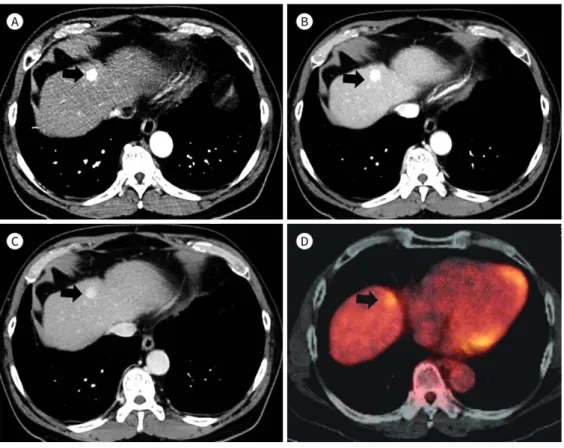
Imaging features of HCCs and cHCC-CCAs are shown in Table 2. Of the 84 HCCs, 58 (69%) showed a type I enhancement pattern (Fig. 2), 14 (17%) showed a type II enhancement pattern, and 12 (14%) showed a type III enhancement pattern. Of the 9 cHCC-CCA patients, no patients showed type I enhancement pattern, 5 (56%) showed type II enhancement pattern (Fig. 3), and 4 (44%) showed type III enhancement pattern (Fig. 4). Type I enhancement pattern was more common in the HCC group, while the type II/III enhancement patterns were more common in the cHCC-CCA group. Enhancement patterns of HCC and cHCC-CCA showed significant dif- ferences (p < 0.001). A significant difference was observed in the presence of the capsular ap- pearance between HCC and cHCC-CCA (p = 0.006). Forty (48%) of the 84 HCC patients showed a capsular appearance. No capsular appearance was observed in the cHCC-CCA patients. How-
A
C
B
D
Fig. 2. Hepatocellular carcinoma with type I enhancement pattern in a 60-year-old man.
A. Arterial-phase CT image shows typical early enhancement of the mass (arrow).
B, C. Portal venous and delayed phase images show washout of the mass (arrows) relative to the liver.
D. Fused PET/CT shows no definite abnormal fluorodeoxyglucose uptake into the mass.
Table 2. Imaging Findings of HCC and cHCC-CCA Cases
HCC cHCC-CCA p-Value
Enhancement pattern < 0.001
Type I 58 (69) 0 (0)
Type II 14 (16.7) 5 (55.6)
Type III 12 (14.2) 4 (44.4)
Tumor size (cm)* 6.2 ± 3.2 6.3 ± 4.6 1.576
Tumor number 0.498
Single 56 (66.7) 7 (77.8)
Multiple 28 (33.3) 2 (22.2)
Capsular appearance 40 (47.6) 0 (0) 0.006
Capsule retraction 10 (11.9) 2 (22.2) 0.301
Vessel invasion 7 (8.3) 1 (11.1) 0.778
PET 0.095
Positive status 51 (60.7) 8 (88.9)
Negative status 33 (39.3) 1 (11.1)
Except where indicated, data are numbers of patients, with percentages in parentheses.
*Data are means ± standard deviations.
cHCC-CCA = combined hepatocellular carcinoma-cholangiocarcinoma, HCC = hepatocellular carcinoma
ever, no significant differences were found in tumor size, tumor number, capsule retraction, and vessel invasion between HCC and cHCC-CCA (p > 0.05).
Further, 60.7% (51/84) of HCCs and 88.9% (8/9) of cHCC-CCAs were PET-positive. The cHCC- CCA had a higher PET-positive rate than did the HCCs, but the difference was not significant (p = 0.095). Of the 59 HCCs with type I enhancement pattern, 37 (64%) were PET-positive. None of the cHCC-CCAs was found to have type I enhancement pattern. Among 19 cases with type II enhancement pattern, 21% (3/14) of HCCs were PET-positive and 80% (4/5) of cHCC-CCAs were PET-positive. cHCC-CCAs showed a significantly higher PET-positive rate (p = 0.020) in cases with type II enhancement pattern. Among the 16 cases with type III enhancement pattern, 92% (11/12) of HCCs and 100% (4/4) of cHCC-CCAs were PET-positive (p = 0.551) (Fig. 5).
In addition, cHCC-CCA showed a significantly higher 18F-FDG uptake (mean SUV max 6.14
± 2.84) than did HCC (mean SUV max 3.90 ± 2.69) (p = 0.048). The mean SUV max of 18F-FDG uptake in HCCs with type I enhancement pattern was 3.87 ± 2.83. For those with type II en- hancement pattern, mean SUV max of 18F-FDG uptake in cHCC-CCA (mean SUV max 4.54 ± 2.07) was significantly higher than that of HCCs (mean SUV max 2.60 ± 0.21) (p = 0.002).
For those with type III enhancement pattern, there was no significant difference in the mean SUV max values of 18F-FDG uptake between cHCC-CCA (mean SUV max 8.15 ± 2.48) and HCC (mean SUV max 5.58 ± 2.76) (p = 0.135).
A
C
B
D
Fig. 3. Combined hepatocellular cholangiocarcinoma with type II enhancement pattern in a 58-year-old man.
A. Arterial-phase CT image shows typical early enhancement of the mass (arrow).
B, C. Portal- and delayed-phase images show prolonged enhancement of the mass (arrows) without washout.
D. Fusion PET/CT image shows definite fluorodeoxyglucose uptake into the mass (arrow) relative to the liver.
DISCUSSION
HCC is the most common primary liver cancer and one of the most prevalent causes of death due to malignant tumors. Early diagnosis of HCC has been facilitated by advances in the image
Fig. 5. Distribution of enhancement pattern and fluorodeoxyglucose uptake in 84 HCC and 9 cHCC-CCA cases.
cHCC-CCA = combined hepatocellular carcinoma-cholangiocarcinoma, HCC = hepatocellular carcinoma, SUV = standardized uptake value
HCC (n = 84) cHCC-CCA (n = 9)
Type I enhancement
HCC (n = 58)
Type II enhancement HCC (n = 58) cHCC-CCA (n = 5)
Type III enhancement HCC (n = 12) cHCC-CCA (n = 4)
PET (+) HCC 63.7% (n = 37) Mean SUVmax = 3.87
PET (+) HCC 21% (n = 3) Mean SUVmax = 2.61
PET (+) cHCC-CCA 80% (n = 4) Mean SUVmax = 4.54
PET (+) HCC 92% (n = 11) Mean SUVmax = 5.58
PET (+) cHCC-CCA 100% (n = 4) Mean SUVmax = 8.15 A
C
B
D
Fig. 4. Combined hepatocellular carcinoma-cholangiocarcinoma with type III enhancement pattern in a 68-year-old man.
A. A hypovascular mass (arrow) is seen on an arterial phase image.
B, C. Portal- and delayed-phase images show persistent poor enhancement of the mass (arrows).
D. Fusion PET/CT image shows definite fluorodeoxyglucose uptake into the mass relative to the liver.
modalities of multiphasic liver CT and MRI (19). HCC can often be diagnosed with noninvasive image modalities such as multiphasic liver CT and liver MRI. However, some cases of intrahe- patic CCA and cHCC-CCA show marked enhancement on the arterial-phase of multiphasic CT that resembles the typical radiologic findings of HCC (9-12).
cHCC-CCA is an important differential diagnosis for other primary liver tumors, although it is rarely encountered clinically. Substantial effort has been invested in improving the preoper- ative performance of radiological findings to differentiate cHCC-CCA from HCC. The correct diagnosis of this tumor on multiphasic liver CT will contribute to the selection of the appropri- ate therapeutic methods, such as transcatheter arterial chemoembolization, RFA, or radical re- section. The type I enhancement pattern is highly specific for HCC in patients with cirrhosis or other HCC risk factors (20-22). However, the type II enhancement pattern is nonspecific, as it can also be observed in those with dysplastic nodules, HCC, and non-HCC malignancies such as intrahepatic CCA and cHCC-CCA (11, 12, 23). In this study, the type I enhancement pattern was seen in only HCCs (58 cases, 69.05% of HCC) and the type II enhancement pattern was observed in both HCC (17%) and cHCC-CCA (56%).
An awareness of the different imaging characteristics of HCC and other liver cancers is im- portant for pre-study planning. An understanding of their features on cross-sectional imaging would provide great value in determining the most appropriate treatment plan and the predi- cation of the prognosis of each patient. However, the role of 18F-FDG in the diagnosis of HCC is controversial (24). This limited sensitivity is due to variable tumor FDG uptake and high back- ground metabolic activity in the normal liver (25, 26). In particular, well-differentiated and early HCC might remain undetected using FDG PET. Indeed, the overall sensitivity for detec- tion of primary HCC is reported to be only 50% to 65% (14, 15). On the other hand, the sensitivi- ty of 18F-FDG PET/CT for detecting CCA is higher than that for detecting HCC (84–94%) (16, 17).
Moreover, mass-forming intrahepatic CCA shows intense FDG uptake, whereas hilar CCA shows only mild uptake (27). Only one report describes the 18F-FDG PET/CT findings of cHCC- CCA (15). The report revealed that 18F-FDG PET/CT shows high FDG uptake for cHCC-CCA.
Whether FDG PET/CT can be used for differential diagnosis of cHCC-CCA and HCC is uncer- tain. The aim of this study was to evaluate multiphasic CT and 18F-FDG PET/CT findings for differential diagnosis of combined cHCC-CCA from HCC.
In our study, cHCC-CCAs had a significantly higher PET-positive rate (p = 0.020) than did HCCs only in cases with a type II enhancement pattern. No significant difference was observed in the PET-positive rates for nodules with type III enhancement pattern. Our results revealed that FDG PET/CT could contribute to differential diagnosis between HCCs and cHCC-CCAs with type II enhancement pattern Ijichi et al. (28) reported that the PET-positive rate differed significantly according to enhancement pattern and tumor size. They proposed that the sen- sitivity of FDG PET for detecting HCCs was significantly associated with tumor differentiation, tumor size, and microvascular invasion. In our study, the HCCs with type II enhancement pat- tern had a lower positive rate of PET than did those with type I/II enhancement pattern (p = 0.008). We surmise that HCCs with type II enhancement pattern were well-differentiated for early HCCs, although we did not evaluate tumor differentiation.
Significant differences were also shown for the capsule appearance between HCC and cHCC- CCA (p = 0.006). The presence of a fibrous capsule is a characteristic finding of classic HCC (29).
This characteristic histologic finding demonstrated a good correlation with the capsule ap- pearance on multiphasic liver CT (30). The capsule appearance is a reliable indicator in the di- agnosis of HCC (20). This result suggests that capsule appearance can be a diagnostic clue for differentiating between HCC and cHCC-CCA.
This study had several limitations. First, it was retrospective, which may have limited the data quality. However, because cHCC-CCA is rare, prospective studies may be difficult. Second, the sample size was relatively small and drawn from a single facility. Moreover, the proportion of cHCC-CCAs was small. However, our study evaluated pathologically confirmed HCC and cHCC-CCA on contrast-enhanced multiphasic CT and 18F-FDG PET/CT. Additional studies us- ing a larger patient sample size will be necessary to verify our conclusions.
In conclusion, In this study, the PET-positive rate of cHCC-CCA was significantly higher than that of HCC in lesions with type II enhancement pattern. The 18F-FDG PET/CT can be used as a clue for the differentiation of cHCC-CCA from HCC in lesions with early arterial enhance- ment without delayed washout on multiphasic CT. Therefore, if there are no typical imaging features of HCC or CCA on multiphasic CT and FDG uptake is visible on 18F-FDG PET/CT, his- tologic confirmation is recommended.
Author Contributions
Conceptualization, P.J.G., J.G.; data curation, P.J.G., P.J.C.; formal analysis, P.J.G., P.J.C.; investigation, P.J.C.; methodology, P.J.G., P.J.C., J.S.; project administration, P.J.G.; resources, P.J.G., J.S.; supervision, J.S., P.J.G.; validation, P.J.G.; visualization, K.H., P.J.C.; writing—original draft, P.J.C., P.J.G.; and writ- ing—review & editing, all authors.
Conflicts of Interest
The authors have no potential conflicts of interest to disclose.
REFERENCES
1. Joo I, Kim H, Lee JM. Cancer stem cells in primary liver cancers: pathological concepts and imaging find- ings. Korean J Radiol 2015;16:50-68
2. Lin G, Toh CH, Wu RC, Ko SF, Ng SH, Chou WC, et al. Combined hepatocellular cholangiocarcinoma: prog- nostic factors investigated by computed tomography/magnetic resonance imaging. Int J Clin Pract 2008;
62:1199-1205
3. Brunt E, Aishima S, Clavien PA, Fowler K, Goodman Z, Gores G, et al. cHCC-CCA: consensus terminology for primary liver carcinomas with both hepatocytic and cholangiocytic differentation. Hepatology 2018;68:
113-126
4. Akiba J, Nakashima O, Hattori S, Tanikawa K, Takenaka M, Nakayama M, et al. Clinicopathologic analysis of combined hepatocellular-cholangiocarcinoma according to the latest WHO classification. Am J Surg Pathol 2013;37:496-505
5. Sasaki M, Sato H, Kakuda Y, Sato Y, Choi JH, Nakanuma Y. Clinicopathological significance of ‘subtypes with stem-cell feature’ in combined hepatocellular-cholangiocarcinoma. Liver Int 2015;35:1024-1035 6. Shibahara J, Hayashi A, Misumi K, Sakamoto Y, Arita J, Hasegawa K, et al. Clinicopathologic characteristics
of hepatocellular carcinoma with reactive ductule-like components, a subset of liver cancer currently clas- sified as combined hepatocellular-cholangiocarcinoma with stem-cell features, typical subtype. Am J Surg Pathol 2016;40:608-616
7. Bota S, Piscaglia F, Marinelli S, Pecorelli A, Terzi E, Bolondi L. Comparison of international guidelines for noninvasive diagnosis of hepatocellular carcinoma. Liver Cancer 2012;1:190-200
8. Song DS, Bae SH. Changes of guidelines diagnosing hepatocellular carcinoma during the last ten-year pe- riod. Clin Mol Hepatol 2012;18:258-267
9. Kim SA, Lee JM, Lee KB, Kim SH, Yoon SH, Han JK, et al. Intrahepatic mass-forming cholangiocarcinomas:
enhancement patterns at multiphasic CT, with special emphasis on arterial enhancement pattern--corre- lation with clinicopathologic findings. Radiology 2011;260:148-157
10. Shetty AS, Fowler KJ, Brunt EM, Agarwal S, Narra VR, Menias CO. Combined hepatocellular-cholangiocarci- noma: what the radiologist needs to know about biphenotypic liver carcinoma. Abdom Imaging 2014;39:
310-322
11. Fukukura Y, Taguchi J, Nakashima O, Wada Y, Kojiro M. Combined hepatocellular and cholangiocarcinoma:
correlation between CT findings and clinicopathological features. J Comput Assist Tomogr 1997;21:52-58 12. Nishie A, Yoshimitsu K, Asayama Y, Irie H, Aibe H, Tajima T, et al. Detection of combined hepatocellular and
cholangiocarcinomas on enhanced CT: comparison with histologic findings. AJR Am J Roentgenol 2005;
184:1157-1162
13. Iglehart JK. The new era of medical imaging--progress and pitfalls. N Engl J Med 2006;354:2822-2828 14. Khan MA, Combs CS, Brunt EM, Lowe VJ, Wolverson MK, Solomon H, et al. Positron emission tomography
scanning in the evaluation of hepatocellular carcinoma. J Hepatol 2000;32:792-797
15. Wudel LJ Jr, Delbeke D, Morris D, Rice M, Washington MK, Shyr Y, et al. The role of [18F]fluorodeoxyglucose positron emission tomography imaging in the evaluation of hepatocellular carcinoma. Am Surg 2003;69:
117-124; discussion 124-126
16. Kim JY, Kim MH, Lee TY, Hwang CY, Kim JS, Yun SC, et al. Clinical role of 18F-FDG PET-CT in suspected and potentially operable cholangiocarcinoma: a prospective study compared with conventional imaging. Am J Gastroenterol 2008;103:1145-1151
17. Kluge R, Schmidt F, Caca K, Barthel H, Hesse S, Georgi P, et al. Positron emission tomography with [18F]flu- oro-2-deoxy-D-glucose for diagnosis and staging of bile duct cancer. Hepatology 2001;33:1029-1035 18. Loyer EM, Chin H, DuBrow RA, David CL, Eftekhari F, Charnsangavej C. Hepatocellular carcinoma and intra-
hepatic peripheral cholangiocarcinoma: enhancement patterns with quadruple phase helical CT--a com- parative study. Radiology 1999;212:866-875
19. Centers for Disease Control and Prevention (CDC). Hepatocellular carcinoma - United States, 2001-2006.
MMWR Morb Mortal Wkly Rep 2010;59:517-520
20. Khan AS, Hussain HK, Johnson TD, Weadock WJ, Pelletier SJ, Marrero JA. Value of delayed hypointensity and delayed enhancing rim in magnetic resonance imaging diagnosis of small hepatocellular carcinoma in the cirrhotic liver. J Magn Reson Imaging 2010;32:360-366
21. Marrero JA, Hussain HK, Nghiem HV, Umar R, Fontana RJ, Lok AS. Improving the prediction of hepatocellu- lar carcinoma in cirrhotic patients with an arterially-enhancing liver mass. Liver Transpl 2005;11:281-289 22. Forner A, Vilana R, Ayuso C, Bianchi L, Solé M, Ayuso JR, et al. Diagnosis of hepatic nodules 20 mm or small-
er in cirrhosis: prospective validation of the noninvasive diagnostic criteria for hepatocellular carcinoma.
Hepatology 2008;47:97-104
23. Rimola J, Forner A, Reig M, Vilana R, De Lope CR, Ayuso C, et al. Cholangiocarcinoma in cirrhosis: absence of contrast washout in delayed phases by magnetic resonance imaging avoids misdiagnosis of hepatocel- lular carcinoma. Hepatology 2009;50:791-798
24. Hatano E, Ikai I, Higashi T, Teramukai S, Torizuka T, Saga T, et al. Preoperative positron emission tomogra- phy with fluorine-18-fluorodeoxyglucose is predictive of prognosis in patients with hepatocellular carcino- ma after resection. World J Surg 2006;30:1736-1741
25. Torizuka T, Tamaki N, Inokuma T, Magata Y, Sasayama S, Yonekura Y, et al. In vivo assessment of glucose metabolism in hepatocellular carcinoma with FDG-PET. J Nucl Med 1995;36:1811-1817
26. Lee JD, Yang WI, Park YN, Kim KS, Choi JS, Yun M, et al. Different glucose uptake and glycolytic mecha- nisms between hepatocellular carcinoma and intrahepatic mass-forming cholangiocarcinoma with in- creased 18F-FDG uptake. J Nucl Med 2005;46:1753-1759
27. Kim YJ, Yun M, Lee WJ, Kim KS, Lee JD. Usefulness of 18F-FDG PET in intrahepatic cholangiocarcinoma. Eur J Nucl Med Mol Imaging 2003;30:1467-1472
28. Ijichi H, Shirabe K, Taketomi A, Yoshizumi T, Ikegami T, Mano Y, et al. Clinical usefulness of 18F-fluorodeoxy- glucose positron emission tomography/computed tomography for patients with primary liver cancer with special reference to rare histological types, hepatocellular carcinoma with sarcomatous change and com- bined hepatocellular and cholangiocarcinoma. Hepatol Res 2013;43:481-487
29. Kojiro M. Histopathology of liver cancers. Best Pract Res Clin Gastroenterol 2005;19:39-62
30. Lim JH, Choi D, Park CK, Lee WJ, Lim HK. Encapsulated hepatocellular carcinoma: CT-pathologic correla- tions. Eur Radiol 2006;16:2326-2333
간세포암종과 혼합성 간세포암종-담관암종에서 다위상 전산단층촬영술 소견과 18F-FDG PET/CT에서 섭취율 차이에 대한 분석
박재춘1 · 박정구1* · 정규식1 · 강 희1 · 전성민2
목적 간세포암종과 혼합성 간세포암종-담관암종의 다위상 전산단층촬영술 소견과 18F-fluo- rodeoxyglucose 양전자방출단층촬영(이하 FDG PET/CT)에서 섭취율 차이를 연구하여 이들 의 감별 진단에 유용성이 있는지를 알아보고자 하였다.
대상과 방법 2007년 1월에서 2016년 4월까지 조직학적으로 간세포성 암종으로 진단된 84명과 혼합성 간세포암종-담관암종으로 진단된 9명의 환자를 대상으로 하였다. 조영증강 양상은 유 형 I (동맥기 조영증강과 지연기 조영유실), 유형 II (지연기 조영유실이 없는 동맥기 조영증강), 유형 III (저혈관성 병변, 침투성 양상 혹은 변연부 조영증강)로 구분하였고, PET/CT 소견은 FDG 섭취 여부에 따라서 양성과 음성으로 분류하였다.
결과 혼합성 간세포암종-담관암종(89%)은 간세포암종(61%)보다 PET/CT 섭취 양성률이 높 았으나 통계적으로 유의한 차이는 보이지 않았다(p = 0.095). 유형 I 조영증강양상을 보이는 58개의 간세포암종 중 37예(64%)가 PET/CT에서 양성이었다. 유형 II 조영증강양상을 보이 는 19예 중 간세포암종 3예(21%)에서 PET/CT에서 양성을 보였고 혼합 간세포암종-담관암종 4예(80%)에서 PET/CT 양성을 보였다. 유형 II 조영증강양상을 보이는 경우 혼합 간세포암- 담관암종이 간세포암종보다 PET/CT 양성률이 유의하게 높았다(p = 0.020). 유형 III 조영증 강 양상을 보이는 16예 중 간세포암 11예(91.6%), 혼합 간세포암-담관암종 4예(100%)에서 PET/CT 양성을 보였다. 유형 III 조영증강양상을 보이는 경우 간세포암과 혼합 간세포암-담 관암종의 PET/CT 양성률의 유의한 차이는 보이지 않았다.
결론 유형 II 조영증강 양상을 보이는 경우 간세포암종과 혼합성 간세포암종-담관암종의 감 별진단에 전산단층촬영술과 병행하는 18F-FDG PET/CT가 도움이 될 것으로 보인다.
고신대학교 의과대학 고신대학교 복음병원 1영상의학과, 2핵의학과