Smile is the key component of Esthetics and has an important role in the determination of first impression of a person. Gingival architecture plays major role in creating smile. Conditions like gingival recession in esthetic zone poses challenge to a clinician. Gingival recession is defined as an apical displacement of soft tissues related to the cementoenamel junction.1
Periodontal reconstructive surgery consists of a variety of mucogingival procedures including root coverage, tooth exposure, crown exposure, vestibular deepening, papilla reconstruction, ridge augmentation, and ridge preservation.
While the primary goal of these procedures is to benefit periodontal health through the reconstruction of lost hard and soft tissues or by preventing additional loss, enhancing the esthetics by improving smile.
Predictable root coverage is one of the goals of periodontal
plastic surgery; enabling periodontal surgeon to solve common patient complaints associated with root exposure such as esthetic complaints, root hypersensitivity, shallow root carious lesions and cervical abrasions.2-7
Microsurgical technique has been a recent advancement in periodontal plastic surgery. The use of magnification has not only improved aesthetics but also shortened the period of healing due to minimal surgical trauma.3,4 Microsurgery is a minimally invasive technique that is performed with magnification and microsurgical instruments and suture materials.3 The combination of small instruments and delicate surgical technique allows for extremely fine and accurate incisions, gentle tissue handling, and precise approximation of wound margins.5 Keeping this goal in mind, present case report is a wonderful example of a periodontal plastic microsurgery for coverage of a denuded root.
Microsurgical Approach for Root Coverage of Receding Gingiva in the Esthetic Zone
Ranjana Mohan, Rohit Jain
Department of Periodontics, Teerthanker Mahaveer Dental College and Research Center, Moradabad, India
CC This is an open-access article distributed under the terms of the Creative Commons Attribution Non-Commercial License (http://creativecommons.org/licenses/by-nc/3.0) which permits unrestricted noncommercial use, distribution, and reproduction in any medium, provided the original work is properly cited.
Copyright © 2013 by the Korean Society for Microsurgery. All Rights Reserved.
Facial esthetics and smiling are key components in nonverbal communication and have an important role in determination of the first impression of a person. The various components of the smile in dental esthetics include Gingival scaffold, lip framework, and Teeth. The periodontist creates a smile by performing various periodontal plastic microsurgery procedures for management of mucogingival problems. A 25-year-old patient reported to the Department of Periodontology at Teerthanker Mahaveer Dental College and Research Center, Moradabad, Northern India, with the chief complaint of long looking teeth in the upper jaw, making him conscious while smiling. Miller class I gingival recession with Maxillary left canine (23) was diagnosed. Periodontal plastic microsurgery employing double papilla grafting with connective tissue graft harvested from the palate in order to cover denuded root was performed using microsurgical instruments and microsuturing with 6-0 suturing material under magnification. Healing was uneventful, with achievement of 100% root coverage of denuded root after three months. The patient was highly impressed and satisfied with his enhanced smile.
Key Words: Esthetics, Gingival recession, Microsurgery, Double papilla graft, Connective tissue graft, Microsuturing
Received September 3, 2013 Revised October 17, 2013 Accepted October 21, 2013
Correspondence to: Ranjana Mohan Department of Periodontics, Teerthanker Mahaveer Dental College and Research Center, Delhi Road, Moradabad-244001, Uttar Pradesh, India
Tel: +91-591-2360046 Fax: +91-591-2476823
E-mail: [email protected]
ARMS
Case Report Editor’s Pick !!CASE REPORT
A 29-year-old male patient reported to the Department of Periodontology, Teerthanker Mahaveer Dental College and Research Center with the complaint of long looking teeth and increasing sensitivity in upper jaw.
Detailed history was taken and gingival and periodontal status was recorded. Intraoral examination revealed class I gingival recession with maxillary left canine (23) measuring about 5 mm of denuded root surface. Etiology of gingival recession was faulty brushing technique (Fig. 1).
Following clinical and radiological examination, patient consent was obtained and phase one therapy performed consisting of thorough scaling and root planning. Correct brushing method was demonstrated.
Case was evaluated after a month. Patient maintained his oral hygiene as per the instructions given to him. Micro- surgical approach for the management of gingival recession was planned on maxillary left canine (23). The study was approved by an institutional review board.
Microsurgical instrument set was arranged for periodontal microsurgery to be performed for maxillary left canine (23).
All the measurements of gingival recession were made with University of North Carolina (NC, USA)-15 periodontal probe.
Surgical technique
1. Recipient site (gingival recession)
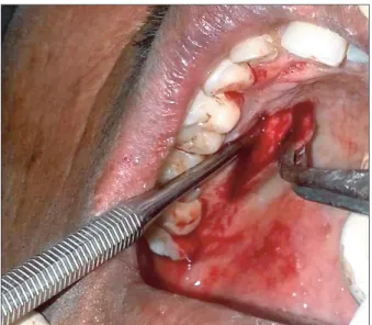
After administration of local anesthesia, initial V-shaped beveled incisions were made in the area of the deformity on maxillary left canine to eliminate marginal tissue (0.5 to 1.0 mm). The horizontal incision was made at 90 degree angle to the gingival surface. Horizontal incision was continued as crevicular incision along the marginal gingiva of the involved tooth. Two flared vertical incision were given at both the ends of the horizontal incisions and are extended beyond the mucogingival junction into the mucosal tissue to permit adequate mobility of the flap (Fig. 2). All the incisions were made with microsurgical knife using ×4.0 magnification Galilean dental surgical loupes. Full thickness flap was elevated using micro-periosteal elevator. Root planing followed by conditioning of the root was performed with citric acid.
2. Donor site (harvesting a graft from palate)
A subepithelial connective tissue graft of masticatory mucosa was harvested from the palatal aspect of the maxillary premolars by the use of a ‘trap door’ approach (Fig. 3).8 Flap was re-placed after procuring the graft and sutured using 4-0 sutures. Advantage of this method is that wound remains closed and heals with primary intention without causing any discomfort to the patient.
Harvested connective tissue graft (Fig. 4) was sutured on the recession area of recipient site with 6-0 resorbable suturing
Fig. 1. Preoperative state of the maxillary left canine. Note the gingival recession as shown by graduated periodontal probe. About 5 mm of root exposure was assessed.
Fig. 2. Preparation of recipient site. Full thickness flap is dissected using mesial and distal vertical incisions extending beyond muco- gingival junction.
material (Fig. 5). Double papilla flap was approximated using 6-0 and placed over the graft covering it advancing coronally and sutured with sling sutures (Fig. 6). Tin foil was placed over the surgical site followed by periodontal dressing.
Antibiotic therapy (amoxicillin 500 mg, three times a day, Novamox; Cipla, Mumbai, India) and suitable analgesic was prescribed for five days. Tooth-brushing was discontinued for the first two weeks at the surgical site. Chemical plaque
control was advised.
Patient was called for suture removal after two weeks.
Following removal of sutures and clinical evaluation of surgical wound was done. Clinically wound healing was satisfactory and uneventful. At the end of three months there was complete coverage of denuded root at the surgical site (Fig. 7).
Fig. 4. Harvested free connective tissue graft. Connective tissue graft is measured and trimmed to fi t on the prepared recipient bed on the recession area in order to cover the entire denuded root.
Fig. 5. Suturing of connective tissue graft on the denuded root surface.
A connective tissue graft has been positioned and sutured with resorbable sutures over the denuded root, just apical to the cemento- enamel junction.
Fig. 6. Suturing of double gingival fl ap over the connective tissue graft.
Following suturing of two fl aps together, the entire unit was advanced and sutured, in order to cover the connective tissue graft coronal to the cement enamel junction.
Fig. 3. Harvesting of connective tissue graft from palate by trap door approach. Connective tissue graft was harvested from the posterior palate. The premolar region has the greatest thickness of palatal connective tissue. During harvest of the graft, the integrity of the palatal mucosa was maintained. The mucosa was sutured back in place to cover the donor site thereby reducing post-operative bleeding and discomfort.
DISCUSSION
Gingival recession is a common clinical entity posing various problems and challenges to a clinician. Since last six decades there has been a great evolution in the management of mucogingival problems. Recently with the introduction of magnification in periodontal practice, dental operating microscope and magnifying loupe along with the microsurgical instruments and microsurgical procedures in periodontal practice, there has been a great scope for honing the conventional surgical skills and achieving promising results especially while performing delicate periodontal plastic surgeries in esthetic zone.
In 1968, Cohen and Ross9 introduced double papilla flap in periodontal therapy. In 1982, Miller10,11 documented a soft tissue graft technique to cover a denuded root surface, observing a high rate of success and classified gingival recession in 1985. Langer and Langer12 demonstrated the technique of procuring subepithelial connective tissue graft from palate. The key concept of this technique was the double surface blood supply to the donor site. In 1986, Tarnow13 published a new technique of semilunar coronally repositioned flap. During mid 1980s and 2000s several root coverage procedures were evolved, such as semilunar coronally repositioned flap and supraperiosteal envelope
technique. Various techniques for root coverage were in practice showing varied results. Burkhardt and Hürzeler,3 Burkhardt and Lang4 utilized surgical microscope for advanced plastic periodontal surgery and stated that due to microsurgical techniques, optimal aesthetics can be obtained in mucogingival surgery. The study on coverage of localized gingival recessions comparing macro and microsurgical techniques concluded that microsurgical approach sub- stantially improved the vascularization of the grafts, and the percentage of root coverage compared to macroscopic approach.
A novel minimally invasive technique for the management of gingival recession employing double papilla flap with connective tissue graft under magnification and using microsurgical instruments has proved to be the best in terms of achieving complete root coverage, appropriate thickness of keratinized tissue, and better color match, There has been a faster wound healing due to reduced surgical trauma with the use of microsurgical instruments and micro sutures which is vital for the treatment of root coverage. Patient was satisfied by impressive and promising results enhancing esthetics, with confident smile on the face.
REFERENCES
1. Carvalho PF, da Silva RC, Cury PR, Joly JC. Modified coronally advanced flap associated with a subepithelial connective tissue graft for the treatment of adjacent multiple gingival recessions. J Periodontol 2006;77:1901-6.
2. Bittencourt S, Ribeiro Edel P, Sallum EA, Sallum AW, Nociti FH, Casati MZ. Semilunar coronally positioned flap or subepithelial connective tissue graft for the treatment of gingival recession: a 30-month follow-up study. J Periodontol 2009;80:1076-82.
3. Burkhardt R, Hürzeler MB. Utilization of the surgical microscope for advanced plastic periodontal surgery. Pract Periodontics Aesthet Dent 2000;12:171-80.
4. Burkhardt R, Lang NP. Coverage of localized gingival recessions:
comparison of micro- and macrosurgical techniques. J Clin Periodontol 2005;32:287-93.
5. Camargo PM, Melnick PR, Kenney EB. The use of free gingival grafts for aesthetic purposes. Periodontol 2000 2001;27:72-96.
6. Fürhauser R, Florescu D, Benesch T, Haas R, Mailath G, Watzek G.
Evaluation of soft tissue around single-tooth implant crowns: the pink esthetic score. Clin Oral Implants Res 2005;16:639-44.
7. Ross SE, Crosetti HW, Gargiulo A, Cohen DW. The double papillae repositioned flap--an alternative. I. Fourteen years in retrospect.
Int J Periodontics Restorative Dent 1986;6:46-59.
8. Harris RJ, Harris AW. The coronally positioned pedicle graft with Fig. 7. Clinical postoperative view at three months. Complete root
coverage on maxillary left canine; with an increased thickness of gingival tissue have been achieved. A good esthetic outcome with proper color match has been obtained.
inlaid margins: a predictable method of obtaining root coverage of shallow defects. Int J Periodontics Restorative Dent 1994;14:
228-41.
9. Cohen DW, Ross SE. The double papillae repositioned flap in periodontal therapy. J Periodontol 1968;39:65-70.
10. Miller PD Jr. Root coverage using a free soft tissue autograft following citric acid application. Part 1: technique. Int J Periodontics
Restorative Dent 1982;2:65-70.
11. Miller PD Jr. A classification of marginal tissue recession. Int J Periodontics Restorative Dent 1985;5:8-13.
12. Langer B, Langer L. Subepithelial connective tissue graft technique for root coverage. J Periodontol 1985;56:715-20.
13. Tarnow DP. Semilunar coronally repositioned flap. J Clin Periodontol 1986;13:182-5.