I. 서론
치은퇴축은 변연치은이 백악법랑경계보다 치근쪽 으로 변위된 것이라고 정의내릴 수 있다1). 치은이 퇴 축되고 치근이 노출되면 심미적인 문제점과 치근과 민증, 치근우식을 유발할 수 있다. 이러한 치은퇴축 의 원인으로는 과도한 또는 부적절한 칫솔질에 의한 외상, 치태 침착에 의한 염증성 반응2-5)등이 있으며 그외 여러 가지 요소가 영향을 줄 수 있다. 이러한 다 른 소인으로 치조골 열개6), 치아의 위치이상7), 교정 적 치아 이동8), 좁고 얇은 변연치은9), 높은 소대 및 근육부착10), 수복치료 및 치주 치료에 연관된 의원성 요인11)등이 있다.
1968년 Sullivan과 Atkins12)는 이러한 치은퇴축을 4가지 유형으로 분류하면서 깊고 넓은 형태의 치근 피개가 가장 어렵다고 하였고, 1983년 Holbrook와 Ochsenbein13), 1992년 Pini Prato등14)은 술전 퇴축 부위의 크기가 폭이 3mm보다 넓고 깊이가 5mm보 다 큰 경우는 성공률이 떨어진다고 하였다. 1985년 Miller15)는 Sullivan과 Atkins 분류의 단점을 보완하 여 치간골이나 연조직의 상실 여부와 퇴축된 변연 조직의 치은 점막 경계부까지의 연장 여부로 Ⅰ급
부터 Ⅳ급까지 분류하면서 Ⅰ급과 Ⅱ급은 완벽한 치근피개를 얻을 수 있지만 Ⅲ급은 부분적으로만 가능하며 Ⅳ급의 경우 치근피개를 기대하기 어렵다 고 하였다.
이와 같이 술전 퇴축부위의 크기에 따라 예후가 달라지며 퇴축부위의 분류에 가장 많이 사용되는 방 법은 Miller의 분류법이다. 그러나 이전의 치은퇴축 의 성공률을 살펴본 여러 문헌에서는 주로 퇴축부위 의 폭과 깊이에 따른 피개율을 다루었고 Miller의 분 류에 따른 피개율에 대해서는 그 자료가 미흡한 편 이다.
치근피개를 위한 방법에도 여러 외과 술식이 개발 되어 변위판막술16-18), 유리치은이식술12-13,19-23), 상피 하 결합조직 이식술24-28), 치주조직유도재생술29-30)등 이 있다. 측방변위판막술은 1956년 Grupe &
Warren16)에 의해 소개된 방법으로 만족스러운 결과 를 보이나 국소적인 치은퇴축에 제한적으로 사용될 수 있고 치관변위판막술도 Miller의 분류Ⅰ급의 경우 에서 한정적으로 가능하다. 유리치은이식술의 경우 광범위한 부위의 피개가 가능하나 색조화면에서 심 미적으로 만족스럽지 못한 단점이 있다. 치주조직유 도재생술은 긴 부착상피로 치유되는 다른 방법들에
상피하 결합조직 이식술을 이용한 치근피개 술식의 임상적 평가
최경희ㆍ백정원·김창성ㆍ최성호·조규성ㆍ김종관ㆍ채중규 연세대학교 치과대학 치주과학교실, 구강과학연구소
대한치주과학회지 : Vol. 32, No. 3, 2002
*이 논문은 1995년도 연세대학교 학습연구비로 이루어졌음.
비해 신부착 획득이 가능하고 5mm 이상의 깊은 치 은퇴축의 치료에 좋은 결과를 나타내지만 부가적인 비용이 들고 술식에 민감하며 얕은 전정과 소대가 있는 경우에는 예후가 감소한다. 이러한 단점을 보 완하고자 1985년 Langer & Langer25)는 1974년 Edel31)이 상피층을 포함하지 않은 결합조직을 각화 치은의 넓이 증가에 사용한 것을 응용하여 부분층 판막 아래 상피하 결합조직을 이식한 후 판막을 치 관쪽으로 변위시키는 방법을 제안하였다. 이 방법은 56 증례에서 4년 동안 2-6mm의 치은 피개를 보여 성 공적인 치은 피개 효과와 심미적으로도 우수한 결과 를 보고하였다. 그 이후 이러한 술식을 변형하여 임 상적으로 우수한 결과를 보고하였는데 1986년 Nelson26)은 결합조직 이식편을 유경판막 하방에 이 식하여 91%의 치근피개를 보고하였고, 1992년 Harris27)도 Nelson과 같은 유경판막을 이용하여 97.4%의 치근피개를 보이며 그 효과를 확인하였다.
또한 1984년 Raetzke32)와 1994년 Allen33)도 상피하 결합조직 이식술을 변형한 방법인 envelope tech- nique과 Tunnel approach로 각각 80%, 84%의 치근 피개를 보고하였다.
이식편의 치유는 치관쪽부위의 긴 접합상피에 의 한 부착과 근첨부위의 결합조직부착으로 이루어지 는데 결합조직에 의한 부착은 노출된 치근면의 가장 근첨부위에서만 이루어진다. 4mm까지의 얕은 퇴축 에서의 동물실험에서 퇴축부위의 약 25%에 해당하 는 부분만이 결합조직부착을 보였다고 보고된 바 있
고34,35) 이보다 더 많은 퇴축의 경우 신부착은 더욱
적게 이루어졌다고 하였다36). 상피하 결합조직 이식 의 경우 변위판막술보다 결합조직 이식편에 의해 상 피의 근단이동이 억제되지만 신부착의 의한 치유는
제한적이다37). Magnusson등38)과 Beaumont등39)은 긴 접합상피가 정상의 치주조직에 비해 치태 감염에 대한 저항능력이 떨어지지 않는다고 하였으나 치태 가 치은퇴축을 일으킬수 있는 원인요소이므로3,4,5)구 강위생능력에 따라 치근피개의 예후가 달라질 수 있 다. 또한 구강위생상태가 좋은 경우 Borghetti와 Gardella40)가 언급한 creeping attachment에 의해 이 차적 치근피개가 일어날 수 있으며 이는 술후 1년까 지 지속되므로 1년 이후의 결과를 알아보는 것도 필 요하리라 사료된다.
이에 본 연구에서는 이러한 연구 결과를 바탕으로 상피하 결합조직 이식술에 의한 치근피개의 결과를 Miller의 분류에 따라 임상적으로 평가해보고 또한 술 후 6개월과 18개월후의 결과를 비교해 보고자 한다.
II. 연구 대상 및 방법
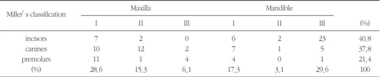
1. 연구 대상연세대학교 치과병원 치주과에 내원하여 상피하 결합조직 이식술을 시행한 48명의 환자의 98개 치아 를 대상으로 하였다. 환자들은 심미적인 문제나 치 근과민증을 호소하였다. 환자들의 연령 분포는 15- 58세로 평균 연령은 33.4세였으며, 성별 분포는 남자 19명(39.6%)과 여자 29명(61.4%)으로 구성되어 있 었다. 상피하 결합조직 이식술을 시행한 치아는 전 치 40개(상악 9개, 하악 31개), 견치 37개(상악 24개, 하악 13개), 소구치 21개(상악 16개, 하악 5개)로 모 두 98개 치아로 구성되어 있었으며, Miller 분류Ⅰ 45 개(45.9%), Ⅱ 18개 (18.4%), Ⅲ 35개(35.7%)였다 (Table 1).
Table 1. Distribution of 98 recession sites in 48 patients treated with subepithelial connective tissue grafts
Miller’s classification Maxilla Mandible
Ⅰ Ⅱ Ⅲ Ⅰ Ⅱ Ⅲ (%)
incisors 7 2 0 6 2 23 40.8
canines 10 12 2 7 1 5 37.8
premolars 11 1 4 4 0 1 21.4
(%) 28.6 15.3 6.1 17.3 3.1 29.6 100
2. 연구 방법
1) 술전 평가
모든 환자는 구강 위생 교육과 치석 제거술을 시행 하고 1개월 후에 임상 검사를 시행한 후 수술하였다.
치은 퇴축은 치주낭 탐침자를 이용하여 백악-법랑 경계(cemento-enamel junction: CEJ)에서 치은연까 지의 거리로 측정하였고, 치근단 방사선 사진으로 치 간 인접부위 치간골 상실 여부를 평가하였다.
2) 치은 퇴축 분류
Miller의 분류를 사용하였고 다음과 같다.
Class Ⅰ: 변연 조직 퇴축이 치은 점막 경계부까 지 연장되지 않은 경우. 치간골이나 연조직 상실은 없다.
Class Ⅱ: 치은 점막 경계부까지 퇴축이 연장된 경 우. 치간골이나 연조직 상실은 없다.
Class Ⅲ: 치은 점막 경계부까지 퇴축이 연장된 경 우. 치간골이나 연조직이 법랑-백악 경계부까지 상 실되어 있으나, 퇴축된 연조직 변연보다는 치관쪽에 존재한다.
Class Ⅳ: 치은 점막 경계부까지 퇴축이 연장된 경 우. 치간골과 연조직이 상실되어 퇴축 변연보다 치 간골과 연조직 변연이 더 치근단쪽으로 존재한다.
3) 수술 방법
부분층 판막 아래 상피하 결합조직을 이식한 후 판 막을 치관쪽으로 변위시키는 Langer and Langer24)의 방법을 이용하였으며 다음과 같다.
((11)) 수수혜혜부부 형형성성 II
백악 법랑 경계에서 피개될 치아의 선각에서 부분 층 판막을 형성한다. 절개는 적절한 수용부를 형성 하기 위해 인접치의 치간 선각 부위까지 연장한다.
치주낭 상피가 제거되고 노출된 치근면은 활택하게 한다.
((22)) 공공여여부부 형형성성
구개측에 치은연에서부터 5-6mm 떨어진 부위에 첫 번째 수평절개를 하고 그보다 1.5-2mm 치관방향 으로 두 번째 수평절개를 한다. 수평절개의 양쪽 끝 에 수직 절개를 하고 부분층 판막 window를 형성하 여 구개골 위에 약간의 결합조직이 있는 골막을 남 겨두고 이식체를 제거한다.
((33)) 수수혜혜부부 형형성성 IIII
이식편을 적용시키고 박리된 치은판막을 조심스 럽게 덮고, sling suture로 밀착 봉합하여 준다. 환자 에게 0.12% chlorhexidine gluconate를 처방하고 잇 솔질을 하지 말라고 교육한다. 술후 7-10일 후 봉합 을 제거한다. 3주후부터 정상적인 구강위생술을 시 행하도록 한다.
4) 통계학적 분석
술전과 술후 6개월, 18개월때의 치은퇴축량의 비 교하기 위해 ANOVA를 시행하였고, 술후 6개월시의 Miller 분류별 피개율과 술후 18개월시의 Miller 분류 별 피개율도 ANOVA를 시행하여 비교하였다. 유의 수준은p<0.05였다.
Table 2. Mean recession at baseline, 6, 18 months following treatment(mm)
Miller’s classification Baseline 6M 18M
Mean±S.D Range Mean±S.D Range Mean±S.D Range
Ⅰ 3.2±0.9 2-6 0.5±0.6* 0-2 0.2±0.4* 0-1
Ⅱ 5.2±1.6 3-9 1.0±1.0* 0-2 0.9±1.0* 0-2
Ⅲ 4.3±1.8 2-9 1.6±1.1* 0-4 1.8±1.3* 0-5
*statistically significantly different from baseline values (p<0.05) 6M= 6 months follow-up, 18M= 18 months follow-up
III. 연구 결과
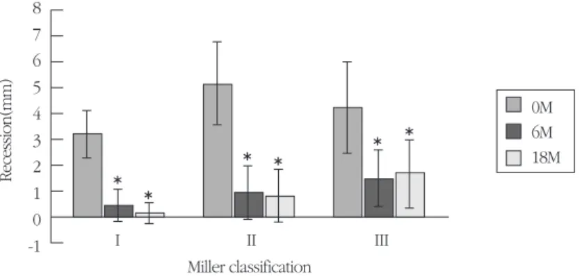
1. 술전·후 치은퇴축량의 변화48명의 98개 치아에서 술전 치은퇴축은 2-9mm로 평균 3.9±1.6mm였으며 Miller 분류별로 보면 다음 과 같다(Table 2, Figure 1).
2. 치근 피개율
6개월 follow-up 총 평균 피개율은 76.2±24%였으 며 18개월 follow-up의 경우는 75±25.2%였다(Table 3, Figure 2). 이중 완전 피개를 보이는 치아는 6개월
시 93개 중 39개로 41.9%, 18개월 시는 51개 치아중 20개로 39.2%에서 100%치근피개를 보였다(Table 3, Figure 3).
이를 Miller 분류에 따라 살펴보면 6개월 follow-up 시 Ⅰ급, Ⅱ급의 경우 각각 84.9%, 82.5%의 피개율 을 보이는 반면, Ⅲ급의 경우 62.3%로 낮았으며 18 개월의 경우에도 Ⅰ급, Ⅱ급이 각각 92.2%, 84.3%인 반면 Ⅲ급은 59.5%로 낮은 결과를 보였다(Table 3, Figure 4).
IV. 총괄 및 고찰
치은퇴축은 변연치은이 근단 부위로 변위된 것으 Figure 1. Mean recession (mm) at baseline, 6, 18 months following treatment
*statistically significantly different from baseline values (p<0.05) 0M=baseline, 6M=6months follow-up, 18M=18months follow-up
0M 6M 18M
Miller classification
Recession(mm)
I II
8 7 6 5 4 3 2 1 0
-1 III
Table 3. Mean % root coverage 6, 18 months following treatment as well as percentage of sites with com- plete coverage
6M 18M
Miller’s classification sites sites of complete % average sites sites of complete % average
coverage (%) coverage coverage (%) coverage
Ⅰ 42 26 (61.9%) 84.9% 15 11 (73.3%) 92.2%
Ⅱ 17 8 (47.1%) 82.5% 12 6 (50%) 84.3%
Ⅲ 34 5 (14.7%) 62.3% 24 3 (12.5%) 59.5%
Total 93 39 (41.9%) 76.2% 51 20 (39.2%) 75%
6M= 6 months follow-up, 18M= 18 months follow-up
sites of complete coverage: number of teeth which were covered completely
% average coverage: mean percent root coverage
로 치주질환 같은 염증이나 개개인에 따른 구강내의 해부학적 구조와 칫솔질 또는 치과진료와 같은 외부 적 요인에 의해서도 영향을 받는 것으로도 보고되고 있다. 이러한 치은퇴축은 환자에게 심미적으로 불편 함을 느끼게 할뿐만 아니라 치근과민증이나 치근우 식을 유발할 수도 있다.
치근피개 술식에는 측방변위판막술, 치관변위판막 술, 유리치은이식술, 상피하 결합조직 이식술, 치주 조직유도재생술 등이 있다. 1993년 Jahnke등41)은 상
면서 각각 80%, 43%로 상피하 결합조직 이식술의 성 공률이 더 높다고 하였다. 또한 1997년 Paolantonio 등42)도 각각 85%, 53%로 상피하 결합조직 이식술의 성공률이 높다고 보고하였다. 치주조직유도재생술 을 이용한 치근피개 술식과의 비교는 Pini Prato등43) 과 Ricci등44)에 의해서 보고되었는데 두 술식간의 치 근피개 효과는 비슷하나 임상부착수준 획득은 치주 조직유도재생술이 우수하고 각화 치은 증가는 상피 하 결합조직 이식술이 우수하다고 하였다.
Figure 4. Root coverage (%) according to Miller’s classification
*statistically significantly different from group I and II value(p<0.05) 6M= 6 months follow-up, 18M= 18 months follow-up
6M 18M
Miller classification
Root coverage(%)
I II III
120 100 80 60 40 20 0
Figure 2. Mean percent root coverage at 6 months and 18 months follow-up
No statistically significant difference between two groups (p=0.75) 6M= 6 months follow-up
18M= 18 months follow-up Period
Roof coverage(%)
6M 18M
100 80 60 40 20 0
Figure 3. Percent of completely covered teeth at 6 months and 18 months follow-up
6M= 6 months follow-up 18M= 18 months follow-up
Period
Teeth(%)
6M 18M
40
30
ty가 적은 것, 경제성, 심미성 등을 고려해야 하는데, 유리치은이식술은 공여부의 불편감이 크고, 이식 부 위에서의 주변조직과의 색조조화가 자연스럽지 못 한 단점이 있으며 치주조직재생술은 경제적인 부담 과 2차수술이 필요하다는 단점이 있다. 이에 본 연구 에서는 상피하 결합조직 이식술을 사용하였다.
1985년 Langer and Langer25)는 상피하 결합조직을 부분층 판막 아래 넣는 방법을 소개하여 2-6mm의 치근 피개를 보였다. 상피하 결합조직을 이용한 방 법에는 Langer and Langer에 의한 방법 외에도 Raetzke에 의한 Envelope technique32), Allen에 의한 Tunnel approach33), Nelson의 bilaminar technique26) 등이 있으며, 각각 80%, 84%, 91%의 피개율을 보고 하였다.
본 연구에서는 Langer and Langer의 방법을 이용 하여 상피하 결합조직 이식술을 시행하였을 때 6개 월, 18개월에서의 치근피개량을 Miller 분류에 따라 임상적으로 평가하고자 하였다. 48명의 환자 98개 치아에서 시행하였으며 상악의 경우는 견치가 24개 (24.5%)로 가장 많았고 하악의 경우는 전치가 31개 (31.6%)로 가장 많은 수를 차지하였으며 특히 하악 전치의 경우 Miller 분류 III급의 경우가 하악 전치중 74%(23개)를 차지하였다(Table 1). 술전 치은퇴축은 2-9mm로 평균 3.9mm였다. Miller 분류별로 살펴보 면 I, II, III급에서 각각 3.2mm, 5.2mm, 4.3mm였다 (Table 2, Figure 1).
술후 총 평균 피개량은 6개월 follow-up시 76.2%, 18개월 follow-up시 75.0%였으며 두 군간에 통계적 으로 유의할 만한 차이는 보이지 않았다. 완전 피개 를 보이는 치아는 6개월, 18개월시 각각 총 41.9%, 39.2%였으며 Miller 분류별로 살펴보면 6개월시 I, II, III급에서 61.9%, 47.1%, 14.7%로 I급에서 III급으로 갈수록 완전 피개를 보이는 치아의 수가 감소하였고 18개월시에도 각각 73.3%, 50%, 12.5%로 I급에서의 완전 피개율이 가장 높았다. 그러나 Miller는 치간골 이나 연조직이 상실된 III급의 경우, 부분 피개만이 가능하며 완전피개가 되지 않는다고 하였는데, 본 연 구에서는 약 10%의 경우에서 완전피개를 보였다. 총 평 균 피 개 량 은 Nelson(91%), Harris(97.4%),
Allen(84%) 등이 보고한 피개율보다 다소 낮은 결과 로 이들 연구는 주로 slight(1-3mm) to moderate(4- 6mm)나 또는 3mm이하의 얕은 퇴축의 경우로 Miller 분류 Ⅰ급이나 Ⅱ급에 해당되는 경우인 반면 본 연 구에서는 Ⅲ급의 치은퇴축까지 모두 포함시켰기 때 문이라고 생각된다. 분류에 따라 살펴보면 1985년 Miller15)는 치은퇴축을 Ⅰ급부터 Ⅳ급까지 분류하면 서 Ⅰ급과 Ⅱ급은 완벽한 치근피개를 얻을 수 있지 만 Ⅲ급은 부분적으로만 가능하며 Ⅳ급은 치근피개 를 기대하기 어렵다고 하였다. 이번 연구에서도 6개 월 follow-up시 Ⅰ급, Ⅱ급의 경우 각각 84.9%, 82.5%의 피개율을 보이는 반면, Ⅲ급의 경우 62.3%
로 낮았으며 18개월의 경우에도 Ⅰ급, Ⅱ급이 각각 92.2%, 84.3%인 반면 Ⅲ급은 59.5%로,Ⅰ급, Ⅱ급의 퇴축에 비해 Ⅲ급의 경우 통계적으로 유의할 만한 차이로 낮은 피개율을 보였다(Table 3, Figure 4). 이 는 치근피개에 있어 치간 인접부위 치주조직의 유지 가 중요하다는 것을 확인시켜주는 결과이다. III급 치은퇴축의 경우를 제외한 I급, II급의 경우만으로 평 균 피개량을 살펴보면 6개월시 84.2%, 18개월시 88.7%로 Nelson이나 Allen 등 다른 연구에서 보고된 바와 비슷한 결과를 보였다.
이상의 연구 결과에서 상피하 결합조직 이식술은 치간 인접부위 치주조직이 유지되어있는 Miller I급, II급 치은퇴축에서 임상적으로 우수한 결과를 보였 으며 술후 18개월에도 술후6개월시보다 치은퇴축이 더 진행되지 않고 유지됨을 알 수 있었다.
V. 결론
노출된 치근면은 심미적인 문제점과 치근과민성, 치근우식을 유발할 수 있다. 이러한 치은 퇴축을 피 개하려는 많은 노력이 이루어져왔으며 여러 가지 술 식이 개발되어지고 소개되어져 왔다. 그 중에서 상 피하 결합조직 이식술은 주변 조직과의 색조 조화나 공여부의 적은 불편감, 풍부한 혈액공급, 예측가능성 의 장점이 있으며 여러 연구에서 높은 치근피개율이 보고되어져 왔다.
본 연구에서는 연세대학교 치과대학 치주과에 내
원하여 상피하 결합조직 이식술을 시행한 48명의 환 자의 98 증례에 대하여 6개월, 18개월간 조사하여 다 음과 같은 결론을 얻었다.
1. 총 평균 피개량은 6개월 follow-up시 76.2±
24%, 18개월 follow-up시 75±25.2%였으며 기 간별로 통계학적으로 유의한 차이는 없었다 (p<0.05).
2. 완전 피개를 보이는 치아는 6개월 follow-up시 41.9%, 18개월 follow-up시 39.2%였다.
3. Miller 분류에 따른 피개량은 6개월 follow-up시
Ⅰ급 84.9±20.7%, Ⅱ급 82.5±17.7%, Ⅲ급 62.3±24.5%로 Ⅲ급 치은퇴축에서 Ⅰ급, Ⅱ급 치은퇴축에 비해 통계학적으로 유의한 차이를 보이며 적은 피개율을 나타냈다(p<0.05).
4. 18개월 follow-up시의 Miller 분류에 따른 피개 량은 Ⅰ급 92.2±13.5%, Ⅱ급 84.3±17.4%, Ⅲ 급 59.5±25.2%로 Ⅲ급 치은퇴축에서 Ⅰ급, Ⅱ 급 치은퇴축에 비해 통계학적으로 유의한 차이 를 보이며 적은 피개율을 나타냈다(p<0.05).
이상으로 미루어 보아 상피하 결합조직 이식술은
Ⅰ급, Ⅱ급의 치은퇴축에서 임상적으로 예측성있는 치근피개 술식으로 사료된다.
VI. 참고 문헌
1. American Academy of Periodontology. Glossary of periodontal terms. 3rd edn. Chicago:
American Academy of Periodontology, 1992.
2. Khocht A, Simon G, Person P, Denepitiya JL.
Gingival recession in relation to relation to histo- ry of hard toothbrush use. J Periodontol 1993;64:900-905
3. Serino G, Wennstrom JL, Lindhe J, Ennerath L.
The prevalence and distribution of gingival recession in subjects with high standard of oral hygiene. J Clin Periodontol 1994;21:57-63
of periodontal disease in man; prevalence, severity, and extent of gingival recession. J Periodontol 1992;63:489-495
5. Joshipura KJ, Kent RL, Depaola PF. Gingival recession: intraoral distribution and associated factors. J Periodontol. 1994;65:864-871
6. Lost C. Depth of alveolar bone dehiscences in relation to gingival recession. J Clin Periodontol 1984;11:583-589
7. Kallestal C, Uhlin S. Buccal attachment loss in Swedish adolescents. J Clin Periodontol 1992;19:458-491
8. Foushee DG, Moriarty JD, Simpson DM. Effects of mandibular orthognathic treatment on mucogingival tissues. J Periodontol 1985;56:727- 733
9. Muller HP, Eger T. Gingival phenotypes in young male adults. J Clin Periodontol 1992;19:458-491
10. Trott JR, Love B. An analysis of localized reces- sion in 766 Winnipeg high school students.
Dental Practice 1966;16:209-213
11. Gorman WJ. Prevalence and etiology of gingival recessions. J Periodontol 1967;38:316-322 12. Sullivan HC, Atkins JH. Free autogenous gingival
grafts Ⅲ. Utilization of grafts in the treatment of gingival recession. Periodontics 1968;6:152-159 13. Holbrook T, Ochsenbein C. Complete coverage
of the denuded root surface with a one stage gingival graft. Int J Periodont Rest Dent 1983;3:9 14. Pini Prato G, Tinti C, Vincenzi G, Magnani C,
Cortellini P, Clauser C. Guided tissue regenera- tion versus mucogingival surgery in the treat- ment of human buccal gingival recession. J Periodontol 1992;53:919-928
15. Miller PD. A classification of marginal tissue recession. Int J Periodont Rest Dent 1985;5:9-13 16. Grupe H, Warren R. Repair of gingival defects
1956;27:92
17. Guinard EA, Caffesse RG. Treatment of localized gingival recessions Ⅲ. Comparison of results obtained with lateral sliding and coronally repo- sitioned flaps. J Periodontol 1978;49:351-356 18. Harris RJ, Harris AW. The coronally positioned
pedicle graft with inlaid margins: A predictable method of obtaining root coverage of shallow defects. Int J Periodont Rest Dent 1982;25:229- 241
19. Miller PD. Root coverage using a free soft tissue autograft following citric acid application. part
Ⅰ. Technique. Int J Periodont Rest Dent 1982;2:65-70
20. Miller PD. Root coverage using a free soft tissue autograft following citric acid application. part
Ⅲ. A successful and predictable procedure in areas of deep-wide recession. Int J Periodont Rest Dent 1985;5:14-37
21. Borghetti A, Gardella J. Thick gingival autograft for the coverage of gingival recession: A Clinical evaluation. Int J Periodont Rest Dent 1990;10:217-229
22. Tolmie PN, Rubins RP, Buck GS, Vagianos V, Lanz JC. The predictability of root coverage by way of free gingival autograft and citric acid application: An evaluation by multiple clinicians.
Int J Periodont Rest Dent 1991;11:261-277 23. Paul L, Michaelides, Suzan G, Wilson. An auto-
genous gingival graft technique. Int J Periodont Rest Dent 1994;14:113-125
24. Langer B, Calagna LJ. The subepithelial connec- tive tissue graft. A new approach to the enhancement of anterior cosmetics. Int J Periodont Rest Dent 1982;2:22-34
25. Langer B, Langer L. Subepithelial connective tis- sue graft technique for root coverage. J Periodontol. 1985;56:715-720
26. Nelson SW. The subpedicle connective tissue
graft: A bilaminar reconstructive procedure for the coverage of denuded root surfaces. J Periodontol 1987;58:95-102
27. Harris RJ. The connective tissue and partial thickness double pedicle graft: A predictable method of obtaining root coverage. J Periodontol 1992;63:477-486
28. Jahnke PV, Sandifer JB, Gher ME, Gray JL, Richardson CA. Thick free gingival and connec- tive tissue autografts for root coverage. J Periodotol 1993;64:315-322
29. Tinti C, Vincenzi G, Cortellini P, Pini Prato GP, Clauser C. Guided tissue regeneration in the treatment of human facial recession. A 12 case report. J Periodontol 1992;63:554-560
30. Trombelli L, Schincaglia G, Scapoli C, Calura G.
Healing response of human buccal gingival recessions treated with expended polytetrafluo- roethylene membranes: A retrospective report. J Periodontol 1995;66:14-22
31. Edel A. Clinical evaluation of connective tissue grafts used to increase the width of keratinized gingiva. J Clin Periodontol 1974;1:185-196 32. Raetzke PB. Covering localized areas of root
exposure employing the "Envelope" Technique.
J Periodontol 1985;56:397-402
33. Allen AL: Use of the supraperiosteal envelope in the soft tissue grafting for root coverageⅡ.
Clinical results. Int J Periodont Rest Dent 1994;14:302-315
34. Wilderman M, Wentz F. Repair of dentogingival defect with a pedicle flap. J Periodontol 1965;36:218-226
35. Caffesse RG, Kon S, Castelli WA, Nasjleti C.
Revascularization following the lateral sliding flap procedure. J Periodontol 1984;55:352-358 36. Cortellini P, DeSanctis M, Pini Prato GP, Baldi C,
Clauser C. Guided tissue regeneration procedure using a fibrin-fibronectin system in surgically
induced recessions in dogs. Int J Periodont Rest Dent 1991;11:151-163
37. 정현철, 최성호, 조규성, 채중규, 김종관. 성견의 실험적 상피하 결합조직 이식시의 치주조직의 치유. 대한치주과학회지, 1997;27:379-394 38. Magnusson I, Runstad L, Nyman S, Lindhe J. A
long junctional epithelium- A locus minoris resistentiae in plaque infection. J Clin Periodontol 1983;10:333-340
39. Beaumont R, O'Leary T, Kafrawy A. Relative resistance of long junctional epithelial adhesions and connective tissue attachments to plaque- induced inflammation. J Periodontol 1984;55:213-223
40. Borghetti, A., Gardella, J.: Thick gingival auto- graft for the coverage of gingival recession: A clinical evaluation. Int J Periodont Rest Dent, 10:217, 1990.
41. Jahnke PV, Sandifer JB, Gher ME, Gray JL, Richardson AC. Thick free gingival and connec-
tive tissue autografts for root coverage. J Periodontol 1993;64:315-322
42. Paolantonio M, Di Murro C, Cattabriga A, Cattabriga M. Subpedicle connective tissue graft versus free gingival in the coverage of exposed root surfaces. A 5-year clinical study. J Clin Periodontol 1997;24:51-56
43. Pini Prato G, Clauser C, Magnani C, Cortellini P.
Resorbable membranes in the treatment of human buccal recession. A 9 case report. Int J Periodont Rest Dent 1995;15:258-267
44. Ricci G, Silvestri M, Tinti C, Rasperini G. A clini- cal/statistical comparison between the subpedi- cle connective tissue graft method and guided tissue regeneration technique in root coverage.
Int J Periodont Rest Dent 1996;16:539-545 45. 박철, 임성빈, 정진형. 독립된 결합조직 이식술로
치은퇴축 치료시 치근 피개에 관한 임상적 연구.
대한치주과학회지, 2000;30:651-660
-Abstract-
A Clinical Results of Subepithelial Connective Tissue Graft for Root Coverage
Kyung-Hee Choi, Jeong-Won Paik, Chang-Sung Kim, Seong-Ho Choi, Kyoo-Sung Cho, Chong-Kwan Kim, Jung-Kiu Chai
Department of Periodontology, College of Dentistry, Yonsei University Oral Science Research Center
Exposed root surfaces can cause esthetic problems, hypersensitivity, and root caries. Numerous efforts have been tried to cover the recessed root surfaces, and various techniques have been developed and introduced.
Among these, subepithelial connective tissue graft which shows high coverage rate in various researches, has the advantage of good color match, less discomfort to the donor site, rich vascularity, and high predictability.
Following results were obtained after investigating 6 and 18 months post operatively, 98 cases of subepithe- lial connective tissue graft from 48 patients who underwent subepithelial connective tissue graft procedure in the department of periodontology, college of dentistry, Yonsei university.
1. The total average root coverage of Miller class I, II & III were 76.2?24% at 6 months follow-up and 75?25.2% at 18 months follow-up with no statistically significant difference between the follow-up peri- ods.(p<0.05)
2. The percentage of teeth showing complete coverage were 41.9% at 6 months follow-up and 39.2% at 18 months follow-up.
3. At 6 months follow-up, Miller classification I showed 84.9?20.7%, class II showed 82.5?17.7%, and class III showed 62.3?24.5% of coverage. In class III recession, statistically significantly less root coverage was observed compared to class I & II.(p<0.05)
4. At 18 months follow-up, Miller classification I showed 92.2?13.5%, class II showed 84.3?17.4%, and class III showed 59.5?24.5% of coverage. In class III recession, statistically significantly less root coverage was observed compared to class I & II.(p<0.05)
In conclusion, subepithelial connective tissue graft for class I and II recession can be used as a clinically pre- dictable treatment modality for root coverage.
Key words : Gingival recession, Root coverage, Subepithelial connective tissue graft