Introduction
Radiological examination is essential for the accurate diagnosis and treatment planning for patients with mandibu- lar diseases. Computed tomography (CT) has been a useful tool for examining the mandible and for evaluating adja- cent tissues, as well as for detecting possible tumor lesions, inflammatory tissues, or pathologic bony changes.1
CT, first introduced by Hounsfield2in 1972, improved
the quality of radiological diagnosis in various clinical fields, along with the development of computer technology.
Not long after the spiral CT first appeared in the early 1990s, multi-detector CT (MDCT) loaded with many detectors began to dominate the market. As a result, radia- tion doses and exposure times required for image proces- sing have decreased.
In the dental field, the newly developed cone-beam com- puted tomography (CBCT) presented better resolution with lower exposure to radiation, as compared with conventional CT. Furthermore, CBCT now utilizes state-of-the-art imag- ing techniques, such as conveying 3-dimensional volumetric information as real images, thereby securing its role as a promising tool for the diagnosis of dentomaxillofacial lesions.3-5
Comparison of effective dose for imaging of mandible between multi-detector CT and cone-beam CT
Dae-Kyo Jeong, Sang-Chul Lee, Kyung-Hoe Huh, Won-Jin Yi*, Min-Suk Heo, Sam-Sun Lee*, Soon-Chul Choi
Department of Oral and Maxillofacial Radiology and Dental Research Institute, School of Dentistry, Seoul National University, Seoul, Korea
*Department of Oral and Maxillofacial Radiology, Dental Research Institute, and BK21 Craniomaxillofacial Life Science, School of Dentistry, Seoul National University, Seoul, Korea
ABSTRACT
Purpose : The aim of this study was to compare the effective dose for imaging of mandible between multi-detector computed tomography (MDCT) and cone-beam computed tomography (CBCT). An MDCT with low dose technique was also compared with them.
Materials and Methods : Thermoluminescent dosimeter (TLD) chips were placed at 25 organ sites of an anthropo- morphic phantom. The mandible of the phantom was exposed using 2 different types of MDCT units (Somatom Sensation 10 for standard-dose MDCT, Somatom Emotion 6 for low-dose MDCT) and 3 different CBCT units (AZ3000CT, Implagraphy, and Kavo 3D eXaM). The radiation absorbed dose was measured and the effective dose was calculated according to the ICRP 2007 report.
Results : The effective dose was the highest for Somatom Sensation 10 (425.84μSv), followed by AZ3000CT (332.4 μSv), Somatom Emotion 6 (199.38 μSv), and 3D eXaM (111.6 μSv); it was the lowest for Implagraphy (83.09 μSv).
The CBCT showed significant variation in dose level with different device.
Conclusion : The effective doses of MDCTs were not significantly different from those of CBCTs for imaging of mandible. The effective dose of MDCT could be markedly decreased by using the low-dose technique. (Imaging Sci Dent 2012; 42 : 65-70)
KEY WORDS : Multidetector Computed Tomography; Cone-Beam Computed Tomography; Thermoluminescent Dosimetry;
Mandible
*This study was supported by grant no 04-2010-0039 from the Seoul National Uni- versity Dental Hospital Research Fund.
Received September 27, 2011; Revised December 5, 2011; Accepted January 5, 2012 Correspondence to : Prof. Min-Suk Heo
Department of Oral and Maxillofacial Radiology, School of Dentistry, Seoul National University, 28 Yeongeon-dong, Jongno-gu, Seoul 110-744, Korea
Tel) 82-2-2072-3016, Fax) 82-2-744-3919, E-mail) [email protected]
Copyright ⓒ 2012 by Korean Academy of Oral and Maxillofacial Radiology
This is an Open Access article distributed under the terms of the Creative Commons Attribution Non-Commercial License (http://creativecommons.org/licenses/by-nc/3.0) which permits unrestricted non-commercial use, distribution, and reproduction in any medium, provided the original work is properly cited.
Imaging Science in Dentistry∙pISSN 2233-7822 eISSN 2233-7830
There have been studies on the CBCT dosimetry as well as the usefulness of CBCT in clinical practice. In particular, there has been much interest that CBCT would be more appropriate in the dental area compared with conventional CT because CBCT requires considerably lower doses.6-8 Chau and Fung6documented that of 3 types of diagnostic tools including spiral CT, conventional CT, and CBCT for dental implant prosthesis, spiral CT required the highest dose, while CBCT required the lowest dose. However, MDCT was not evaluated in their study.
Carrafiello et al9compared the effective doses between 64-slice MDCT, CBCT, and panoramic units. Although their study concluded that the effective dose of MDCT was 9 times higher than that of CBCT, it left much to be decided since the experiment was performed simply by exposing the X-ray beams on the same anatomical area, without considering the real clinical environment wherein MDCT operates. Meanwhile, Suomalainen et al10 reported no significant difference in effective dose between MDCT and CBCT. However, they did not consider the actual clin- ical situations. Kim et al11demonstrated that the absorbed and effective doses of CBCT were lower than that of con- ventional CT. However, their study was performed not with MDCT but with conventional CT, which required a higher dose than MDCT in most cases. Tack et al12indicated that low-dose MDCT was the imaging method of choice in patients with suspected chronic sinusitis.
MDCT has always been a secondary option in dental field in spite of its superior image quality due to the high level of x-ray exposure. In the early years of CBCT deve- lopment, the source of x-rays for CBCT was identical to other dental radiographic equipments, and thus was expect- ed to obtain dependable images with much lower x-ray doses compared with MDCT. However, since early CBCT devices produced significantly higher noise levels than medical CT scanners, recent CBCT units have replaced conventional sources with higher-capacity ones. With the invention of multi-detector CT which obtains its images after only one tube rotation, the dose gap between MDCT and CBCT has been diminished.
The purpose of this study was to compare the effective dose for mandibular imaging between MDCT and CBCT considering the clinical situations.
Materials and Methods
An Alderson radiation therapy phantom (Radiologic Sup- port Devices Inc., Long Beach, CA, USA), a human phan- tom torso structure which included the oral and maxillo-
facial areas, was used to measure x-ray dose (Fig. 1). The phantom consisted of 32 horizontal sections, each one with a thickness of 2.5 cm, and included the major anatomical
Fig. 1.Alderson radiation therapy phantom.
Fig. 2. Thermoluminescent dosimeter (TLD) chips are inserted into each of the organ structures.
structures of the craniofacial and visceral organs. Each organ structure contained a number of 5 mm-diameter holes that had thermoluminescent dosimeter (TLD) chips installed (Fig. 2). The dosimetry procedure was performed with a TLD chip (TLD-100 Li F chip, Harshaw Chemical Co., Cleveland, OH, USA) and a Harshaw model 3500 TLD reader (Harshaw Chemical Co., Cleveland, OH, USA).
This study evaluated the doses for the following MDCT units: Somatom Sensation 10 (Siemens AG, Erlangen, Ger- many) and Somatom Emotion 6 (Siemens AG, Erlangen, Germany). Also, three kinds of CBCT units, AZ3000CT (Asahi Roentgen Co., Kyoto, Japan), KaVo 3D eXam (Kavo Dental GmbH, Biberach/Riss, Germany) and Implagraphy (Vatech Co., Yongin, Korea) were used for this study.
The imaging procedures were designed to mimic the real clinical situations. The MDCT image for the standard dose was taken by following the mandible CT protocol adopted by Seoul National University Dental Hospital.
Briefly, MDCT was begun by taking a scout view and then focusing on the mandibular area. After the height of the scan was adjusted, by operator-controlled collimation, to cover the mandibular area 5 cm in height, the tube poten- tial, tube current, and rotation time were set at 120 kVp, 23.8 mA and 8.4 seconds, respectively. Low-dose MDCT images were taken by following the mandible protocol adopted by Dankook University Dental Hospital. MDCT was also begun by taking a scout view with a scanning width of 25×25 cm and 5 cm in height. Then, the tube potential, tube current, and rotation time were set at 110 kVp, 3.1 mA and 16 seconds, respectively. For CBCT units, AZ3000CT images were taken with a mandibular mode with a FOV of 7.9×7.1 cm. The scanning time was 17 seconds. Implagraphy images were taken with a FOV of 8.0×5.0 cm, and the total scanning time was 19 seconds.
Since there was no mandibular mode in KaVo 3D eXaM, the custom mode was selected in order to set the FOV height for taking the images of mandibular area as with MDCT. The images of both sides were taken with a single
scan at a FOV of 10×5 cm and the scanning time was 26.9 seconds. Table 1 shows the factors for the equipments.
Three TLD chips were installed into each of the 25 tis- sues or organs, including bilateral organs except for skin (Table 2). After taking images of the mandible region, the chips collected from the phantom were inserted into the TLD reader in order to measure the electronic charges. The mean value of the chips was deduced after removing the outliers, which ranged up to more than double the standard deviation.13For bilateral organs, the average value of the 2 organs was adopted. Harshaw calibrated each dosimeter by exposing it to a known quantity of radiation from a Cs- 137 source. Dosimeters were analyzed using an automatic hot gas reader and the raw data were recorded. Individual TLD chip sensitivity was obtained and applied as a correc- tion factor to subsequent exposure and reading of each TLD. The standard deviation of calibrated readings from the supplied TLD 100 chips is stated to be less than ±5%.
After measuring the electronic charges with the TLD reader,
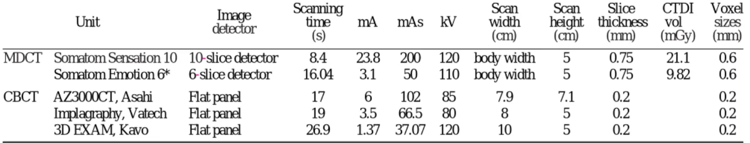
Table 1.Technical factors for MDCT and CBCT imaging of the mandibular area
Image Scanning Scan Scan Slice CTDI Voxel
Unit time mA mAs kV width height thickness vol† sizes
detector
(s) (cm) (cm) (mm) (mGy) (mm)
MDCT Somatom Sensation 10 10-slice detector 8.4 23.8 200 120 body width 5 0.75 21.1 0.6 Somatom Emotion 6* 6-slice detector 16.04 3.1 50 110 body width 5 0.75 9.82 0.6
CBCT AZ3000CT, Asahi Flat panel 17 6 102 85 7.9 7.1 0.2 0.2
Implagraphy, Vatech Flat panel 19 3.5 66.5 80 8 5 0.2 0.2
3D EXAM, Kavo Flat panel 26.9 1.37 37.07 120 10 5 0.2 0.2
*low-dose technique, †CTDI vol: volume computed tomography dose index
Table 2.Location of thermoluminescent dosimeter (TLD) chips in the Alderson radiation therapy (ART) phantom. Three TLDs used were used each level
Location Phantom level
Midbrain 2
Calvarium-right/left: superior 3
Calvarium-right/left: inferior 4
Mandibular ramus-right/left 5
Mandibular body-right/left 6
Submandibular gland-right/left 7
Esophagus 8
Thyroid-right/left 8
Lung-right/left 15
Heart 16
Liver 18
Stomach 21
Kidney-right/left 25
Colon 26
Ovary-right/left 28
Bladder 30
the chips were initialized, first annealing them in a 400�C chamber for 1 hour, then cooling them at room tempera- ture, and finally re-annealing them in a 100�C chamber for 2 hours. The absorbed dose (μGy) was calculated by multi- plying the exposed dose (mR) and a correction factor of 8.69 (the exposed dose was obtained by the TLD reader software program).14-16The absorbed dose for the whole- body bone marrow was calculated by summing up the indi- vidual equivalent doses to the calvarium and the mandible.
The determination of these equivalent doses was based on the distribution of active bone marrow throughout the adult body: the mandible contains 1.3% active marrow and the calvarium contains 11.8% active marrow.4,17For the calvar- ium, the average value of 4 sites in the cranial bone was used. Likewise, for the mandible, the average dose value of the mandibular body and ramus was used.
The doses obtained from TLDs at the different positions within the tissues or organs were averaged to express the average tissue-absorbed dose in micrograys (μGy). These values were used to calculate the equivalent dose (HT) with the following equation: HT==»WR×DT, where the equiva- lent dose (HT) for a tissue or organ is the product of the radiation weighting factor (WR) and the average absorbed dose (DT) measured for the specific tissue or organ. HTwas used to compare the effects of different types of radiation on tissues or organs. Since the radiation weighting factor of x-ray was 1, the values for both the absorbed and equiv- alent doses were the same, however the unit of measure- ment was changed from microgray (μGy) to an equivalent
unit, the microsievert (μSv).
The effective dose (E) is a dose proposed by the Interna- tional Commission on Radiological Protection (ICRP) to estimate radiation damage to an exposed population. It is deduced by multiplying actual organ doses by “tissue weighting factors” depending on an individual organ’s sensitivity. The effective dose represents the amount of radiation dose that the whole body receives, which also provides information on cancer risk, as compared to other organs receiving different doses. The effective dose (E) is calculated as follows: E==»(WT×HT), where E is the product of the ICRP tissue weighting factor (WT) for the type of tissue or body and the human-equivalent dose for tissue (HT). The tissue weighting factor represents the con- tribution that each specific tissue or organ gives to the overall risk of radiation damage. In this study, the ICRP 2007 weighting factors shown in Table 3 were adopted, which included the salivary tissue.
Table 3.Tissue-weighting factors for the calculation of effective dose according to ICRP 2007 recommendations
Tissue WT
Bone-marrow (red), Colon, Lung, Stomach 0.12
Breast, Remainder tissues* 0.08
Gonads 0.08
Bladder, Esophagus, Liver, Thyroid 0.04 Bone surface, Brain, Salivary glands, Skin 0.01
*Remainder tissues: Adrenal glands, extrathoracic (ET) region, gall bladder, heart, kidney, lymphatic nodes, muscle, oral mucosa, pancreas, prostate, small intestine, spleen, thymus
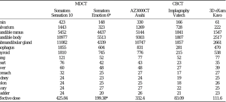
Table 4.Absorbed and effective dose (μGy) of exposure for MDCT and CBCT units for mandibular imaging
MDCT CBCT
Somatom Somatom AZ3000CT Implagraphy 3D eXam
Sensation 10 Emotion 6* Asahi Vatech Kavo
Brain 423 148 330 166 61
Calvarium 1443 323 1269 728 222
Mandible ramus 5452 4437 5144 1841 1547
Mandible body 10977 5513 9303 1807 2517
Submandibular gland 11082 4339 10747 1857 2661
Esophagus 1855 604 831 281 470
Thyroid 1810 745 776 215 538
Lung 121 52 77 52 77
Heart 76 42 43 23 35
Liver 60 48 48 27 39
Stomach 32 25 27 17 27
Kidney 24 23 24 19 25
Colon 24 25 25 18 26
Ovary 24 27 27 22 25
Bladder 24 20 26 21 23
Effective dose 425.84 199.38* 332.4 83.09 111.6
*low-dose technique
Results
Table 4 shows the absorbed doses for each of the tissues and organs. In the intra-machinery comparisons between all the units, the directly exposed regions, such as the man- dible and submandibular gland, showed higher values. In the inter-machinery comparison, Somatom Sensation 10 showed the highest absorbed dose value, especially at the directly exposed regions and their adjacent organs including the mandible ramus, mandible body, and submandibular gland. The lowest value was achieved with Implagraphy which showed the absorbed dose 3-5 times lower than that of Somatom Sensation 10.
The effective doses are presented in Table 4. Of the 5 different units, Somatom Sensation 10 showed the highest value of 425.84μSv, followed by a value of 332.4 μSv for AZ3000CT, and a value of 199.38μSv for Somatom Emo- tion 6. KaVo 3D eXaM and Implagraphy showed similar values of 111.6 and 83.09μSv, respectively.
Discussion
This study was designed to compare the effective dose for mandibular imaging between CBCT and MDCT units.
Since CT images are useful for evaluating the mandibular area, many previous studies have been conducted utilizing conventional or spiral CT.17However, the newly developed, cost-effective, and low radiation dose-producing CBCT began to replace conventional CT for mandibular evalua- tion.18,19Since then, most studies have focused on CBCT itself and its comparisons with spiral CT or conventional tomography. Meanwhile, the remarkable progress of MDCT technology has produced commercially available devices with more detectors, allowing for faster scanning times and low-dose exposures compared with conventional CT.
However, there have been few studies on comparisons between MDCT and CBCT. Therefore, this study was designed exclusively to compare MDCT and CBCT, accord- ing to the scanning protocol for mandible used in clinical examination. Our results were contrary to those of the pre- vious studies that reported that MDCT showed a signifi- cantly higher absorbed dose than CBCT.6,10,19,20
The effective dose was the highest for Somatom Sensa- tion 10, which was not significantly different from that of AZ3000CT. This value was also 4 times the effective dose of Kavo 3D eXaM, a CBCT device, and 5 times that of Implagraphy, a CBCT unit requiring the lowest effective dose. This implied wide variations of effective doses among CBCT units. Somatom Emotion 6 showed a lower dose
than AZ3000CT. The difference in the effective doses between CBCT units was also confirmed by a study by Ludlow et al21 which reported that dental CBCT units required a higher effective dose than 64-slice MDCT, espe- cially when taken with a high FOV. Of possible factors such as the tube potential, tube current, exposure time and detector sensitivity, the scanning time and tube current would be the most important factors of all the factors sug- gested by our study.
In this study, the AZ3000CT mandibular mode had a relatively high tube current. The scanning time of Impla- graphy was about 19 seconds shorter than that of KaVo 3D exam, and its tube current was the lowest (9.5 mA).
These might be the reasons why Impragraphy required such a low effective dose.
Since the pre-set optimal conditions of x-ray dose vary among different units, the total dose cannot be estimated only by tube potential, tube current and scanning time. In image taking, image quality is also an important considera- tion along with radiation exposure. Although image quality was not evaluated in our study, there has been no argument about the fact that MDCT provided superior image quality compared with CBCT.22Mulkens et al23showed that with modern technologies, low-dose CT of the sinuses in chil- dren yield a good diagnostic image quality with an effective dose comparable to that used for standard radiography.
This study offered the results inconsistent with those of the previous studies that asserted that MDCT requires a higher dose than CBCT. The effective dose of MDCT was lower than that of some CBCT units for imaging of man- dible by using the low-dose technique.
There has always been reluctance to use MDCT as the method of choice despite its superior contrast and resolu- tion, as well as precise diagnoses and treatment plans, because there is a putative fear of its seemingly higher radiation exposure. Our study, however, provided direct evidence that MDCT was appropriate for the evaluation of the mandibular area in the dental field.
This study had a limitation due to its small sample size.
More experimental data and further studies on clinical comparison of various CT units would be needed to verify our results. The quality control of the low-dose technique should be further validated.24
In conclusion, the effective dose of MDCT for imaging of mandible was not significantly different from that of CBCT which showed the highest value. CBCT showed significant variations in dose level among different devices.
The effective dose of MDCT could be markedly decreased by using the low-dose technique.
References
1. Nakagawa Y, Kobayashi K, Ishii H, Mishima A, Asada K, Ishibashi K. Preoperative application of limited cone beam computerized tomography as an assessment tool before minor oral surgery. Int J Oral Maxillofac Surg 2002; 31 : 322-6.
2. Hounsfield GN. Computerized transverse axial scanning (to- mography). 1. Description of system. Br J Radiol 1973; 46 : 1016-22.
3. Ludlow JB, Davies-Ludlow LE, Brooks SL. Dosimetry of two extraoral direct digital imaging devices: NewTom cone beam CT and Orthophos Plus DS panoramic unit. Dentomax- illofac Radiol 2003; 32 : 229-34.
4. Ludlow JB, Davies-Ludlow LE, Brooks SL, Howerton WB.
Dosimetry of 3 CBCT devices for oral and maxillofacial radio- logy: CB Mercuray, NewTom 3G and i-CAT. Dentomaxillofac Radiol 2006; 35 : 219-26.
5. Lee JN, Han WJ, Kim EK. Absorbed and effective dose from newly developed cone beam computed tomography in Korea.
Korean J Oral Maxillofac Radiol 2007; 37 : 93-102.
6. Chau AC, Fung K. Comparison of radiation dose for implant imaging using conventional spiral tomography, computed tomography, and cone-beam computed tomography. Oral Surg Oral Med Oral Pathol Oral Radiol Endod 2009; 107 : 559-65.
7. Dula K, Mini R, van der Stelt PF, Sanderink GC, Schneeberger P, Buser D. Comparative dose measurements by spiral tomo- graphy for preimplant diagnosis: the Scanora machine versus the Cranex Tome radiography unit. Oral Surg Oral Med Oral Pathol Oral Radiol Endod 2001; 91 : 735-42.
8. Hirsch E, Wolf U, Heinicke F, Silva MA. Dosimetry of the cone beam computed tomography Veraviewepocs 3D compared with the 3D Accuitomo in different fields of view. Dentomax- illofac Radiol 2008; 37 : 268-73.
9. Carrafiello G, Dizonno M, Colli V, Strocchi S, Pozzi Taubert S, Leonardi A, et al. Comparative study of jaws with multis- lice computed tomography and cone-beam computed tomo- graphy. Radiol Med 2010; 115 : 600-11.
10. Suomalainen A, Kiljunen T, Kaser Y, Peltola J, Kortesniemi M. Dosimetry and image quality of four dental cone beam computed tomography scanners compared with multislice computed tomography scanners. Dentomaxillofac Radiol 2009; 38 : 367-78.
11. Kim SY, Han JW, Park IW. Comparison of cone beam CT and conventional CT in absorned and effective dose. Korean J Oral Maxillofac Radiol 2008; 38 : 7-15.
12. Tack D, Widelec J, De Maertelaer V, Bailly JM, Delcour C, Gevenois PA. Comparison between low-dose and standard-
dose multidetector CT in patients with suspected chronic sinusi- tis. AJR Am J Roentgenol 2003; 181 : 939-44.
13. Groves AM, Owen KE, Courtney HM, Yates SJ, Goldstone KE, Blake GM, et al. 16-detector multislice CT: dosimetry estimation by TLD measurement compared with Monte Carlo simulation. Br J Radiol 2004; 77 : 662-5.
14. Avendanio B, Frederiksen NL, Benson BW, Sokolowski TW.
Effective dose and risk assessment from detailed narrow beam radiography. Oral Surg Oral Med Oral Pathol Oral Radiol Endod 1996; 82 : 713-9.
15. Frederiksen NL, Benson BW, Sokolowski TW. Effective dose and risk assessment from computed tomography of the maxil- lofacial complex. Dentomaxillofac Radiol 1995; 24 : 55-8.
16. Frederiksen NL, Benson BW, Sokolowski TW. Effective dose and risk assessment from film tomography used for dental implant diagnostics. Dentomaxillofac Radiol 1994; 23 : 123-7.
17. Underhill TE, Chilvarquer I, Kimura K, Langlais RP, McDavid WD, Preece JW, et al. Radiobiologic risk estimation from den- tal radiology. Part I. Absorbed doses to critical organs. Oral Surg Oral Med Oral Pathol 1988; 66 : 111-20.
18. Mozzo P, Procacci C, Tacconi A, Martini PT, Andreis IA. A new volumetric CT machine for dental imaging based on the cone-beam technique: preliminary results. Eur Radiol 1998; 8 : 1558-64.
19. Loubele M, Bogaerts R, Van Dijck E, Pauwels R, Vanheusden S, Suetens P, et al. Comparison between effective radiation dose of CBCT and MSCT scanners for dentomaxillofacial applications. Eur J Radiol 2009; 71 : 461-8.
20. Schulze D, Heiland M, Thurmann H, Adam G. Radiation expo- sure during midfacial imaging using 4-and 16-slice computed tomography, cone beam computed tomography systems and conventional radiography. Dentomaxillofac Radiol 2004; 33 : 83-6.
21. Ludlow JB, Ivanovic M. Comparative dosimetry of dental CBCT devices and 64-slice CT for oral and maxillofacial radio- logy. Oral Surg Oral Med Oral Pathol Oral Radiol Endod 2008;
106 : 106-14.
22. Liang X, Jacobs R, Hassan B, Li L, Pauwels R, Corpas L, et al. A comparative evaluation of Cone Beam Computed Tomo- graphy (CBCT) and Multi-Slice CT (MSCT) Part I. On subjec- tive image quality. Eur J Radiol 2010; 75 : 265-9.
23. Mulkens TH, Broers C, Fieuws S, Termote JL, Bellnick P.
Comparison of effective doses for low-dose MDCT and radio- graphic examination of sinuses in children. AJR Am J Roent- genol 2005; 184 : 1611-8.
24. Vassileva J, Stoyanov D. Quality control and patient dosime- try in dental cone beam CT. Radiat Prot Dosimetry 2010; 139 : 310-2.