89
Immune Network
Introduction
Rheumatoid arthritis (RA) is a systemic disease marked by synovial inflammation and hyperplasia that often destroys involved joints (1,2). Infiltration of the synovium by antibody secreting plasma cells is a hallmark of the disease (3,4). Immunoglobulins are heteromeric proteins consisting of two heavy and two light chains, each of which is divided into a variable domain (V) that defines the antigen specificity of the molecule. V domains contain 3 hypervariable intervals of the complementary-determining regions (CDR1, CDR2, and CDR3) that are divided into 3 relatively constant domains of the framework regions (FR1,
FR2, and FR3)(5-7). The VH-DH-JH and VL-JL and non-germline-encoded (N) nucleotides additionally define CDR3. The CDRs are usually directly involved in antigen binding, with CDR3 at the center of antigen binding sites (6).
Previous studies showed high levels of non- ger- mline encoded nucleotides (N regions) in VJ joins of kappa light chain from RA synovia and unsorted cells of RA peripheral blood (PB)(8,9). N-nucleotides enhance antibody diversity by encoding additional amino acid residues in CDR3. This finding may be due to positive antigenic selection or rearrangement abnormality from the bone marrow or lymph nodes.
In order to clarify these matters, we separated IgM+ IgD+ B cells, which come from bone marrow without contacting antigen, and IgM-IgD- B cells (presum- ably IgG+ or IgA+) in order to compare CDR3 and N-nucleotides between RA and normal individuals.
We choose the IgM+ IgD+ B cells subset as a precursor of GC B cells and the IgM-IgD- cells
in B Cell Subsets in Rheumatoid Arthritis: Evidence for Over-expression of TDT in B Lineage
Choong Won Lee1 and S. Louis Bridges, Jr2
1Division of Rheumatology, Department of Internal Medicine, Wallace Memorial Baptist Hospital, Busan, Korea, and 2Division of Immunology and Rheumatology, University of Alabama at Birmingham, Birmingham, Alabama, U.S.A
ABSTRACT
Background: Unusually high amounts of N region addition and CDR3 length diversity were found in immunoglobulin (Ig) light chain Vk and Jk joins in patients with rheumatoid arthritis (RA). We sought to determine whether this finding is due to excessive activity of the enzyme responsible for N region addition (terminal deoxy- nucleotidyl transferase [TdT]) in B lineage cells in bone marrow or from positive antigenic selection of B cells with long CDR3 lengths. Methods: We used FACS to isolate IgM+/IgD+ B cells (predominantly naive) and IgM-IgD- B cells (pre- dominantly class-switched) B cells from peripheral blood of a patient with RA known to have enrichment for long Vk CDR3s and from that of two normal controls. RT-PCR of VkIII transcripts was performed, followed by sequencing of individual cDNA clones.
We analyzed the CDR3 lengths and N region additions in 97 clones. Results: There was enrichment for long CDR3 lengths (11 or 12 amino acids) in both IgM+/IgD+ and IgM-IgD- B cells in RA compared to B cell subsets in the normal controls. The IgM+/IgD+ B cell subset in RA was markedly enriched for N region addition and was similar to that seen in the IgM-/IgD- subset. Conclusion: These data suggest that enrichment for N region addition and long CDR3 lengths in RA may result from unusually high or prolonged activity of TdT in bone marrow. (Immune Network 2003;3(2):89-95)
Key Words: Rheumatoid arthritis, N region, CDR3
Correspondence to: Choong Won Lee, Division of Rheumatology, Department of Internal Medicine, Wallace Memorial Baptist Hospital, Gumjung-gu P.O Box 100, Namsan-dong, Geumjeong-gu, Busan 609-340, Korea. (Tel) 051-580-1257, (Fax) 051-583-7114, (E-mail) choong@wmbh.co.kr
subset as GC derived memory B cells. To clarify whether abnormal N-nucleotides and CDR3 in RA comes from rearrangement abnormality or positive antigen selectivity, we compared N-nucleotides and CDR3 in those B cells subsets through sequencing of the V light chain.
Materials and Methods
Patient and control characteristics and cell separation of peripheral blood mononuclear cells (PBC). PBC from patient AS, a 46-year-old black female with a 10-year history of RF-positive RA, and from two normal healthy individuals, a 35-year-white female SB and a 37-year old Asian male CL, were isolated by Ficoll- Isopaque density centrifugation. PBC were incubated with anti-CD19-microbeads for 15 min at 4oC. CD19+ B cells were enriched by magnetic cell separation using the MiniMACS system (Miltenyi Biotec, Au- burn, CA, USA). B cells were enriched 98% by a single step of magnetic sorting for analysis by immunofluorescence staining with anti-human CD19 FITC (PharMingen, San Diego, CA, USA). Cells of the CD19-enriched fraction were incubated with anti-human IgM-PE (PharMingen, San Diego, CA, USA)) and anti-human IgD-FITC for 20 min on ice, then sorted into IgM+IgD+ and IgM-IgD- fractions on a FACS advantage SE (Becton Dickson, Franklin lakes, NJ, USA).
Generation of cDNA and PCR amplification of Vκ -containing transcripts. Total RNA was isolated from each IgM+IgD+ subset and from IgM-IgD- subset B cells using Tri-zol reagent (Molecular Research Center, Inc., Cincinnati, OH, USA). Oligo d(T)- primed first-strand cDNA was generated from total RNA using SuperScript II for RNase H- Reverse transcriptase kit (GibcoBRL, Gaithersburg, MD, USA).
PCR amplifications were performed on 2-μl aliquots of first-strand cDNA, using Taq DNA polymerase.
PCR conditions were: 35 cycles of denaturation at 94oC for 2 min, annealing at 55oC for 30 sec, extension at 72oC for 1 min, with a final extension at 72oC for 7 min. PCR amplifications were designed to Cκ sequence as an anti-sense 3' primer (LSK-19: 5'- GCGCCGTCTAGAATTAACACTCTCCCCTGTTGA A-3') and the leader and FR1 of germline Humkv325 as a sense 5' primer (LSK-16: 5'- CCACCGGAGAGC TCGTGTTGACGCAGTCTCCA-3'). To control for possible contamination, a mock PCR reaction mixture, lacking a template and containing products of the first strand cDNA reaction without reverse transcriptase were prepared. None of the controls contained amplified product as assessed by ethidium-stained agarose gel electrophoresis.
Cloning, transformation and sequencing of PCR products.
Aliquots of PCR products were subcloned with pGEM-T vector using TA cloning kit (Promega, Madison, WI, USA). Plasmids were transformed into DH5α E.Coli by 37oC heat shock. For colonies that were digested with EcoR1 restriction enzyme, plas- mid DNA was obtained and sequenced by dye terminator cycle sequencing using an automated sequencer (Applied Biosystems, Inc., Model ABI373, Foster, CA, USA).
Sequence analysis. Sequences were analyzed with a sequence aliment (DNAPLOT) and SAW (Sequences Analysis Workshop) software (10). Nucleotide ex- changes in Vκ were considered for the analysis of somatic mutation. Levels of somatic hypermutation were assessed by comparing the number of nu- cleotides from the codon 20 of FR1 throughout the CDR3 domains (6). Non-germline encoded nucleo- tides at the Vκ-Jκ joint, which represent N-region insertion, and P nucleotides, which are palindromic to terminal nucleotides of the coding sequence, were not counted as somatic mutations (11).
Statistical analysis. The results are presented as the
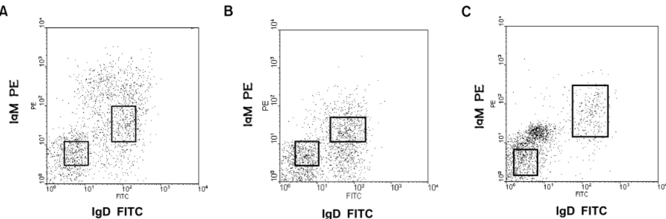
Figure 1. Fluorescence analysis and gate for sorting of B cell subsets derived from peripheral lymphocytes of the rheumatoid arthritis (A) and normal controls (B and C).
IgD FITC
IgMPE IgMPE
IgD FITC IgD FITC
IgMPE
A B C
mean±SD. Differences in the amounts of somatic mutation, N-region addition, and CDR3-length heterogeneity between RA patients and normal individuals were analyzed using the Chi-square test, Fisher exact test (two-tailed), or Student t-test, as appropriate.
Results
Vκ transcripts from PBC of IgM+IgD+ B cells had no, or fewer, somatic mutations than those from IgM-IgD- B cells.
Following enrichment of CD19+ B cell by micro- beads magnetic cell separation, IgM+IgD+ and IgM- IgD- B cells were purified by fluorescence activated cell sorting (Fig. 1). The levels of somatic mutation, as reflected by divergence from germline, among the transcripts from the different samples are shown in Fig. 2. The transcripts from IgM+IgD+ B cells of the RA patient and normal individuals (AS, CL and SB, n=46) were significantly less mutated (0.54±0.12%) than their transcripts from IgM-IgD- B cells (4.51±
0.42%, n=52) (P<0.0001). Somatic mutation level of the IgM-IgD- B cells (memory cells) in RA was also significantly higher than those in the two normal controls (P=0.04).
Twenty-seven (59%) of 46 Vκ transcripts from IgM+IgD+ B cells were completely germline in the Vκ region, compared with none of 52 colonies from IgM-IgD- B cells.
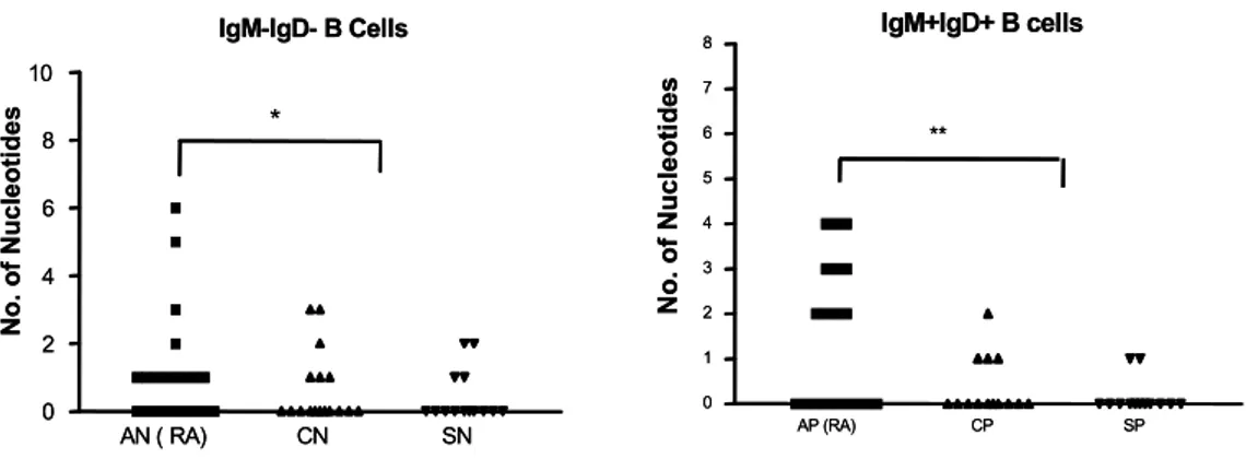
N-region addition. The nucleotide sequences of N regions of each clone in RA are shown in Fig. 3. The percentages of clones with at least one nucleotide of N region addition in IgM+IgD+ B cells were: SB 2 of 13 (15%), CL 3 of 13 (23%), and AS 10 of 19 (53%). Those of N region addition in IgM-IgD- B cells were: SB 4 of 14 (29%), CL 7 of 17 (41%), and AS 12 of 21 (57%)(Table I). The nucleotides of N-region addition in the RA patient (1.3±0.3, n=40) were significantly higher than those of the two normal controls (0.4±0.1, n=58)(P=0.0003), as in previous report (9)(Fig. 3). The nucleotides of N region addition in IgM+IgD+ B cells in the RA patient (1.5±0.4) were significantly higher than those of the two normal individuals (0.3±0.1) (P=0.0004).
But those in IgM-IgD- B cells did not differ significantly between RA and normal controls.
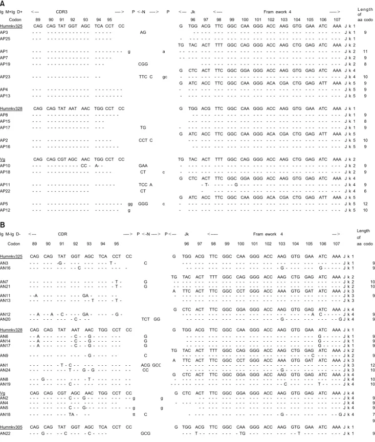
CDR3 lengths of Vκ transcripts. The Vκ repertoires of the RA patient are compared with those of the normal controls in Table I and Fig. 4, in order to demonstrate CDR3 length. Clones with unusually long CDR3s (one 11 and two 12 amino acid codons) were found in 3 of 40 (7.5%) transcripts from the RA patient, but in none of 58 transcripts from the normal controls. One 11 and one 12 amino acid codons were found in 19 transcripts of IgM+IgD+ B cells (naive cells) in RA (Fig. 5). One 12 amino acid codons was also found in 21 transcripts of IgM- IgD- B cells in RA. Each of 3 long CDR3s came from Humkv325, Vg, and Humkv328 Vκ gene seg- ments.
Germline derivation of κ light chains amplified with Humkv325 and Cκ primers. The majority of transcripts amplified with Humkv325 and Cκ primers were derived from Humkv325 (A27) (36%). Most of the remaining clones were derived from two other members of the Vκ III family, 29% from Humkv328 (L2) and 21% from Vg (L6). One clone, AN22, was
Figure 2. Somatic mutation in IgM+IgD+ and IgM-IgD- B cells between rheumatoid arthritis (AP, AN) and normal controls (CP, CN, SP and SN). *P=0.04: AN versus CN & SN.
CP SP AP(RA) CN SN AN(RA)
0 10 20
IgM+IgD+ IgM-IgD-
*
No. of Mutated Nucleotide
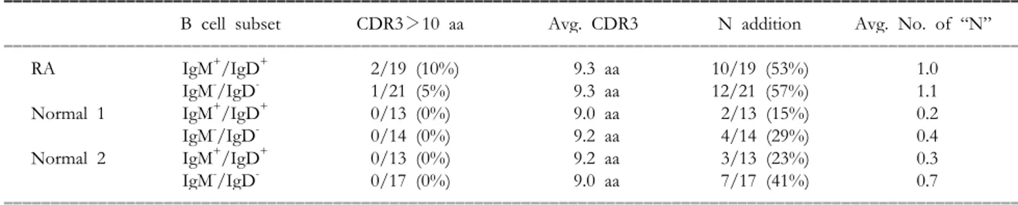
Table I. N region addition and CDR3 length in IgM+IgD+ and IgM-IgD- cells in the rheumatoid arthritis (RA) and normal controls.
ꠚꠚꠚꠚꠚꠚꠚꠚꠚꠚꠚꠚꠚꠚꠚꠚꠚꠚꠚꠚꠚꠚꠚꠚꠚꠚꠚꠚꠚꠚꠚꠚꠚꠚꠚꠚꠚꠚꠚꠚꠚꠚꠚꠚꠚꠚꠚꠚꠚꠚꠚꠚꠚꠚꠚꠚꠚꠚꠚꠚꠚꠚꠚꠚꠚꠚꠚꠚꠚꠚꠚꠚꠚꠚꠚꠚꠚꠚꠚꠚꠚꠚꠚꠚꠚꠚꠚꠚꠚꠚꠚꠚꠚꠚꠚꠚꠚꠚꠚꠚꠚꠚꠚꠚꠚꠚꠚꠚ
B cell subset CDR3>10 aa Avg. CDR3 N addition Avg. No. of “N”
ꠏꠏꠏꠏꠏꠏꠏꠏꠏꠏꠏꠏꠏꠏꠏꠏꠏꠏꠏꠏꠏꠏꠏꠏꠏꠏꠏꠏꠏꠏꠏꠏꠏꠏꠏꠏꠏꠏꠏꠏꠏꠏꠏꠏꠏꠏꠏꠏꠏꠏꠏꠏꠏꠏꠏꠏꠏꠏꠏꠏꠏꠏꠏꠏꠏꠏꠏꠏꠏꠏꠏꠏꠏꠏꠏꠏꠏꠏꠏꠏꠏꠏꠏꠏꠏꠏꠏꠏꠏꠏꠏꠏꠏꠏꠏꠏꠏꠏꠏꠏꠏꠏꠏꠏꠏꠏꠏꠏ
RA IgM+/IgD+ 2/19 (10%) 9.3 aa 10/19 (53%) 1.0
IgM-/IgD- 1/21 (5%) 9.3 aa 12/21 (57%) 1.1
Normal 1 IgM+/IgD+ 0/13 (0%) 9.0 aa 2/13 (15%) 0.2
IgM-/IgD- 0/14 (0%) 9.2 aa 4/14 (29%) 0.4
Normal 2 IgM+/IgD+ 0/13 (0%) 9.2 aa 3/13 (23%) 0.3
IgM-/IgD- 0/17 (0%) 9.0 aa 7/17 (41%) 0.7
ꠏꠏꠏꠏꠏꠏꠏꠏꠏꠏꠏꠏꠏꠏꠏꠏꠏꠏꠏꠏꠏꠏꠏꠏꠏꠏꠏꠏꠏꠏꠏꠏꠏꠏꠏꠏꠏꠏꠏꠏꠏꠏꠏꠏꠏꠏꠏꠏꠏꠏꠏꠏꠏꠏꠏꠏꠏꠏꠏꠏꠏꠏꠏꠏꠏꠏꠏꠏꠏꠏꠏꠏꠏꠏꠏꠏꠏꠏꠏꠏꠏꠏꠏꠏꠏꠏꠏꠏꠏꠏꠏꠏꠏꠏꠏꠏꠏꠏꠏꠏꠏꠏꠏꠏꠏꠏꠏꠏ aa: amino acid.
derived from the VκIII gene segment Humkv305 (A11). None of the other potentially functional members of the Vκ III family were represented in this analysis. Seven clones (7%) were derived from Vκ I gene segment Vd (DPK8), 012 (DPK9), and 018(DPK1), and six (6%) from the single member of the Vκ IV family DPK24 (B3). The Jκ4 and Jκ1 were frequently used in RA and normal controls. The incidence of Jκ in 30 sequences in RA were 11 of Jκ 1(36%), 11 of Jκ 4 (36%), 8 of Jκ 2 (27%), 6 of Jκ 5 (20%), and 4 of Jκ 3 (13%). The incidence of Jκ in 58 sequences in normal controls were 22 of Jκ 4 (38%), 16 of Jκ 1 (28%), 13 of Jκ 3 (22%), 4 of Jκ 3 (7%), and 3 of Jκ 5 (5%).
Analysis of clones from the IgM-IgD- B cells (memory B cells) in normal controls revealed two sets of 4 closely related sequences. SN 17, SN 32, CN30, and CN34 were completely identical to each other.
Analysis from the IgM-IgD- B cells in RA revealed two sets of 5 related sequences. AN6, AN14, and AN17 were completely identical and AN2 and AN5 were completely identical to each other. But none of
IgM+IgD+ B cells of RA and normal controls revealed an identical sequence.
Discussion
During B cell development, a series of highly regulated gene rearrangements generates a function antibody molecule. In the pro-B cell stage, the heavy chain locus undergoes rearrangement. Variable heavy- chain domains are encoded by variable (VH), diversity (DH), and joining (JH) gene segments, which undergo sequential somatic rearrangement to become jux- taposed in the genomic DNA (5). Initially, one or more DH gene segments are rearranged to a JH gene segment, followed by rearrangement of the VH gene segment to the DH-JH join. After heavy chain rearrangement, the kappa light chain segment under- goes rearrangements of VL and JL gene segments to generate light chain variable domains, presumably in the pre-B cell stage (7).
During the pro-B cell stage, the enzyme respon- sible for N-region addition, TdT is present in the nucleus of the cell. Although functional TdT has not been shown to be present at the time of kappa light chain rearrangement, N region addition is clearly present in kappa light chains from normal individuals and patients with RA (10-12,16). Therefore, TdT appears to be active throughout all stages of imm- unoglobulin gene rearrangements in human B cells.
Most PB B cells express IgM throughout their life and these cells are distinguished into IgM+IgD+ and IgM+IgD- B cells (14,15). Sequence analysis of the V genes expressed by the B-cell subset revealed most IgM-only cells while IgM-IgD- cells were mutated, but the IgM+IgD+ cells in PB had no, or few somatic mutations (13,14). Therefore, IgM+IgD+ B lymphocytes establish the naive, unmutated antibody repertoire, and if a somatically mutated subset exists among IgD-expressing PB B cells, it must be small
Figure 3. N-region addition in IgM+IgD+ and IgM-IgD- B cells between rheumatoid arthritis (AP, AN) and normal controls (CP, CN, SP and SN). **P=0.0004: AP versus CP & SP, *P=not significant: AN versus CN & SN.
IgM-IgD- B Cells
AN ( RA) CN SN
0 2 4 6 8 10
*
No. of Nucleotides
IgM-IgD- B Cells
AN ( RA) CN SN
0 2 4 6 8 10
*
No. of Nucleotides
IgM+IgD+ B cells
AP (RA) CP SP
0 1 2 3 4 5 6 7 8
**
No. of Nucleotides
IgM+IgD+ B cells
AP (RA) CP SP
0 1 2 3 4 5 6 7 8
**
No. of Nucleotides
Figure 4. Percentage of Vκ clones containing CDR3 length of 11 and 12 amino acids between rheumatoid arthritis (AS) and normal controls (SB, CL).
0 2 4 6 8 10 12
%
IgM+IgD IgM-IgD AS (RA) CL SB
fraction of CD27-expressing B cells (16). The mem- ory B-cell compartment in humans consists of so- matically mutated, class-switched, which represent
IgM-IgD- such as IgG+ and IgA+ PB cells, and IgM bearing B cells (17).
If B cells succeed in generating a functional,
Figure 5. Nucleotide sequences of CDR3 regions of clones in IgM+IgD+ B cells (A) and those of clones in IgM-IgD- B cells (B) in rheumatoid arthritis.
B
Ig M-Ig D- <--- CDR ----> P <-N ----> P<--- Jk <--- Fram ework 4 ---> Length
of
Codon 89 90 91 92 93 94 95 96 97 98 99 100 101 102 103 104 105 106 107 aa codo
Humnkv325 CAG CAG TAT GGT AGC TCA CCT CC G TGG ACG TTC GGC CAA GGG ACC AAG GTG GAA ATC AAA J k 1
AN3 - - - - - - -G - - - - - - - - - - T - C - - - - - - - - - - - - - - - - - - - - - - - - - - - - - - - - - - - - J k 1 9 AN16 - - - - - - - - - - - C - - - - - - - - - - - - - - - - - - - - - - - - - - - - - - - G - - - - - - - G - - - J k 1 9
TG TAC ACT TTT GGC CAG GGG ACC AAG CTG GAG ATC AAA J k 2 AN7 - - - - - - - - - - - - - - - - - - - T - G - - - - - - - - - - - - - - - - - - - - - - - - - - - - - - - - - - J k 2 10 AN21 - - - - - - - - - - - - - - - - - - - T - G - - - - - - - - - - - - - - - - - - - - - - - - - - - - - - - - - - J k 2 10
A TTC ACT TTC GGC CCT GGG ACC AAA GTG GAT ATC AAA J k 3
AN11 - -A - - - - - - - - - GA - - - - - - - - - - - - - - - - - - - - - - - - - - - - - - - - - - - - - - - - - J k 3 9 AN13 - - - - - - - - - - - - - - T - - - - T - - - - - - - - - - - - - - - - - - - - - - - - - - - - - - - - - - - - - J k 3
G CTC ACT TTC GGC GGA GGG ACC AAG GTG GAG ATC AAA J k 4
AN12 - - A - - A - C - - - - GA - - - - - G - - - - - - - - - - - - - - - - - - - - - - - - - - - - - - A C - - - - - J k 4 9 AN20 - - - - - - - - - - C - - - TCT GG - - - - - - - - - - - - - - - - - - - - - G - - - - - - - - - - - - G J k 4 9
Humnkv328 CAG CAG TAT AAT AAC TGG CCT CC G TGG ACG TTC GGC CAA GGG ACC AAG GTG GAA ATC AAA J k 1
AN6 - - A - - - - - - - C - - G - - - - - - - G - - - - - - - - - - - - - - - - - - - - - - - - - - - - - G - - - - - J k 1 9 AN14 - - A - - - - - - - C - - G - - - - - - - G - - - - - - - - - - - - - - - - - - - - - - - - - - - - - G - - - - - J k 1 9 AN17 - - A - - - - - - - C - - G - - - - - - - G - - - - - - - - - - - - - - - - - - - - - - - - - - - - - G - - - - - J k 1 9
TG TAC ACT TTT GGC CAG GGG ACC AAG CTG GAG ATC AAA J k 2 AN9 - - - - - - - - - - - - - G - - - - - - C - - - - - - - - - - - - - - - - - - - - - - - - - - - - - C - - - - - - J k 2 9
A TTC ACT TTC GGC CCT GGG ACC AAA GTG GAT ATC AAA J k 3
AN1 - - - - - - - T - C - - - - - - - - - - - - - ACG GCC - - - - - - - - - - - - - - - - - - - - - - - - - - - - - - - - - - - - J k 3 12 AN24 - - - - - - - - - T - - G - G - - - - - - - - CC - - - - - - - - - - - - - - - - - - - - - G - - - - - - - - - - - - - J k 3 10
G CTC ACT TTC GGC GGA GGG ACC AAG GTG GAG ATC AAA J k 4
AN8 - - - G - - - - - - - - - T - - - - - - - - - - - - - - - - - - - - - - - - - - - - - - - - - - - - - - - - - - - - J k 4 10 AN19 - - - - - - - - - C - - - - - - - - - - - - - - - - - - - - - - - - - - - - - - - - - - - - - - - C - - - - - - T - - - - - J k 4 10
Vg CAG CAG CGT AGC AAC TGG CCT CC G CTC ACT TTC GGC GGA GGG ACC AAG GTG GAG ATC AAA J k 4
AN2 - - - - - - - - - C - - G - - - - - - - - - - g g - - - - - - - - - - - - - - - - - - - - - - - - - - - - - - - - J k 4 9 AN4 - - - - - - - - - - - - - - - - - - - - - - - - - - - - - - - - - - - - - - - - - - - - - - - - - - - - - - - J k 4 9 AN5 - - - - - - - - - C - - G- - - - - - - - - - - -g g - - - - - - - - - - - - - - - - - - - - - - - - - - - - - - - - J k 4 9 AN18 - - - - - - - - - TA - - - tt C - - - - - - - - - - - - - - - - - - - - G - - - - - - - - - - - - G J k 4 7 9
Humnkv305 CAG CAG TAT GGT AGC TCA CCT CC G TGG ACG TTC GGC CAA GGG ACC AAG GTG GAA ATC AAA J k 1
AN22 - - - G - - - - C - - - - C - - - - GCG - - - T - - - - - - - - - TG - - - - - - - - - - - T - - - - - - - - - J k 1 9
A
Ig M+Ig D+ <--- CDR3 ----> P <-N ----> P <--- Jk <---- Fram ework 4 ---> L e n g th
of
Codon 89 90 91 92 93 94 95 96 97 98 99 100 101 102 103 104 105 106 107 aa codo
Humnkv325 CAG CAG TAT GGT AGC TCA CCT CC G TGG ACG TTC GGC CAA GGG ACC AAG GTG GAA ATC AAA J k 1
AP3 - - - - - - - - - - AG - - - - - - - - - - - - - - - - - - - - - - - - - - - - - - - - - - - J k 1 9 AP25 - - - - - - - - - - - - - - - - - - - - - - - - - - - - - - - - - - - - - - - - - - - - - - - J k 1
TG TAC ACT TTT GGC CAG GGG ACC AAG CTG GAG ATC AAA J k 2 AP1 - - - - - - - - - - g a - - - - - - - - - - - - - - - - - - - - - - - - - - - - - - - - - - - - - - J k 2 11 AP7 - - - - - - - - - - - - - - - - - - - - - - - - - - - - - - - - - - - - - - - - - - - - - - - - J k 2 9 AP19 - - - - - - - - - - CGG - - - - - - - - - - - - - - - - - - - - - - - - - - - - - - - - - J k 2 8
G CTC ACT TTC GGC GGA GGG ACC AAG GTG GAG ATC AAA J k 4 AP23 - - - - - - - - - TTC C gc - - - - - - - - - - - - - - - - - - - - - - - - - - - - - - - - - - - - - J k 4 10
G ATC ACC TTC GGC CAA GGG ACA CGA CTG GAG ATT AAA J k 5 9
AP4 - - - - - - - - - - - - - - - - - - - - - - - - - - - - - - - - - - - - - - - - - - - - - - - J k 5 9 AP13 - - - - - - - - - - - - - - - - - - - - - - - - - - - - - - - - - - - - - - - - - - - - - - - J k 5 9
Humnkv328 CAG CAG TAT AAT AAC TGG CCT CC G TGG ACG TTC GGC CAA GGG ACC AAG GTG GAA ATC AAA J k 1
AP8 - - - - - - - - - - - - - - - - - - - - - - - - - - - - - - - - - - - - - - - - - - - - - J k 1 9 AP15 - - - - - - - - - - - - - - - - - - - - - - - - - - - - - - - - - - - - - - - - - - - - - - J k 1 8 AP17 - - - - - - - - - - TG - - - - - - - - - - - - - - - - - - - - - - - - - - - - - - - - - - - - - J k 1 9
G ATC ACC TTC GGC CAA GGG ACA CGA CTG GAG ATT AAA J k 5 AP2 - - - - - - - - - - CCT C - - - - - - - - - - - - - - - - - - - - - - - - - - - - - - - - - - - - J k 5 10 AP16 - - - - - - - - - - - - - - - - - - - - - - - - - - - - - - - - - - - - - - - - - - - - - J k 5 9
Vg CAG CAG CGT AGC AAC TGG CCT CC TG TAC ACT TTT GGC CAG GGG ACC AAG CTG GAG ATC AAA J k 2
AP10 - - - - - - CC - A- - GAA - - - - - - - - - - - - - - - - - - - - - - - - - - - - - - - - - - - - - J k 2 9 AP18 - - - - - - - - - - CT c - - - - - - - - - - - - - - - - - - - - - - - - - - - - - - - - - - - - - J k 2 9
G CTC ACT TTC GGC GGA GGG ACC AAG GTG GAG ATC AAA J k 4 AP11 - - - - - - - - - - - TCC A - T- - - - - - G - - - - - - - - - - - - - - - - - - - - - - - - J k 4 9 AP22 - - - - - - - CT - - - - - - - - - - - - - - - - - - - - - - - - - - - - - - - J k 4 6
G ATC ACC TTC GGC CAA GGG ACA CGA CTG GAG ATT AAA J k 5 AP5 - - - - - - - - - - gg GGG c - - - - - - - - - - - - - - - - - - - - - - - - - - - - - - - - - - - - - J k 5 12 AP12 - - - - - - - - - - g - - - - - - - - - - - - - - - - - - - - - - - - - - - - - - - - - - - - - J k 5 10
non-autoreactive, antigen receptor, they come into the peripheral B cell pool as naive cells. Upon encountering antigen, the antibodies expressed by a B cell are modified by class-switch recombination and somatic hypermutation (18,19). Somatic hypermu- tation appears to B cells proliferating within the microenvironment of germinal center (GC)(20,21).
Thus somatic mutation of V-region gene is a hall- mark of GC B cell and their descendants.
Vκ transcripts from IgM-IgD- B cells of a patient with RA is higher somatically mutated than those from normal individuals. The V genes of human PB IgM-IgD- lymphocytes, which are thought to be GC-derived memory B cells, are highly diversified by somatic mutation and independent of the age of an individual (13,22). We also found high levels of somatic mutation among Vκ transcripts derived from the IgM- IgD- B cells of the two normal individuals (3.93%
and 3.74%), similar to the 3.9% reported for Vκ gene of IgM-IgD- B cells of a 67-year-old female (13).
But the somatic mutation level of those cells was significant higher in the RA patient than in the normal controls (5.5%, P=0.04). Differences in the proportions of different B cell subsets between RA and normal individuals may explain the increase in somatic mutation in IgM-IgD- B cells in the RA patient. RA may have a higher proportion of circulating memory B cells that have been exposed to antigen, which are more likely than nave cells to express mutated antigen receptor.
Abnormal CDR3 regions encoding 11 or 12 amino acids of Vκ expressed in IgM-IgD- B cells in RA. We found that Vκ transcripts of the RA patient contain distinctively long CDR3 regions (Fig. 4). Data from other investigators show that Vκ CDR3 regions with more than 10 codons were very rare in normal individuals (6,12,13,23). Most data showed less than 5% of CDR3 domains of 11 amino acids from normal PB. Nobody has reported CDR3 domains of 12 amino acids in Vκ transcripts. Our data also showed no CDR3 domains of 11 or 12 amino acids among the 59 Vκ transcripts of two normal controls.
However, there were two CDR3 domains of 12 amino acids and one of 11 amino acids expressed among the 40 Vκ transcripts (7.5%) of the RA patient. Our previous data from unsorted peripheral B cells in the same RA patient (AS) showed two CDR3 domains of 10 sequences with 11 amino acids (9). Interestingly, two of 3 abnormally long CDR3 domains in RA were expressed in IgM-IgD- B cells (memory B cells).
Three factors influence the number of nucleotides at the Vκ-Jκ join and thus determine the CDR3 length: the rearrangement site of the Vκ gene segment, that of the Jκ gene segment, and the
presence of N nucleotides. Since abnormally long CDR3 was expressed in IgM+IgD+ B cells (naive B cell), the mechanism responsible for generation of CDR3 regions of unusual length in RA maybe unusual Vκ and Jκ rearrangement or abnormal N region addition in bone marrow. But we couldn't exclude peripheral antigen selection because of the 2 abnormally long CDR3 in IgM-IgD- B cells in RA.
N-region addition is extensively higher in IgM+IgD+ B cells in RA than those in normal individuals. Insertion of non-germline encoded nucleotides (N-regions) during Ig gene rearrangement was first described as a mechanism for the generation of junctional diversity in heavy chain CDR3 (24,25). A previous study with unsorted PBC in normal individuals and the same RA patient has shown N-region addition was present in kappa light chains with a similar proportion (9). In our present study with sorted IgM+IgD+ B cells in RA, N-region addition was extensively higher than in normal controls. There are several possible explanations for the presence of non-germline en- coded nucleotides at the V-J junctions of light chains.
Most likely, TdT in an amount sufficient to introduce N regions into the light chains junction is expressed from the pro-B cell to pre-B cell stage (26,27).
Alternatively, light chain rearrangement can potentially precede heavy chain rearrangement in normal pro-B cells that express TdT (28). Thus, this high proportional N-nucleotides addition of IgM+IgD+ B cells in RA may result from abnormal regulation of TdT or light chain rearrangement in bone marrow level.
In conclusion, these data suggest that enrichment for N region addition and long CDR3 lengths in RA may be from unusually high or prolonged activity of TdT in bone marrow B lineage cells.
Acknowledgments
We thank Stephanie Bayer for technical assistance.
References
1. Zvaifler NJ: The immunopathology of joint inflammation in rheumatoid arthritis. Adv Immunol 16;256-336, 1973 2. Harris ED Jr: Rheumatoid arthritis. Pathophysiology and
implications for therapy. N Engl J Med 322;1277-1288, 1990 3. Mellors RC, Heimer R, Corcos J, Korngold L: Cellular origin
of rheumatoid factor. J Exp Med 110;875-886, 1959 4. Smiley JD, Sachs C, Ziff M: In vitro synthesis of immuno-
globulin by rheumatoid synovial membrane. J Clin Invest 47;624-632, 1968
5. Tonegawa S: Somatic generation of antibody diversity. Nature 302;575-581, 1983
6. Kabat EA, Wu TT: Identical V region amino acid sequences and segments of sequences in antibodies of different specificities. Relative contributions of VH and VL genes, minigenes, and complementarity-determining regions to binding of antibody-combining sites. J Immunol 147;1709- 1719, 1991
7. Max EE: Immunoglobulins: molecular genetics. In: Paul WE
(ed). Fundamental Immunology, 3 rd ed. Raven Press, New York, p315-382, 1993.
8. Lee SK, Bridges SL, Jr., Koopman WJ, Schroeder HW Jr:
The immunoglobulin kappa light chain repertoire expressed in the synovium of a patient with rheumatoid arthritis.
Arthritis Rheum 35;905-913, 1992
9. Bridges SL, Jr., Lee SK, Johnson ML, Johnson ML, Lavelle JC, Fowler PG, Koopman WJ, Schroeder HW Jr: Somatic mutation and CDR3 lengths of immunoglobulin kappa light chains expressed in patient s with rheumatoid arthritis and normal individuals. J Clin Invest 96;831-841, 1995
10. Elgavish RA, Schroeder HW Jr: SAW: a graphical user interface for the analysis of immunoglobulin variable domain sequences. Biotechniques 15;1066-1071, 1993
11. Lafaille JJ, DeCloux A, Bonneville M, Takagaki Y, Tonegawa S: Junctional sequences of T cell receptor gamma delta genes:
implications for gamma delta T cell lineages and for a novel intermediate of V-(d)-J joining. Cell 59;859-870, 1989 12. Victor KD, Capra JD: An apparently common mechanism
of generating antibody diversity: length variation of the VL-JL junction. Mol Immunol 31;39-46, 1994
13. Klein U, Kppers R, Rajewsky K: Human IgM+ IgD+ B cell, the major B cell subset in the peripheral blood, express V kappa genes with no or little somatic mutation throughout life. Eur J Immunol 23;32272-3277, 1993
14. Chapple MR, MacLennan IC, Johson GD: A phenotypic study of B lymphocyte subpopulations in human bone marrow. Clin Exp Immunol 81;166-172, 1990
15. Rajewsky K: Clonal selection and learning in the antibody system. Nature 381;751-758, 1996
16. Jelinek DF, Splawski JB, Lipsky PE: Human peripheral blood B lymphocyte subpopulations: functional and phenotypic analysis of surface IgD positive and negative subsets. J Immunol 136;83-92, 1986
17. Agematsu K, Nayumo H, Yang FC, Nakazawa T, Fukushima K, Ito S, Sugita K, Moris T, Kobata T, Morimoto C, Komiyama A: B cell subpopulations separated by CD27 and crucial collaboration of CD27+ B cells and helper T cells in
immunoglobulin production. Eur J Immunol 27;2073-2079, 1997 18. Klein U, Goossens T, Fischer M, Kanzler H, Braeuniner A,
Rajewsky K, Kppers R: Somatic hypermutation in normal and transformed human B cells. Immunol Rev 162;261-281, 1998
19. Liu YJ, Malisan F, de Bouteiller O, Gurest C, Lebecque S, Banchereau J, Mills FC, Max EE, Martinez-Valdez H: Within germinal centers, isotype switching of immunoglobulin genes occurs after the onset of somatic mutation. Immunity 4;241-250, 1996
20. Berek C, Berger A, Apel M: Maturation of the immune response in germinal centers. cell 67;1121-1129, 1991 21. Kroses FG, Timens W, Niewenhuis P: Germinal center
reaction and B lymphocytes: morphology and function. Curr Top Pathol 84;103-148.1990.
22. Weber JC, Blaison G, Martin T, Knapp AM: Evidence that the VkIII gene usage is nonstochastic in both adult and newborn peripheral B cells and that peripheral CD5+ adult B cells are oligoclonal. J Clin Invest 93;2093-2105, 1994 23. Kurosawa Y, Tonegawa S: Organization, structure, and
assembly of immunoglobulin heavy chain diversity DNA segments. J Exp Med 155;201-218, 1982
24. Alt FW, Baltimore D: Joining of immunoglobulin heavy chain gene segments: implications from a chromosome with evidence of three D-JH fusions. Proc Natl Acad Sci USA 79;4118-4122, 1982
25. George JF Jr, Schroeder HW Jr: Developmental regulation of Dβ reading frame and junctional diversity in TCRβ transcripts from human thymus. J Immunol 148;1230-1239, 1992
26. Nishimoto N, Kubagawa H, Cooper MD: Comparison of pre-B cell differentiation in normal and X-linked agamma- globulinemia (XLA) individuals. Fed Proc 5;1346a (Abstract), 27. Kubagawa H, Cooper AJ, Burrows PD: Light-chain gene 1991
expression before heavy chain rearrangement in pre-B cells transformed by Epstein-Barr virus. Proc Natl Acd Sci USA 86;2356-2360, 1989