http://dx.doi.org/10.5763/kjsm.2015.33.1.1 pISSN 1226-3729 eISSN 2288-6028
체외 충격파 치료를 이용한 견관절 석회화 건염의 치료 효과
국민건강보험 일산병원 정형외과
1, 연세대학교 의과대학 정형외과학교실
2이윤태1ㆍ박준영2ㆍ성사현2ㆍ박상훈1
The Effect of Extracorporeal Shock Wave Therapy for Calcific Tendinitis of the Shoulder
Yun-Tae Lee
1, Jun-Young Park
2, Sa-Hyun Soung
2, Sang-Hoon Park
11
Department of Orthopedic Surgery, National Health Insurance Service, Ilsan Hospital, Ilsan,
2
Department of Orthopedic Surgery, Yonsei University College of Medicine, Seoul, Korea
To evaluate the functional and radiologic outcomes of extracorporeal shock wave therapy (ESWT) in shoulders with chronic calcific tendinitis. We report a retrospective study to compare the outcome after ESWT (group l, 15 cases) with the effect of medication treatment (group 2, 15 cases) in patients with chronic calcific tendinitis. Patients were aged 42 to 58 years, mean of 48 years and treated with extracorporeal shock waves or medication from September 2012 to May 2014. The ESWT was performed six cycles of shock waves, weekly treatment for the three cycles and the rest cycles after 2 weeks of pause. In the same period, there were 12 women and 3 men treated with medication treatment for calcific tendinitis. The clinical outcomes were evaluated according to Constant and Murley score and pain visual analogue scale. Radiologic evaluation was performed to confirm disintegration of calcific deposits 3 months and 6 months after treatment. Clinical outcomes were significantly improved in ESWT group, and there was significant difference between ESWT group and medication group. In radiographic evaluation, the calcific deposit was significantly decreased in ESWT group. ESWT therapy is more effective to achieve functional improvement and to alleviate pain in the patients with calcific tendinitis of the shoulder.
Keywords: Shoulder, Calcific tendinitis, Extracorporeal shock wave therapy
Received: October 23, 2014 Revised: March 19, 2015 Accepted: May 27, 2015
Correspondence: Sang-Hoon Park
Department of Orthopedic Surgery, National Health Insurance Service, Ilsan Hospital, 100 Ilsan-ro, Ilsandong-gu, Goyang 410-719, Korea
Tel: +82-31-900-0340, Fax: +82-31-900-0343 E-mail: [email protected]
Copyright ©2015 The Korean Society of Sports Medicine
CC
This is an Open Access article distributed under the terms of the Creative Commons Attribution Non-Commercial License (http://creativecommons.org/
licenses/by-nc/4.0) which permits unrestricted non-commercial use, distribution, and reproduction in any medium, provided the original work is properly cited.
서 론
견관절의 석회화 건염은 회전근개 내의 칼슘염 침착에 의한
반응성 석회화 질환으로 견관절 통증과 운동 제한을 초래하는
흔한 질환이다
1,2). 칼슘염 침착은 극상근이 상완골에 부착하는
부위에서 가장 흔하게 발견되며 극하근, 소원형근, 견갑하근
의 순으로 관찰된다
3). 석회화 건염의 원인은 명확하지 않으며,
40–50대의 여성에서 더 흔한 것으로 보고된다. 석회화 건염의
발생률은 초음파, 자기공명영상(magnetic resonance imaging),
X-ray 등의 진단 방법, 무증상 환자의 포함 여부에 따라서
2.7%에서 63%까지 다양하게 보고된다
1,4-6). 석회화 건염은 석
Table 1. Demnographic factors
Parameter Group 1 (n=15)
(ESWT therapy)
Group 2 (n=15)
(conservative treatment) p-value*
Age (y) 48.1±4.2 (42−58) 49.1±4.5 (43−59) 0.331
Gender (male/female) 3/12 3/12 -
Morbidity period (mo) 6.9±2.8 7.1±3.1 0.272
ESWT: extracorporeal shock wave therapy.
*Mann-Whitney test.
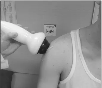
Fig. 1. Extracorporeal shock wave therapy.
회화 침착물의 자발적인 용해로 증상이 자연 호전된다고 하나
7), 석회침착의 용해 기간이 길며 통증을 유발하기 때문에 환자의 일상 생활에 불편을 초래한다.
석회화 건염의 치료는 질환의 자연경과를 앞당기거나 증상 을 완화시키는 목적으로 이루어지며 현재 다양한 방법을 이용 한 보존적 치료를 시행하고 있다. 비스테로이드성 소염제의 경구 투여나 견봉하 간격으로 스테로이드 주사, 주사침을 이용 한 석회 침착 제거
8), 수동적 견관절 관절 운동을 포함하는 물리치료 등의 방법이 있으나
9), 그 효과에 한계가 있는 것으로 보고되고 있다
10-12). 보존적 치료에 반응하지 않는 환자에서는 개방적 술식 또는 관절경을 이용한 수술적 제거술이 증상 호전에 도움이 되기도 한다
2,13).
본 연구는 견관절 석회화 건염 환자에 대한 체외 충격파 치료의 임상적, 방사선학적 효과에 대해 알아보고자 한다.
연구 방법
1. 환자 선정
견관절의 석회화 건염 주소로 본원 외래를 내원한 환자 중 2013년 9월부터 2014년 5월까지 체외 충격파 치료 또는 약물 치료를 받은 30명을 후향적으로 분석하였다. 연구 대상으 로 선정된 환자들은 체외 충격파 치료를 시행한 제 1군과 약물 치료를 시행한 제 2군으로 분류하였다. 제1군은 15명(남자 3명, 여자 12명)으로 42세에서 58세까지의 연령분포로 평균연령은 48.1세였다. 제2군은 15명(남자 3명, 여자 12명)으로 43세에서 59세까지 연령분포를 보이고, 평균연령은 49.1세였다. 최소 3개월 이상 1년 이내의 이환 기간을 갖는 환자를 대상으로 하였으며, 제1군은 6.9개월(범위, 3–12개월), 제2군은 7.5개월 (범위, 3–12개월)이었다(Table 1).
2. 치료 방법
체외 충격파 치료는 6회에 걸쳐 진행되었다. 충격파는 0.03
mJ/mm
2의 강도로 분당 120회, 총 1,200회 시행하였다. 1주 간격으로 3회 치료를 시행한 뒤 2주간의 휴지기를 두고 다시 3회의 치료를 시행하였다. 휴지기는 환자의 치료 준수(com- pliance)를 높이고, 회전근개 부위의 손상을 막기 위해 2주간 시행하였다. 체외 충격파는 동일한 시술자에 의해 시행되었고, 석회성 건염의 위치는 체외 충격파 조사 전 초음파를 통하여 확인하였다 . 모든 환자는 앉은 자세에서 체외 충격파를 시행받 도록 하였다(Fig. 1). 약물치료는 비스테로이드성 소염제 (aceclofenac)를 하루 2회씩 2개월간 투여하였고, 위장관계 부 작용 예방을 위하여 H2 차단제(H2-blocker)를 같이 투여하였다.
3. 치료 후 분석 방법
견관절의 기능 회복을 평가하기 위해 두 군의 환자들에게
치료 전과 치료 후 6개월 뒤 Constant and Murley Scale (CMS)을
계산하였고 동통의 정도를 파악하기 위해 pain visual analog
scale (VAS)를 측정하였다. CMS는 견관절의 기능을 평가하는
표준화된 임상 방법으로 총 100점 만점으로 통증 정도, 일상
Table 2. Clinical outcomes between ESWT therapy and conservative treatment
Outcome
Group 1 (n=15) (ESWT therapy)
Group 2 (n=15)
(conservative treatment) p-value
Preop Last F/U Preop Last F/U
CMS score 26 72 (47−92) 29 45 (23−78) <0.05
VAS score 8.8 2.3 8.3 5.2 <0.05
ESWT: extracorporeal shock wave therapy, Preop: preoperative, F/U: follow up, CMS: Constant and Murley Scale, VAS:
visual analog scale.
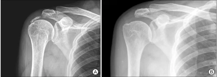
Fig. 2. (A) Initial plain X-ray of calcific tendinitis patient. (B) After 6 months, follow-up X-ray of extracorporeal shock ave therapy therapy patient for calcific tendinitis.
생활 수행능력의 주관적 요소(35점)과 운동범위, 근력의 객관 적 요소(65점)으로 구성된다. VAS는 동통을 주관적인 평가에 기초하여 나타내며 통증이 없는 경우를 0점으로 하고 극심한 통증을 10점으로 정하였고, 치료 전과 치료 이후 6개월에 조사 하였다. 방사선학적 치료 평가를 위하여 치료 전과 치료 후 3개월 및 6개월에 단순방사선촬영을 시행하여 석회화 결절의 크기를 확인하였다. 통계적 검증은 SPSS ver.17.0 (SPSS Inc., Chicago, IL, USA)를 이용하였다. Demographic factor는 Mann- Whitney test로, 치료 전 후 결과는 paired t-test를 이용하여 p-value<0.05인 변수는 유의한 차이가 있는 것으로 분석하였다.
결 과
CMS 점수는 체외 충격파 치료를 시행한 제1군에서 치료 전 평균 26점에서 치료 후 평균 72점(범위, 47–92점)으로 증가 하였고 약물 치료를 시행한 제 2군에서는 치료 전 평균 29점에 서 치료 후 평균 45점(범위, 23–78점)으로 호전되어 체외충격 파 치료를 시행한 경우 더 월등한 견관절 기능의 회복을 보였
다 . VAS 점수도 두 군에서 모두 감소하였으나 제1군에서 치료 전 평균 8.8점(범위, 6–10점)에서 치료 후 평균 2.3점(범위, 0–5점)으로 감소하였고 제2군에서는 치료 전 평균 8.3점(범위, 5–10점)에서 치료 후 평균 5.2점(범위, 2–10점)으로 체외 충격 파 치료 시행 시 더 나은 통증의 감소를 보였다. 이는 모두 통계학적으로 유의하였다(Table 2).
방사선학적 평가에서 제2군에서는 총 15명의 환자 중 약물 치료 3개월 뒤 3명, 6개월 뒤 7명이 방사선 촬영에서 석회화 결절의 감소가 관찰된 것에 비해 제1군에서는 총 15명 중 체외 충격파 치료 3개월 뒤 7명, 6개월 뒤 13명에서 석회화 결절의 감소가 관찰되었다. 체외 충격파 치료 시 방사선학적 석회화 결절의 감소도 더 뚜렷한 것으로 나타났다(Fig. 2).
체외충격파 치료 이후 건 파열이나 골 부종과 같은 심각한
임상적 부작용은 없었다.
고 찰