427 Copyright © 2012 The Korean Society of Cardiology
Korean Circulation Journal
Introduction
Coronary artery spasm is usually a transient mechanism for epi- sodes of variant angina, involving single epicardial coronary artery in most cases. However, sometimes coronary artery spasm occurs in more than one artery and it may last much longer than usual an- gina. Serious cardiac conditions, such as myocardial infarction or malignant arrhythmias, may ensue in proportion to the extent and du- ration of ischemia. Prolonged coronary artery spasm as an under- lying cause for acute myocardial infarction has been recognized
1-7)
since the possibility was first suggested nearly 50 years ago.
8) How- ever, global ischemia induced by simultaneous multiple coronary artery spasm, with clinical presentation of cardiogenic shock, has been rarely reported. Only recently was such a case published for the
Case Report
http://dx.doi.org/10.4070/kcj.2012.42.6.427
Print ISSN 1738-5520 • On-line ISSN 1738-5555
Cardiogenic Shock From Global Myocardial Ischemia Induced by Simultaneous Multivessel Coronary Spasm
Jihye Ahn, MD, Bo-sung Kim, MD, Hyekyong Park, MD, Kyungil Park, MD, and Young Dae Kim, MD
Department of Internal Medicine, Dong-A University School of Medicine, Busan, Korea
Coronary artery spasm is an uncommon, but well recognized, etiology for acute myocardial infarction. However, cardiogenic shock with myocardial infarction resulting from simultaneous multiple coronary artery spasm has been rarely reported, and not in Korea. Recently, we experienced such a case in a 50-year-old Korean man without previous diagnosis of variant angina. The patient, hospitalized for blood sug- ar control, developed severe chest pain accompanying ST-segment elevation in multiple leads. The patient immediately received cardiac catheterization because of cardiogenic shock. Coronary angiogram revealed the severe and simultaneous spasm of three major epicardial ar- teries, which was promptly relieved by an intracoronary administration of isosorbide dinitrate. This case highlights the need to rule out the potential mechanism of coronary spasm even in the most severe episodes of acute coronary syndrome. (Korean Circ J 2012;42:427-430) KEY WORDS: Shock, cardiogenic; Myocardial infarction; Coronary vasospasm.
Received: September 6, 2011 Revision Received: October 25, 2011 Accepted: November 4, 2011
Correspondence: Young Dae Kim, MD, Department of Internal Medicine, Dong-A University School of Medicine, 26 Daesingongwon-ro, Seo-gu, Busan 602-715, Korea
Tel: 82-51-240-5020, Fax: 82-51-248-1510 E-mail: [email protected]
• The authors have no financial conflicts of interest.
This is an Open Access article distributed under the terms of the Creative Commons Attribution Non-Commercial License (http://creativecommons.
org/licenses/by-nc/3.0) which permits unrestricted non-commercial use, distribution, and reproduction in any medium, provided the original work is properly cited.
first time in the English-language literature.
9) To our knowledge, a case has never been reported in Korea.
Case
A 50-year-old male was admitted to our hospital for optimal con- trol of blood sugar. The patient had been diabetic for 1 year and was being treated with an oral hypoglycemic agent. A recent blood sugar profile indicated a poorly controlled state, with glycated hemoglo- bin of 9.6%. The patient had a 30-year smoking history. In the previ- ous month, the patient had been hospitalized via the emergency de- partment to evaluate acute chest pain and minimally elevated serum cardiac troponin I. Coronary angiography conducted under the im- pression of unstable angina revealed an intermediate lesion at mid- left anterior descending artery (LAD). The patient was treated with medical therapy. On discharge, the patient received a beta-adrener- gic blocker and isosorbide dinitrate, but ceased taking the nitrate se- veral days later because of severe headache.
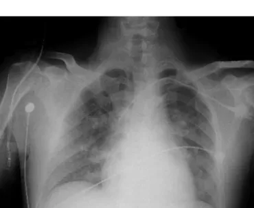
On the 5th hospital day in the morning, the patient developed ex- cruciating chest pain followed by drop of systolic blood pressure to 70 mm Hg. Heart rate was 69 beats/min, and breathing rate was 28/
min with 98% oxygen saturation on nasal oxygen (3 L/min). The pa-
tient was acutely ill looking, with a pale expression and profuse per-
spiration. Physical examination revealed fine rales on the bilateral
lung field. However, cardiac murmur, friction rub, hepatomegaly, and
peripheral edema were not present. An electrocardiogram showed