Acute respiratory viral infections are a major cause of morbidity in pediatric pa- tients.1Although they are most often self-limited and confined to the upper res- piratory tract, they lead to a substantial number of upper or lower respiratory tract complications. Therefore, accurate and rapid diagnosis of respiratory virus infection is essential for the initiation of early treatment and the prevention of viral spread.
Several studies evaluating polymerase chain reaction (PCR)-based methods for the detection and typing of respiratory viruses were reported.2,3The adenovirus
Identification of Adenovirus, Influenza Virus, Parainfluenza Virus, and Respiratory Syncytial Virus by Two Kinds of Multiplex Polymerase Chain Reaction (PCR) and a Shell
Vial Culture in Pediatric Patients with Viral Pneumonia
Jong-Han Lee,
1Jin-Kyong Chun,
2Dong Soo Kim,
2Yongjung Park,
1Jong Rak Choi,
1and Hyon-Suk Kim
1Departments of 1Laboratory Medicine and 2Pediatrics, Yonsei University College of Medicine, Seoul, Korea.
Purpose:Early identification of causative agents in lower respiratory infection of pediatric patients can reduce morbidity and prevent an overuse of antimicrobials.
Two kinds of multiplex polymerase chain reaction (PCR) and a commercial shell vial viral culture were performed to identify causative agents in pediatric patients.
Materials and Methods:Nasopharyngeal aspirates of 220 children diagnosed with viral pneumonia were obtained. Two kinds of multiplex PCR (SeeplexTMRV detection kit, and LabopassTMRV detection kit), and a shell vial culture by R-Mix were performed. Results: Positive samples from 220 total samples by two multiplex PCRs were 52.7% and 46.4%, respectively. We also cultured 103 sam- ples that showed positive results of the adenovirus, influenza virus, parainfluenza virus, and respiratory syncytial virus (RSV) by two multiplex PCR. The RSV was most frequently detected in 53.0% (Seeplex) and 51.7% (Labopass) of patients.
The detection rate of adenovirus (AdV) was 10.3% and 12.1%, influenza virus (IFV) A and B was 12.5% and 3.4%, and parainfluenza virus (PIFV) 1, 2, and 3 were 2.9% and 2.6%. Shell vial cultures showed concordant results with each multiplex PCR by 96.1% and 77.7%, respectively. Sequencing results were 90%
consistent with multiplex PCR. Conclusion:Multiplex PCR showed more posi- tivity than the shell vial culture and it can be an effective primary test. Other com- plementary efforts such as viral cultures and sequencing analysis could be considered, according to clinical and laboratory conditions.
Key Words: Multiplex PCR, respiratory virus, virus culture, viral pneumonia
Received: September 22, 2009 Revised: December 3, 2009 Accepted: December 24, 2009
Corresponding author: Dr. Hyon-Suk Kim, Department of Laboratory Medicine, Yonsei University College of Medicine, 250 Seongsan-ro, Seodaemun-gu, Seoul 120-752, Korea.
Tel: 82-2-2228-2443, Fax: 82-2-364-1583 E-mail: [email protected]
∙The authors have no financial conflicts of interest.
© Copyright:
Yonsei University College of Medicine 2010 This is an Open Access article distributed under the terms of the Creative Commons Attribution Non- Commercial License (http://creativecommons.org/
licenses/by-nc/3.0) which permits unrestricted non- commercial use, distribution, and reproduction in any medium, provided the original work is properly cited.
INTRODUCTION
(AdV), influenza virus (IFV), parainfluenza virus (PIFV), and respiratory syncytial virus (RSV) have been identified as significant pathogens in community-acquired and noso- comial respiratory infections.4,5Recently, some respiratory viral infections caused by IFV, enterovirus (EnV), or AdV may now benefit from specific antiviral treatment.6-8
The aim of this study is to evaluate and suggest clinical usefulness of two kinds of multiplex PCR for identifying causative viruses in pediatric viral pneumonia, which is the most severe lower respiratory infection in children.
Sample collection
Nasopharyngeal aspirates from 220 pediatric patients during a period of 6 months were obtained by a mucus extractor (Sewoon Inno-Vision Medical, Seoul, Korea) and transferred to each vial of the universal transport medium (Diagnostic Hybrids, Inc., Athens, OH, USA). All 220 pediatric patients were admitted with symptoms of severe lower respiratory infection. For definitions of cases, moderate to severe lower respiratory infections were deter- mined according to the World Health Organization (WHO) recommended surveillance standards, 2nd edition (1999).
Lower respiratory viral infections were diagnosed by an expert pediatrician through physical examinations, chest X-rays, blood tests, erythrocyte sedimentation rate (ESR), and C-reactive protein (CRP), excluding bacterial origin.
The 220 aspiration samples were immediately made aliquot and stored at -75˚C until nucleic acid extraction, viral culture, or sequencing analysis proceeded. All patients’ sam- ples were collected according to the protocols of the Institu- tional Review Board of Yonsei University Health System.
Nucleic acid extraction
QIAamp Viral RNA Mini Kit (QIAGEN, Hilden, Ger- many) with automated QIAcube®(QIAGEN) was used to extract nucleic acid. The 220 nucleic acid extracts were kept in a deep freezer at - 75˚C until analysis.
Multiplex PCR for detection of respiratory virus
SeeplexTMRV detection kit
(http://www.seegene.co.kr/en/index.php)
Nucleic acids extracted from nasopharyngeal aspirates were used for the synthesis of first-strand cDNAs by Mol- oney murine leukemia virus reverse transcriptase (Prome- ga, Madison, WI, USA). The SeeplexTMRV detection kit (Cosmo Genetech, Seoul, Korea) contained A and B sets of primers designed by highly conserved regions of genetic sequences for the 12 respiratory viruses. The SeeplexTMkit
is designed to identify AdV, human metapneumovirus (hMPV), Human coronavirus (HCoV) 229E/NL63, parain- fluenza virus (PIFV) 1, PIFV 2, and PIFV 3, and the Seep- lexTMRV detection kit B is designed to detect IFV A, IFV B, RSV A, RSV B, rhinovirus (RhV), and HCoV OC43/
HKU1. Each PCR was conducted in a final reaction volume of 20 µL containing 3 µL of cDNA, 3 µL of 8-methoxy- psoralen (MOP) solution, 4 µL of 5×RV Primer, and 10 µL of 2×master mix. The PCR protocol was 94˚C for 30 sec, followed by 35 cycles of 60˚C for 1.5 min, and 72˚C for 1.5 min, followed by a 10 min final extension at 72˚C.
The amplified products were separated on a 2% agarose gel stained with ethidium bromide. Each run included a molecular size marker and internal control. Also, Ameri- can Type Culture Collection (ATCC) standard viruses were used for positive control and 10 µL distilled water as negative control.
LabopassTMRV detection kit
(http://www.cosmo4.com/index_eng.html )
Nucleic acids from nasopharyngeal aspirates were also used for the LabopassTM kit. This kit was also designed to detect 12 types of viruses. The final reaction volume was 50 µL composed of 40 µL premixture and 10 µL nucleic acid. Two kinds of PCR protocols were used. AdV and human bocavirus (HboV) operated at 95˚C for 3 min, then 35 cycles of 95˚C for 1 min, 55˚C for 1 min, and 72˚C for 1 min, followed by a 5 min final extension at 72˚C and preserved at 4˚C. HCoV, EnV, PIFV, RhV, and RSV all operated at 42˚C for 60 min, 95˚C for 3 min, then 35 cycles of 95˚C for 1 min, 55˚C for 1 min, and 72˚C for 1 min, followed by a 5 min final extension at 72˚C and preserved at 4˚C. The PCR products were analyzed by identifying bands with a 2% agarose gel stained with ethidium bromide.
Virus culture by R-Mix ReadyCells with antigen staining Samples showing any positive results for AdV, IFV, PIFV, and RSV from the two multiplex PCR kits were performed with shell vial cultures by R-Mix ReadyCells (Diagnostic HYBRIDS, Inc., Athens, OH, USA) according to manu- facturer’s procedures. Firstly, the cryopreserved R-Mix cell reagents were heated in a 37˚C heat block for 4 min, and rinsed with a rinse buffer and then remained for 4 min at room temperature. In the meantime, the re-feed medium and 1.0 mL R-Mix ReadyCells were mixed. Then, 200 µL specimens were inoculated into R-Mix ReadyCells and centrifuged at 700×g for 60 min at room temperature.
After overnight incubation at 35˚C in the incubator, cell monolayers of the shell vial were washed and fixed with acetone, and stained with respiratory virus fluorescent antibodies by D3 DFA (Diagnostic Hybrids, Inc., Athens, OH, USA). If virus-specific fluorescence was noted by
MATERIALS AND METHODS
screening, virus identification was performed using indi- vidual monoclonal antibodies staining (IFV A and B, AdV, PIFV 1,2,3 and RSV). When the initial screening was negative, the vial was re-examined on day 3 and day 5.
Sequencing analysis
Ten samples, which were culture negative but multiplex PCR positive, were proceeded with sequencing analysis to identify these equivocal results. All sequencing analyses were proceeded after repeating PCR then proper primers were prepared for specific viruses identification. An auto- mated sequencing analyzer (Applied Biosystems, Foster City, CA, USA) accompanied with its recommended reagent was used according to the manufacturer’s protocol. Finally, results of all sequences were analyzed by matching those of GenBank data using Basic Local Alignment Search Tool (BLAST) (http://blast.ncbi.nlm.nih.gov/Blast.cgi).
Nucleic acid amplification results by 2 kinds of multiplex PCR
The basic characteristics and major results of 220 pediatric patients are summarized in Table 1. The positive rate of the SeeplexTMRV detection kit was 52.7% (116 positive samples/total 220 samples). SeeplexTMidentified 136 viruses which were the sum of AdV (n = 14, 10.3%), HCoV 229E/NL63 (n = 5, 3.7%), HCoV OC43/HKU1 (n = 4, 2.9%), IFV A (n = 8, 5.9%), IFV B (n = 9, 6.6%), HMPV (n = 2, 1.5%), PIFV 1 (n = 0, 0%), PIFV 2 (n = 0, 0%), PIFV 3 (n = 4, 2.9%), RhV A/B (n = 18, 13.2%), RSV A (n
= 39, 28.7%), and RSV B (n = 33, 24.3%).
The LabopassTMRV detection kit showed 46.4% posi- tivity (102 positive samples/total 220 samples). The Labo- passTMRV detection kit identified 116 viruses which were the sum of AdV (n = 14, 12.1%), HBoV (n = 4, 3.4%), HCoV NL63 (n = 1, 0.9%), HCoV OC43 (n = 2, 1.7%), EV (n = 0, 0%), IFV A (n = 2, 1.7%), IFV B (n = 2, 1.7%), HMPV (n = 22, 19.0%), PIFV 1 (n = 2, 1.7%), PIFV 2 (n = 0, 0%), PIFV 3 (n = 1, 0.9%), RhV A/B (n = 6, 5.2%), and RSV (n = 60, 51.7%). The coinfection (more than 2 virus species identified) rate of the SeeplexTMRV kit was 6.8%
(15/220) and of the LabopassTMRV kit was 5.9% (13/220).
Shell vial cultures with direct immunostaining for identifying AdV, IFV, PIFV, and RSV
If one or more of the viruses were identified from a multi- plex PCR, R-Mix ReadyCell cultures were proceeded with direct immunofluorescence staining. There were 103 samples that showed a positive multiplex PCR of AdV, IFV, PIFV, and RSV. The shell vial culture assay showed 93 positive samples of the total 103 samples (positive rate 90.3%). There were two samples which showed 2 kinds of viruses. A total of 95 positive numbers of the R-Mix culture was noted from 15 AdV, 6 IFV A, 9 IFV B, 2 PIFV, and 63 RSV. Positive viral culture rate from the SeeplexTMRV de- tection kit was 96.1% (99 samples of the total 103 samples) and that of the LabopassTMRV detection kit was 77.7% (80 samples of the total 103 samples).
Fifty samples of the 103 total samples showed the same results by the two kinds of multiplex PCR and viral culture.
However, there were 53 samples which showed discrepant results by each test. The 53 results were divided into 5 groups according to discrepant characters (Table 2). The R-Mix culture which we used in this study targeted only four viruses (AdV, IFV, PIFV, and RSV), and so other viruses could not be isolated. The positive results of multi- plex PCR and R-Mix culture for detecting AdV, IFV, PIFV, and RSV are compared in Fig. 1.
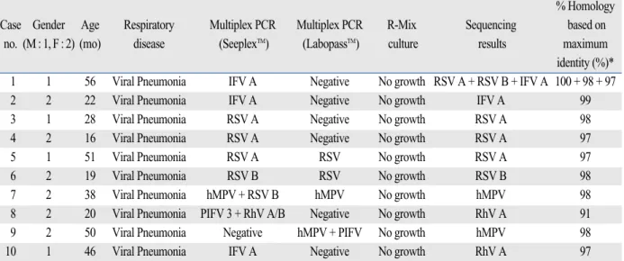
Sequencing results of multiplex PCR positive but viral culture negative samples
Ten samples of culture negative but any multiplex PCR positive were further analyzed by sequencing. The results are showed in Table 3. The corresponding viruses were identified with a homology of 91-100%. Of the ten sequenc- ing results, nine were consistent with multiplex PCR results.
There was one completely discrepant result between multiplex PCR and sequencing analysis (Case No. 10: IFV A versus RhV, Table 3).
Rapid and accurate identification of causative agents in viral
RESULTS
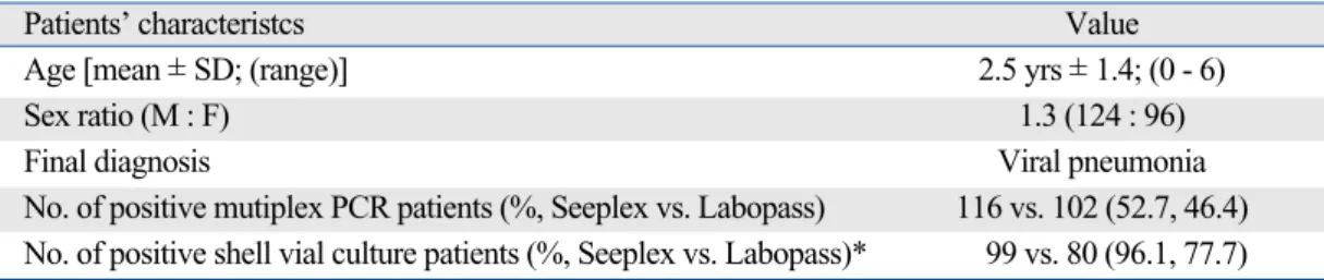
Table 1. Basic Characteristics of 220 Pediatric Patients
Patients’ characteristcs Value
Age [mean ± SD; (range)] 2.5 yrs ± 1.4; (0 - 6)
Sex ratio (M : F) 1.3 (124 : 96)
Final diagnosis Viral pneumonia
No. of positive mutiplex PCR patients (%, Seeplex vs. Labopass) 116 vs. 102 (52.7, 46.4) No. of positive shell vial culture patients (%, Seeplex vs. Labopass)* 99 vs. 80 (96.1, 77.7)
*Total 103 samples were proceeded shell vial cultures.
DISCUSSION
pneumonia of pediatric patients is essential to prevent disease propagation. It is also helpful for early initiation of proper management. An early start of proper anti-viral management can reduce unnecessary overuse of antibac- terial agents. In addition, some respiratory viruses, including IFV, EnV, and AdV, may now benefit from specific antivi-
ral treatment.6-8So, we designed and performed this study to evaluate two kinds of multiplex PCR kits and a commercial shell vial culture method for their clinical efficacy with prospectively collected samples of pediatric patients.
Until now, virus isolation by a cell culture and a direct immunofluorescent antigen staining assay has been the Table 2. Comparison Results of Multiplex PCR Positive Cases
Multiplex PCR Multiplex PCR
R-Mix culture Total no.
(SeeplexTM) (LabopassTM) (103 cases)
Consistent (50) AdV AdV AdV 10
IFV B IFV B IFV B 2
RSV A RSV RSV 38
Inconsistent (36) AdV AdV + IFV A + RhV AdV + IFV A 1
AdV hMPV + PIFV 3 PIFV 3 1
AdV Negative AdV 1
IFV A Negative IFV A 4
IFV A Negative IFV B 1
IFV A Negative No growth 1
IFV A Negative No growth 1
IFV A Negative No growth 1
IFV B Negative IFV B 5
IFV B + RSV A Negative RSV + IFV B 1
IFV B + RSV A RSV RSV 1
hMPV + RSV B hMPV No growth 1
Negative IFV A IFV A 1
Negative MPV + PIFV 1 No growth 1
Negative RSV RSV A + RSV B 2
PIFV 3 AdV AdV 1
PIFV 3 + RhV A/B Negative No growth 1
PIFV 3 + RhV A/B PIFV 1 PIFV 3 1
PIFV 3 + RhV A/B + RSV B AdV AdV 1
RSV A Negative No growth 2
RSV A Negative RSV 4
RSV A RSV No growth 1
RSV A + RSV B Negative RSV 1
RSV B RSV No growth 1
Indeterminate (17) AdV + HCoV OC43 AdV + HCoV OC43 AdV 1
HCoV 229E/NL63 + RSV B RSV RSV 3
HCoV OC43 + RSV A RSV RSV 1
HCoV OC43 + RSV B RSV RSV 1
HCoV 229E/NL63 + RSV A RSV RSV 1
RhV A/B + RSV A RSV RSV 2
RSV A hMPV + RSV RSV 3
RSV A + RSV B RSV + HBoV RSV 1
RSV B hMPV + RSV RSV 2
RSV B RSV + hMPV RSV 2
PCR, polymerase chain reaction; AdV, adenovirus; HCoV, human coronavirus; IFV, influenza virus; PIFV, parainfluenza virus; hMPV, human metapneumovirus; RhV, rhinovirus; RSV, respiratory syncytial virus; HBoV, human bocavirus.
*R-Mix culture could only detect AdV, IFV, PIFV and RSV in this study.
most commonly used method for identifying respiratory viruses.9An enzyme immunoassay may be quicker but it is less sensitive. These conventional methods may be affect- ed by specimen quality, virus type, and technical skill.10-12 The virus culture is still considered the gold standard for respiratory virus detection, but it has limitations in turnaro- und time, specimen transport, and storage conditions in maintaining the infectivity of the virus.11,13To compare the test methods, Choi, et al.14reported that the positive rate of the direct antigen test, viral culture method, and multiplex PCR for detecting AdV, IFV, PIFV, and RSV was 28.4%, 36.2%, and 44.8%, respectively.
Recently, virus identification by immunostaing after shell vial culture is the most widely accepted laboratory standard method of virus testing. But, this method is still not easy for routine testing in clinical laboratories. The nucleic acid amplification method including multiplex PCR is a powerful alternative, but it has some limitations of false positivity and false negativity. The nucleic acid amplification test is faster than the culture method and has
been reported to be more sensitive9,15-17 and can be auto- mated these days. In addition, there are advantages in detecting some viruses which grow poorly in cell cultures such as HMPV.18
A Korean national survey19of respiratory virus testing was performed and thirty-one clinical laboratories respond- ed that they provided respiratory virus testing for clinical diagnosis in Korea. Among the responders, PCR and/or culture were most widely adopted, in 42% of the institutes, rapid immunochromatographic method 29%, immunofluo- rescent antigen assay 23%, and enzyme immunoassay 7%.
We focused on the four major respiratory viruses of AdV, IFV, PIFV, and RSV. The exact concordance rate between the two kinds of multiplex PCR and R-Mix viral culture was 48.5% (50/103). Our results of the two multi- plex PCR showed an equivalent or higher positive rate than in other studies.9,10,14Two kinds of multiplex PCR reagents showed different positivity according to the virus type. The positive rate of the SeeplexTMRV detection kit was higher than the LabopassTMRV kit for IFV and RhV. However, the
Fig. 1. Comparison of multiplex PCRs and R-Mix culture for detecting adenovirus (AdV), influenza virus (IFV), parainfluenza virus (PIFV) and respiratory syncytial virus (RSV) in pediatric patients with viral pneumonia.
Table 3. Sequencing Confirmations of 10 Samples that Showed Multiplex PCR Positive and Culture Negative Results
% Homology Case Gender Age Respiratory Multiplex PCR Multiplex PCR R-Mix Sequencing based on
no. (M : 1, F : 2) (mo) disease (SeeplexTM) (LabopassTM) culture results maximum identity (%)*
1 1 56 Viral Pneumonia IFV A Negative No growth RSV A + RSV B + IFV A 100 + 98 + 97
2 2 22 Viral Pneumonia IFV A Negative No growth IFV A 99
3 1 28 Viral Pneumonia RSV A Negative No growth RSV A 98
4 2 16 Viral Pneumonia RSV A Negative No growth RSV A 97
5 1 51 Viral Pneumonia RSV A RSV No growth RSV A 97
6 2 19 Viral Pneumonia RSV B RSV No growth RSV B 98
7 2 38 Viral Pneumonia hMPV + RSV B hMPV No growth hMPV 98
8 2 20 Viral Pneumonia PIFV 3 + RhV A/B Negative No growth RhV A 91
9 2 50 Viral Pneumonia Negative hMPV + PIFV No growth hMPV 98
10 1 46 Viral Pneumonia IFV A Negative No growth RhV A 97
PCR, polymerase chain reaction; IFV, influenza virus; PIFV, parainfluenza virus; hMPV, human metapneumovirus; RhV, rhinovirus; RSV, respiratory syncytial virus.
*Respiratory viruses were confirmed by the Basic Local Alignment Search Tool (BLAST) program.
LabopassTMRV kit detected more HMPV than the Seepl- exTMRV kit.
Discrepant results between these two types of multiplex PCR and R-Mix culture comprised 51.5% (53/103) of the total. Weinberg, et al.17reported that the PCR method could detect viruses two times more, compared with virus culture.
Our study showed that the concordance rate of Seeplex PCR to the virus culture was 83% (90/103), and that of Labopass PCR, 80.6% (83/103). These results were similar to previous reports, of 83.2% (556/668)17and 80% (40/50).20
The discrepancy might be caused from multiplex PCR limitations. One of the major limitations of PCR detection is false-negative results as a consequence of PCR inhibitors present in clinical samples that are not removed by the extraction process.9Another limitation is the principle of PCRs, which could produce false results if a primer region has nucleotide variation and is unable to detect new types or strains of a virus. This is the reason that direct antigen tests or virus cultures cannot be completely substituted by solitary multiplex PCR tests until now.21
We noticed a 6.8% (15/220) coinfection rate (more than 2 virus types identified) by the SeeplexTMRV kit and 5.9%
(13/220) by the LabopassTMRV kit. Previous studies have suggested that double-virus infections are associated with greater severity of respiratory tract infection.22In our study, we could not find out any remarkable parameters or differ- ence of clinical severity in cases of coinfection.
Ten cases of culture negative but multiplex PCR posi- tive were further evaluated by direct sequencing in our study. Sequencing results confirmed almost all the original results of multiplex PCR.
In conclusion, identification of respiratory viruses by multiplex PCR can be more rapid, an easier method, and show more positive results than the viral culture method.
Hence, multiplex PCR can be the first choice for detection of respiratory viruses in a clinical laboratory. Other comple- mentary efforts such as viral cultures and sequencing methods could be selectively proceeded in selected cases according to each laboratory’s environment.
1. Regamey N, Kaiser L, Roiha HL, Deffernez C, Kuehni CE, Latzin P, et al. Viral etiology of acute respiratory infections with cough in infancy: a community-based birth cohort study. Pediatr Infect Dis J 2008;27:100-5.
2. Eugene-Ruellan G, Freymuth F, Bahloul C, Badrane H, Vabret A, Tordo N. Detection of respiratory syncytial virus A and B and parainfluenzavirus 3 sequences in respiratory tracts of infants by a single PCR with primers targeted to the L-polymerase gene and differential hybridization. J Clin Microbiol 1998;36:796-801.
3. Gilbert LL, Dakhama A, Bone BM, Thomas EE, Hegele RG.
Diagnosis of viral respiratory tract infections in children by using a reverse transcription-PCR panel. J Clin Microbiol 1996;34:140-3.
4. Ljungman P, Ward KN, Crooks BN, Parker A, Martino R, Shaw PJ, et al. Respiratory virus infections after stem cell transplan- tation: a prospective study from the Infectious Diseases Working Party of the European Group for Blood and Marrow Transplan- tation. Bone Marrow Transplant 2001;28:479-84.
5. Whimbey E, Englund JA, Couch RB. Community respiratory virus infections in immunocompromised patients with cancer.
Am J Med 1997;102:10-8.
6. Cooper NJ, Sutton AJ, Abrams KR, Wailoo A, Turner D, Nicho- lson KG. Effectiveness of neuraminidase inhibitors in treatment and prevention of influenza A and B: systematic review and meta- analyses of randomised controlled trials. BMJ 2003;326:1235.
7. De Clercq E. Clinical potential of the acyclic nucleoside phospho- nates cidofovir, adefovir, and tenofovir in treatment of DNA virus and retrovirus infections. Clin Microbiol Rev 2003;16:569-96.
8. Pasquinelli L. Enterovirus infections. Pediatr Rev 2006;27:e14-5.
9. Syrmis MW, Whiley DM, Thomas M, Mackay IM, Williamson J, Siebert DJ, et al. A sensitive, specific, and cost-effective multi- plex reverse transcriptase-PCR assay for the detection of seven common respiratory viruses in respiratory samples. J Mol Diagn 2004;6:125-31.
10. Gröndahl B, Puppe W, Hoppe A, Kühne I, Weigl JA, Schmitt HJ.
Rapid identification of nine microorganisms causing acute respi- ratory tract infections by single-tube multiplex reverse transcrip- tion-PCR: feasibility study. J Clin Microbiol 1999;37:1-7.
11. Kehl SC, Henrickson KJ, Hua W, Fan J. Evaluation of the Hexa- plex assay for detection of respiratory viruses in children. J Clin Microbiol 2001;39:1696-701.
12. Liolios L, Jenney A, Spelman D, Kotsimbos T, Catton M, Wes- selingh S. Comparison of a multiplex reverse transcription-PCR- enzyme hybridization assay with conventional viral culture and immunofluorescence techniques for the detection of seven viral respiratory pathogens. J Clin Microbiol 2001;39:2779-83.
13. Fan J, Henrickson KJ, Savatski LL. Rapid simultaneous diagno- sis of infections with respiratory syncytial viruses A and B, influ- enza viruses A and B, and human parainfluenza virus types 1, 2, and 3 by multiplex quantitative reverse transcription-polymerase chain reaction-enzyme hybridization assay (Hexaplex). Clin Infect Dis 1998;26:1397-402.
14. Choi EH, Lee HJ, Kim SJ, Eun BW, Kim NH, Lee JA, et al. The association of newly identified respiratory viruses with lower respiratory tract infections in Korean children, 2000-2005. Clin Infect Dis 2006;43:585-92.
15. van Elden LJ, van Kraaij MG, Nijhuis M, Hendriksen KA, Dekker AW, Rozenberg-Arska M, et al. Polymerase chain reac- tion is more sensitive than viral culture and antigen testing for the detection of respiratory viruses in adults with hematological cancer and pneumonia. Clin Infect Dis 2002;34:177-83.
16. Weinberg A, Zamora MR, Li S, Torres F, Hodges TN. The value of polymerase chain reaction for the diagnosis of viral respiratory tract infections in lung transplant recipients. J Clin Virol 2002;25:
171-5.
17. Weinberg GA, Erdman DD, Edwards KM, Hall CB, Walker FJ, Griffin MR, et al. Superiority of reverse-transcription polymerase chain reaction to conventional viral culture in the diagnosis of acute respiratory tract infections in children. J Infect Dis 2004;189:706-10.
18. van den Hoogen BG, Osterhaus DM, Fouchier RA. Clinical impact and diagnosis of human metapneumovirus infection.
REFERENCES
Pediatr Infect Dis J 2004;23:S25-32.
19. Kang J, Kim E, Lee K, Lee N, Lee C. Surveillance for respiratory virus testing situation in Korea and epidemiology for the respira- tory viruses detected in 5 university hospitals. Korean J Clin Microbiol 2007;10:102-8.
20. Roh KH, Kim J, Nam MH, Yoon S, Lee CK, Lee K, et al. Com- parison of the Seeplex reverse transcription PCR assay with the R-mix viral culture and immunofluorescence techniques for detec-
tion of eight respiratory viruses. Ann Clin Lab Sci 2008;38:41-6.
21. Leland DS, Ginocchio CC. Role of cell culture for virus detection in the age of technology. Clin Microbiol Rev 2007;20:49-78.
22. Templeton KE, Scheltinga SA, van den Eeden WC, Graffelman AW, van den Broek PJ, Claas EC. Improved diagnosis of the etiol- ogy of community-acquired pneumonia with real-time polymerase chain reaction. Clin Infect Dis 2005;41:345-51.