약학회 지 제 48 권 제 1 호 93-98 (2004)
Yakhak Hoeji Vol. 48, No. 1 ■稱學会확
산화적 스트레스상태에서 B16F10 Murine Melanoma 세포의 항산화효소 활성에 대한 훅축추출물의 효과
김 안 근 # • 차은정 숙명여자대학교 약학대학
(Received February 4, 2004; Revised February 20, 2004)
Effect of Pharbitidis Seed Extract on the Antioxidant Enzyme Activity in B16F10 Murine Melanoma Cells by Oxidative Stress
A n K eun K im # and E un Jung Cha College of Pharmacy, Sookmyung Women's University
Abstract —— The purpose of this study was to evaluate the effect of pharbitidis seed extract (PE) on antioxidant enzymes.
The cytotoxicities of PE were measured by 3-(4,5-dimethylthiazol-2-yl)-2,5-diphenyltetrazolium bromide (MTT) assay. The change of superoxide dismutase (SOD), glutathione peroxidase (GPx), and catalase (CAT) activity assay were measured.
The SOD activities by PE-treated groups were lower than control group's one. In the co-treated with hydrogen peroxide (H20 2) group, SOD activity was higher than H 20 2 treated group's activity. In the case of GPx, GPx activities were increased in both PE-treated and co-treated with H 20 2 group. In the case of CAT, H 20 2 treated group's activityies were very increased.
The CAT activities by PE-treated groups were lower than control group's one, but the activity of co-treated group with H 20 2 was higher than that of control group's one. These results suggest that PE has antioxidant activity.
Keywords □ antioxidant enzyme activity, pharbitidis seed, ROS
살아있는 생물체는 산소를 이용하는 정상적인 대사를 통하여 지속적으로 superoxide radical anion(0 2._), hydrogen peroxide (H20 2), hydroxyl radical(OH-) 등과 같은 반웅성이 큰 reactive oxygen species(ROS)를 생성하는데,1} 이러한 ROS들은 UV ozone, 공기오염, 여러 가지 약물 등과 같은 외부 자극에 의해서 도 그 생성이 중가된다고 알려져 있다.2》인체에 존재하는 항산 화 체계는 여러 가지 항산화 물질들이 관여하는 비효소적 방어 체계와 항산화 효소들이 관여하는 효소적 방어체계로 나눌 수 있 다. 항산화 물질로는 ascorbic acid, a-tocopherol, glutathione, p-carotene, vitamine A 등이 있고,3》중요한 항산화 효소로 는 superoxide dismutase(SOD), glutathione peroxidase(GPx), catalase(CAT), glutathione reductase를 들 수 있다. Free oxygen radicals이 생성되면 SOD, CAT, GPx와 같은 항산화 효 소들은 membrane의 lipid peroxidative damage, sulfhydryl-
*본 논문에 판한 문의는 저자께게로 (전화) 02-710-9561, 9566 (팩스) (E-mail) [email protected]
containing enzymes의 inactivation, integral protein의 cross- linking 등에 관련된 oxygen species를 불활성화 시키거나 제거 함으로써 항산화 작용을 하게 된다.4’5> 그러나 이러한 항산화작 용이 제대로 되지 않으면 R 0S 가 체내에 축적되게 되고 생체내 의 고분자 물질인 불포화 지방산, 단백질, DNA 등과 반응하여 다양한 기전을 통해 세포의 노화와 변형을 초래함으로써 여러 가 지 질병과 밀접한 연관을 갖는 것으로 알려져 있다.6^
본 연구는 산화적 스트레스 상태에서의 항산화 효소활성에 대 한 연구의 일환으로 침습성이 강하고 전이가 빠른 악성 흑색종 세포를 이용하였고 H20 2 스트레스 상태에서 혹축(Pharbitidis Semen, pharbitidis seed)추출물 투여로 인한 효소활성 변회를 즉 정하여 검토하고자 하였다. 또한 MTT Assay를 통하여 이 혹축 추출물이 세포의 중식에 미치는 영향을 관찰하였다.
심험방법
재료 및 시약
RPM I 1640 powder medium, fetal bovine serum(FBS),
94 김인^ . 치은정
antibiotics(penicillin G sodium 10,000 units/m/, streptomycin sulfate 10,000 ^ig/rn/), trypsin-EDTA(0.25% trypsin, 1 mM EDTA • 4Na), sodium pyruvate, trypan blue는 Gibco BRL Life Technologies Inc. 제품을 사용하였고,sodium bicarbonate, N-2- hydroxyethyl piperazine-A^-2-ethane sulfonic acid(HEPES), ethylenediaminetetraacetic acid disodium salt(EDTA), 3-[4,5- dimethylthiazol-2-yl]-2,5-diphenyltetra2olium bromide; thiazolyl blue(MTT), dimethyl sulfoxide(DMSO), P-nicotinamide adenine dinucleotide phosphate reduced form(P-NADPH), glutathione reductase, glutathione reduced form, pyrogallol, sodium azide, txis[hydroxy-methyl]aminomethane(Tris), protein assay kite는 Sigma chemical Co. 제품을 사용하였다.
사용기기
Electronic top loading balance(AC211S, Sartorius), digital pH meter(Mettler Delta 340), labo autoclave(Sanyo), ELISA reader(Dynatech MR5000), C 0 2 incubator(New brunswick science), clean bench(한국 삼공), table top centrifuge(Hanil science industrial Co. Ltd.), microcentrifuge(microl7TR, Hanil science industrial Co. Ltd.), vacuum pump(DOA-V 152- AA, Gast), inverted microscope(01ympus CK2), pipet-aid (Barnstead), vortex mixer(Broomall, PA, USA), magnetic stirrer(Corning), sonicator(2210, Bransonic®), deep freezer (OPR-DFU-128C, Operon), UV/visible spectrophotometer (Ultrospec 2000, Pharmacia Biotech)를 사용하여 실험하였다.
세포 배양
실험에 사용한 세포는 mouse melanoma로부터 유래된 B16F10 cell로 한국 세포주 은행 (Korean cell lines bank)으로부 터 분양 받았다. 10% FBS와 항생제(penicillin G 100 sodium 10,000units/m/, streptomycin sulfate 10,000|ig/m/), Im M sodium pyruvate를 포함히는 RPM I 1640 배지를 배양액으로 하 여 37°C, humidified 5% C 02 incubator에서 배양되었다. 배양액 의 pH는 10 mM HEPES와 2 mg/m/ sodium bicarbonate를 사 용히여 7.2ᅳ7.4로 조정하였다. 25 cm2 tissue culture flask나 75 cm2 tissue culture flask에서 계대 배양하고 confluent되었을 때 trypsin-EDTA 용액을 처리하여 실험에 사용하였다.
훅측 추출물의 준비
한약재인훅축은서울경동시장내의 한약건재상에서 건조상 태로구입하여사용하였다. 흑축을각각 100 於1 둥근플라스크 에넣고 80% 메탄올 3/를 넣은후 3시간씩 3회가열, 추출하고 3 M 여과지를이용하여 여과하였다. 이여과액을 rotary vacuum evaporator에서감압, 농축하였다. 이렇게농축된용액을 freeze-
dryer로 동결 건조하여 그 분말을 사용하였다.
시료의 처리
4 x 10⁵ cells/m/의 B16F10 melanoma cell suspension 을 1007t tissue culture dish에 가한 후, 배양기에서 24시간 동안 안 정화시킨다. 각각의시료를 cell suspension과동량으로가한후 24시간 배양한다. dish에서 배지를 제거하고 PBS로 2회 세척하 여 농도별 sam pled.OxlO6 cells)을' 얻고 상등액을 제거한 pellet 을 -20oC에 저장한다. cell pellet에 cold PBS lm /을 가하고 얼 음 위에서 한 sample 당 15초 간격으로 10초 동안 3회 sonication시킨후, 12000rpm , 40C에서 •간원심분리하고상 등액만을 취하여 enzyme assay sample로 사용하였다. Sample 의 protein양은 bovine serum albumin(BSA)을: standard^. 사용 하여 BCA protein assay에 의해 정량하였다.
Superoxide dismutase의 활성 축정
Superoxide dismutase(SOD) 활성 측정은 pyrogallol auto- xidation의 저해를 관찰하는 Marklun의 방법을 사용하였다.9’10) 50 mM tris-cacodylic acid buffer(pH 8.2, Im M EDTA) 1 m l 에 pyrogall이 stock solution(0.2 mM/10 mM HC1) 약 500 나/를 가히여 1분당 0.015 정도의 흡광도 증가를 나타내도록 조절하여 standard absorbance로 사용하였다. 이 substrate 용액 1 m/에 각 sample 100 p/를 가하고 UV/visible spectrophotometer를 사용 해 420nm에서 tris-cacodylic acid buffer# blank로 하고 1분마 다 10분까지의 흡광도를 측정하고, lag phase에서 직선이 성립 하는 &분•에서 10분까지 1분당 흡광도 변화를 평균내어 효소 활 성을 계산하였다.
Glutathione peroxidase의 휠성 축정
Glutahione peroxidase(GPx)의 활성은 Flohe와 Gunzler의 방 법에의해 spectrophotom eter를사용하여 측정하였다.11’12) GPx 의 반응동안 glutathione(GSSG)은 reduced glutathione(GSH)의 일정 농도에 대해서 제공되는 과잉의 glutathione reductase(GR) 에 의 해 계속 환원되 며 이 때 reduced nicotinamide adenine dinucleotide phosphate(NADPH)의 산화를 관찰하였다. 0.1 M potassium phosphate buffer(pH 7.0,1 mM EDTA) 500 ji/에 각 sample, GR, 10 mM GSH, 1 mM sodium azide# 각각 100 야씩 넣은 후 37°C에서 10분■간 예배양시킨다. 여기에 1.5 mM NADPH lOOp/를 가한 다음 3분 동안 H20 2가 없는 상태에서의 NADPH 소비를 관찰하였다. 1.5 mM H20 2를 넣고 감소한 NADPH의 흡 광도가 직선이 나타나도록 340nm에서 5분•간 흡광도를 측정한다.
Catalase의 활성 측정
Catalase(CAT)의 활성 측정은 hydrogen peroxide의 분해에 따
J. Pharm. Soc. Korea
산화적 스트레스상태에서 B16F10 Murine Melanoma 세포의 항산화효소 활성에 대한 혹 축 추 ^의 효과 95
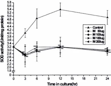
Fig. 1 - Effects of pharbitidis seed on SOD activity. The pharbitidis seed-treated cells and control cells were harvested at the indicated times and SOD activity of th큰 extracts was measured. The results were expressed as the average of triplicate samples with S.D. ***P<0.001, **P<0.01, *P<
0.05 compared with control. Control, untreated; M 50 나g, treated with 50 p.g/m/ pharbitidis seed; M 100 |ig, treated with 100 나g/m/ pharbitidis seed; M 150 [ig, treated with 150 pharbitidis seed; M 200 |ig, treated with 200 \ig/
ml pharbitidis seed.
Fig. 2 - Effects of pharbitidis seed in combination with H20 2 on SOD activity. The pharbitidis seed-treated cells and control cells were harvested at indicated times and SOD activities of the extracts were measured. The results were ex
pressed as the average of triplicate samples with S.D.
***P<0.001, **P<0.01, *P<0.05 compared with control.
Control, untreated; H20 2, exposured to 2.5 mM H20 2;
H20 2+M 50 (ig, treated with 50 [ig/ml pharbitidis seed in combination with 2.5 mM H20 2; H20 2+M 100 jig, treated with 100 [Lg/ml pharbitidis seed in combination with 2.5 mM H20 2; H20 2+M 150 나g, treated with 150 [ig/ml pharbitidis seed in combination with 2.5 mM H20 2; H20 2+
M 200 |ig, treated with 200 pharbitidis seed in combination with 2.5 mM H20 2.
라 감소하는 흡광도를 측정하는 Aebi의 방법을 이용하였다.13) 50 mM phosphate buffer(pH 7.0) 100 m/당 30% H20 2를 넣어 10 mM substrate solution(A240=0.5)을 만든다. 이 substrate solution 3 m/에 각 sample 100 jo/를 가한 후, 43.6 M^cm"1^]
exitinction coefficient1。를 사용하여 UV/visible spectrophoto- meter로 240nm에서 phosphate buffer를 blank로 하고 1 분마다 2분 동안 absorbance를 측정하였다.
세포 생존율 측정
B16F10 melanoma cells을 2.5 x lO 4 cells/m/의 농도로 96- well plate의 well에 100 |i/씩 가한 후, 배양기 (37°C, humidified 5% C02)에서 24시간 동안 안정화시킨다. 각각의 시료 용액을 농 도별로 각 well에 100 야씩 넣어 실험군으로 하고, 각 시료를 녹 인 용매 100 |i/씩 가한 것을 대조군으로 하여 시료 용액을 넣은 후 24시간 배양한다. MTT 용액을 well 당 50|oi씩 넣어 4시간 동안 배양기에 방치한 후 원심 분리하여 상등액을 제거한다. 여 기에 DMSO 원액을 well 당 50p/씩을 가하여 1분간 혼들어주 면서 formazan을 완전히 용해시켜 ELISA reader를 이용하여 흡 광도(570 nm)를 측정한다.
통계처리
그래프와 표의 모든 수치는 각 실험횟수에 대한 평균과 표준 오차로 나타내었으며, 통계적 유의성에 대한 검증은 Student's t-testl■ 사용하여 계산하였다.
결 과 및 고 찰
혹축(Pharbitidis Semen, pharbitidis seed)추출물이 항산화 효 소의 활성에 미치는 영향을 알아보기 위하여 추출물 단독 투여 군과 산화적 스트레스를 유발하는 H202와의 병용 투여군으로 나 누어 각각 SOD, GPx 그리고 CAT의 활성을 측정하였으며,세 포 독성은 MTT assay를 실시하여 측정하여 다음과 같은 결과 를 얻었다.
Superoxide dismutase 활성에 미치는 영향
H202가 없는 상태에서 혹축추출물만 처리한 경우 SOD 활성 이 모든 실험군에서 control보다 활성이 낮게 나타났는데, 3시간 과 6시간 배양 시 control의 65%의 활성을 보이다가 시간이 지 날수록 control 보다 활성이 닛아져 12시간에서는 control의 59%, 24시간에서는 60%의 활성을 나타냈다(Fig. 1). H20 2와의 병용 투여한 경우 control보다는 효소활성이 낮은 것으로 나타났으나 배양 15시간까지는 높은 활성을 나타냈다. H20 2를 단독투여 한 경우는 배양 후 6시간 이후 점차 회복되는 것으로 나타났다 (Fig. 2).
% 김안근. 치'은정
0 3 6 9 12 15 18 21 24
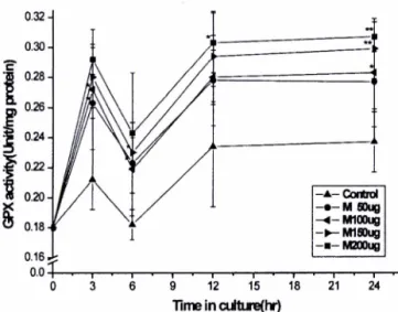
Fig. 3 - Effects of pharbitidis seed on GPx activity. The pharbitidis seed-treated cells and contol cells were harvested at the indicated times and GPx activity of the extracts was measured. The results were expressed as the average of triplicate samples with S.D. ***P<0.001, **P<0.01, *P<
0.05 compared with control. Control, untreated; M 50 나g, treated with 50 p.g/m/ pharbitidis seed; M 100 [Lg, treated with 100 pharbitidis seed; M 150 p.g, treated with 150 \ig/n)l pharbitidis seed; M 200 나g, treated with 200 나g/
ml pharbitidis seed.
Fig. 4 - Effects of pharbitidis seed in combination with H20 2 on GPx activity. The pharbitidis seed-treated cells and control cells were harvested at indicated times and GPx activity of the extracts were measured. The results were expressed as the average of triplicate samples with S.D. ***P<0.001,
**P<0.01, *P<0.05 compared with control. Control, untreated; H20 2, exposured to 2.5 mM H20 2; H202+M 50 (ig, treated with 50 [ig/ml pharbitidis seed in comb
ination with 2.5 mM H20 2; H20 2+M 100 나g, treated with 100 ^g/m/ pharbitidis seed in combination with 2.5 mM H202; H20 2+M 150 나g, treated with 150 [ig/ml pharbitidis seed in combination with 2.5 mM H202; H202+M 200 나g, treated with 200 [ig/ml pharbitidis seed in combination with 2.5 mM H20 2.
Glutathione peroxidase 훨■성에 미치는 영향
GPx의 경우에는 효소의 활성이 가장 많이 중가하였는데 혹축 추출물 단독 투여 시에는 control 군보다 활성이 크게 중가하였 고 처리한 모든 농도에서 배양시간에 따른 활성 변화가 큰 것을 볼 수 있었다. 활성변화양상은 contol과 매우 유사하여 3시간까 지 급격히 중가하던 효소 활성이 &시간 배양군에서는 급격히 감 소하는 양상을 보였고, 다시 12시간까지 중가하다가 24시간에서 는 12시간과 비슷한 활성을 나타내었으며, 모두 control보다 높 은 수준이었다(Fig. 3). H jA 와의 병용 투여 시에는 control이나 H202만을 투여한 군보다도 활성이 중가하였음을 관찰할 수 있 었고 변화 양상도 비슷함을 알 수 있었다(Fig. 4).
Catalase 활성에 미치는 영향
훅축추출물만을 처리한 경우 처리한 모든 농도에서 control보 다 약간 낮은 수준에서 비슷한 활성을 보였고 *3시간에서 최대치, 24시간에 최저치를 나타냈다. 효소활성변화 잉상도 control과 유 사하였다(Fig. 5).
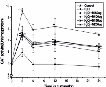
H202만을 투여하였을 때는 CAT의 활성을 크게 상승시켰다.
혹축추출물과 병용 ■투여한 경우는 효소활성이 닛아졌으나 control 보다는 높게 나타났고 효소활성변화양상은 유사하였다(Fig. 6).
H202와 병용 투여한 경우에 있어서는 배양 후 3시간에서 최대 치를 나타내었고, 모든 농도에서 control보다는 높고 H202보다 는 낮은 수준에서 비슷함을 알 수 있다.
Fig. 5 - Effects of pharbitidis seed on CAT activity. The pharbitidis seed-treated cells and contol cells were harvested at the indicated times and CAT activity of the extracts was measured. The results were expressed as the average of triplicate samples with S.D. ***?<0.001, **P<0.01, *P<
0.05 compared with control. Control, untreated; M 50 jig, treated with 50 [ig/ml pharbitidis seed; M 100 jig, treated with 100 p.g/m/ pharbitidis seed; M 150 나g, treated with 150 [ig/ml pharbitidis seed; M 200 나g,treated with 200 [ig/
m/ pharbitidis seed.
{ujaywdBUJAIUn^AJIAnoB
x
&
J. Pharm. Soc. Korea
산화적스트레스상태에서 B16F10 Murine Melanoma 세포의항산화S소활성에대한훅축주출물의효과 97
10- ***
— ★
0-|— «— i— ■— |— >— i— >— I— «— i— •— i— •— I— ■— I— ^
0 3 6 9 12 15 18 21 24
Time in culture(hr)
Fig. 8 - Effects of pharbitidis seed in combination with H20 2 on proliferation of B16 melanoma cells. The cells were treated with various concentration of pharbatidis seed for indicated times. The results were expressed as the average of triplicate samples with S.D. ***P<0.001, **P<0.01, *P<
0.05 compared with control. H20 2, exposured to 2.5 mM H202; H20 2+M 50 [ig, treated with 50 [Lg/ml pharbatidis seed in combination with 2.5 mM H20 2; H20 2+M 100 jig, treated with 100 \ig/ml pharbatidis seed in combination with 2.5 mM H202; H202+M 150 |ig, treated with 150 [ig/
ml pharbatidis seed in combination with 2.5 mM H202;
H20 2 + M 200 나g, treated with 200 |ig/m/ pharbatidis seed in combination with 2.5 mM H20 2.
B16 melanoma cell의 proliferation에 미치는 영향
MTT assay를 통하여 세포 생존율을 측정해 본 결과 0.2 mg/
m/의 농도에서 24시간 배양 시 흑축의 세포 생존율은 각각 95.9%
로 흑축 추출물이 세포에 미치는 영향은 거의 없슴을 알 수 있 다(Fig. 7). 와 병용 투여시, H20 2 단독 투여군의 경우 2.5mM 농도로 처리한 후 24시간 배양시 생존율이 거의 0에 가까운데 비하여 흑축추출물과 병용 투여시 세포 생존율이 단독 투여군에 비하여 6배 정도까지 중가하였다(Fig. 8). MTT assay를 실시한 결과 실험한 전 농도와 전 시간동안 세포 독성을 거의 나타내지 않았다.
위의 결과는 훅축 추출물만을 단독으로 투여한 경우 SOD의 활성은 control에 비해 감소하였고 GPx는 활성의 증가가 나타났 으나, CAT는 c o n tr o l 비교하여 별다른 치이가 없었다. 그러나 H202와 병용 투여한 경우에는 SOD의 활성만이 control보다 낮 았을 뿐 다른 두 효소의 활성은 control보다 증가하였다. H20 2 만을 투여하였을 때 나타나는 결과에서 보는 바와 같이 SOD는 H202가 높을 때 불활성화 되는 것으로 알려져 있다. 그러나 H20 2 와 흑축추출물을 병용 투여하였을 때는 SOD 활성이 어느정도 회복되는 상태를 나타내었다.
이상의 결과들올 살펴볼 때, 흑축 추출물은 H20 2로 인한 산화
0 3 6 9 12 15 18 21 24
Fig. 6 - Effects of pharbitidis seed in combination with H20 2 on CAT activity. The pharbitidis seeds-txeated cells and control cells were harvested at indicated times and CAT activity of the extracts were measured. The results were expressed as the average of triplicate samples with S.D.
***P<0.001, **P<0.01, *P<0.05 compared with control.
Control, untreated; H20 2, exposured to 2.5 mM H202;
H20 2+M 50 |ig, treated with 50 |ig/m/ pharbitidis seed in combination with 2.5 mM H20 2; H20 2+M 100 ^g, treated with 100 |ig/m/ pharbitidis seed in combination with 2.5 mM H20 2; H20 2+M 150 나g, treated with 150 [ig/ml pharbitidis seed in combination with 2.5 mM H202;
H20 2+M 200 나g, treated with 200
[Lg/ml
pharbitidis seed in combination with 2.5 mM H20 2.Fig. 7 - Effects of pharbitidis seed on proliferation of B16 melanoma cells. The cells were treated with various concentration of pharbitidis seed for indicated times. The results were expressed as the average of triplicate samples with S.D.
***P<0.001, **P<0.01, *P<0.05 compared with control.
M 50 \ig, treated with 50 p.g/m/ pharbitidis seed; M 100 jxg, treated with 100 [Lg/ml pharbitidis seed; M 150 jig, treated with 150
[ig/ml
pharbitidis seed; M 200 \ig, treated with 200 ixg/m/ pharbitidis seed.^ c l a
BUWJn^MABae
B
OOJJLKW
'sOA^HqelAlpo
98 김0J e • 치은정
적 스트레스상태에 있는 악성 흑색종 세포에서 주로 GPx나 CAT 와 같은 항산화 효소를 활성화시켜 과산화물을 제거시킴으로써 항산화기능을 나타내는 것으로 사료된다.
결 론
흑축이 항산화 효소의 활성에 미치는 영향을 B16 melanoma cells에서 살펴본 결과, 다음과 같은 결론을 얻을 수 있었다.
1. 혹축추출물만을 단독으로 투여한 군에서는 SOD가 control 보다 낮은 활성을 보였고, H20 2와 병용 투여한 실험 군에서는 H20 2 딘독 투여 군보다는 높으나 control보다는 낮은 활성을 나 타냈다.
2. GPx는 혹축추출물 단독 투여 군과 H20 2 병용 투여 군 모 두에서 control에 비해 높은 활성 중가를 관찰할 수 있었다.
3. 혹축추출물 단독 투여군에서는 CAT가 control과 비슷하거 나 조금 낮은 활성 을 보였으나,산화적 스트레스 상태인 H20 2 와의 병용 투여군에서는 H20 2 단독 투여군보다는 높고 control 보다는 낮게 나타났다.
4. MTT assay를 통하여 이들 추출물이 B16 melanoma cells 의 proliferation에 미치는 영힘을 즉정한 결과, 이들이 미치는 영 향은 매우 작은 것으로 나타났다.
감사의 말씀
본 연구는 숙명여자대학교 2002년도 교내연구비 지원에 의해 수행되었음.
참고문헌
1) Terry D. Oberley, Janice L. Schultz, Ning Li and Larry W Oberley : Antioxidant enzyme levels as a function of growth state in cell culture. Free Radical Biol Med. 19, 53 (1995).
2) Yasuko, Shindo and Takashi, Hashimoto : Time course of changes in antioxidant enzymes in human skin fibroblasts after UV irradiation. J. Dermatol Sci. 14, 225 (1997).
3) Bernadette, E. K., Marianne, D. and Bernhard, R : Pro
tective effect against sunburn of combined systemic ascorbic acid (vitamin C) and a-tocopherol (vitamin E). J. Am.
Acad. Dermatol. 38,45 (1998).
4) Zoltan, Gregus and Curtis D. Klaassen : Mechanisms of toxicity. In Casarett and Doull's toxicology, pp. 39-43 McGraw-Hill Com. (1996).
5) Kenneth S. Ranos, Enrique Chacon and Jn Daniel Acosta : Toxic responses of the heart and vascular systems. In Casarett and Doull's toxicology, pp. 492-495. McGraw-Hill Com. (1996).
6) Lisa A. Brennan, Gerard M. Morris, Gillian R. Wasson, Bernadette M Hannigan and Yvonne A. Barnett : The effect of vitamin C or vitamin E supplementation on basal and H20 2-induced DNA damage in human lymphocytes.
British Journal of Nutrition. 84, 195 (2000).
7) Gavat, V and Voroniuc, 0. : Oxidative ' stress and anti- oxidants in the diet in pathological processes at the level of the cardiovascular system. Rev. Med. Chir. Soc. Med.
N at Iasi. 103, 37 (1999).
8) Elke, Rohrdanz and Regine, Kahl : Alterations of anti- oxidant enzyme expression in response to hydrogen peroxide. Free Radical B iol Med. 24,27 (1998).
9) Stefan, Marklund and Gudrun, Marklund : Involvement of the superoxide anion radical in the autoxidation of pyro - gallol and a convenient assay for superoxide dismutase.
Eur, J. Biochem. 47, 469. (1974).
10) Eugene E Roth, Jn and Harriet S. Gilbert : The pyro- gallol assay for superoxide dismutase: absence in glutath - ione artifact A m i Biochem. 137, 50 (1983).
11) Flohe, Leopold and Wolfgang A. Gunzler : Assays of glutathione peroxidase. Methods in Emymology 105, 93 (1984).
12) Donald E. ftglia and V iliam N. Valentine : Studies on the qauntitative and qualitative characterization of erythro
cyte glutathione peroxidase. J. Lab. & Clin. Med. 70, 158 (1967).
13) Hugo, Abei : Catalase in vitro. Methods in Enzymology 105, 93 (1984).
/. Pharm. Soc. Korea