■藥學合확
백렴 추출물의 Matrix Metalloproteinase-1 발현 저해 효과
조 영호# • 심관섭 . 김진희 . 박성민 . 이범천 • 표형배 . 윤여표* • 박흠대**
한불화장품 기술연구소,* 충북대학교 약학대학, ** 대구대학교 공과대학 생물공학과 (Received November 3,2004; Revised November 26,2004)
Effect of Melothria heterophylla Extract on Expression of Matrix Metalloproteinase-1 in Human Skin Fibroblasts
Young Ho Cho#, Gwan Sub Sim, Jin Hui Kim, Sung Min Park, Bum Chun Lee, Hyeong Bae Pyo, Yeo Pyo Yun* and Hum Dai Park**
R& D Center, H anbul Cosmetics Corporation, 72-7, Yongsung-ri, Samsung-myun, Umsung-kun, Chungbuk 369-830’ Korea
^College of Pharmacy, Chungbuk N ational University, Cheongju 361-763, Korea
**Department of Biotechnology, College of Engineering, Daegu University, Gyeongsan 712-714,Korea
Abstract — Matrix metalloproteinases (MMPs) are known to play an important role in photoaging by mediating the deg
radation of extracellular matrix proteins. In this study, to develop a new anti-aging agent, we investigated the antioxidant activity and the inhibitory effect of Melothria heterophylla extract on expression of MMP-1 in UVA-irradiated human dermal fibroblasts and MMP-1 activity. The M. heterophylla extract was found to scavenge radicals and reactive oxygen species (ROS) with the SC50 values of 13 against 1,1 -diphenyl-2-picrylhydrazyl (DPPH) radicals and 20 [ig/ml against super
oxide radicals in the xanthine/xanthine oxidase system, respectively. UVA-induced MMP-1 expression was reduced about 80% by 100 [Lg/ml of the M. heterophylla extract but MMP-1 mRNA expression was not inhibited. Therefore, we conclude that the M heterophylla extract significantly inhibits MMP-1 expression at the protein level. Also, the M heterophylla extract inhibited MMP-1 activity in a dose dependent manner. From these results, we suggest that the M. heterophylla extract can be used as a new anti-aging agent by antioxidant activity, regulation of UVA-induced MMP-1 production, and inhibition of MMP-1 activity.
Keywords □ Melothria heterophylla, antioxidant, MMP
피부노화는세월이흘러감에따라피할수없는노화현상인 내인성 노화(intrinsic aging)와 외적 노화(extrinsic aging)로 구 분되며, 내인성 노화는유전적 요소에 영향을받기 때문에 인위 적인조절이 어려운반면,외적 노화는환경적인 요소에영향을 받기때문에 인위적인조절이비교적용 이 하 다 피 부 노화에영 향을미치는환경 요소들은기후, 흡연,공해, 자외선등을들수 있으며, 특히 자외선에 의한노화를 광노화(photoaging)라고한 다. 자외선은유해한활성산소종(reactive oxygen species)!- 많 이만들어내고, ROS는피부 세포에서 고분자물질들인 불포화
지방산, 단백질,DNA 둥과반응하여 피부콜라겐등의 결합조
#본논문에 관한문의는 저자에게로
(전화) 043-879-2283 (팩스) 043-881-2128 (E-mail) [email protected]
직형성 파괴, 세포막기능 저해,DNA 변이 촉진, 단백질 작용 변형,세포간에너지 전이 , 신진대사과관련된분자들의 변형등 을유발하는것으로 알려져 있다.2ᅪ} 일반적으로진피충은대다 수의 type I collagen과 약간의 type III collagen, elastin, proteoglycan, fibronectin 등으로구성되어 있다.5) 만성적 일광 손상을 입은 피부에서는 진피 상부쪽 교원질의 비정상적인 elastotic material의침착6ᅵ파 proteoglycan이증가되고진피의주 단백질인콜라겐이현저히 감소되는것으로알려져 있다.71또한, 콜라겐은피부에강도와장력을부여히여외부의 자극이나힘으 로부터 피부를보호하는 역할을하며 진피층의 약 90%를차지 하고있다. 따라서콜라겐의 감소는피부의노화와매우밀접한 관계를가지고있다.8)
Matrix metalloproteinases(MMPs)는훨성중심부에아연을갖 는금속단백질분해효소로서약20여종이상의종류가있는것
358
백렴추출물의항노화 359
으로 알려져 있다. 구조와기능에 따라 interstitial collagenase, gelatinase, stromelysin, membrane type MMP 등으로 구분하 기도한다.9〕특히 MMPs는피부의 keratinocytes, fibroblasts를 비롯한 많은세포들로부터 분비되어 세포외 기질(extracellular matrix; ECM)과기저막(basement membrane; BM )을구성하는 대부분의단백질성분을분해함으로써 피부탄력을유지하는결 합조직을파괴하여 주름과탄력저하및피부처짐의 원인이 되
는것으로알려져 있다.10) 또한, MMPs는세포에서저장되지 않
고불활성화된 proenzyme 및 zymogen 형태로분비되어아미노 말단부위가절단되는등구조적 변형이 일어나활성화되며, 활 성화된 MMP는 a2-macroglobulin이나 TIMPs(tissue inhibitors of metalloproteinases) 같은저해제에 의해 활성이 조절된다.u) Brenneisen 등은 UV 조사와 ROS에의해 피부내의 MMPs 활 성이중가되어 진피층내의콜라겐등과같은세포외기질들의붕
괴에 영향을미치며, MMPs가광노화에매우중요한역할을하
고있음을보고한바있다.1^
생약제인 백렴 (Melothria heterophylla(Lour.) Cogn.)은박과에 속하는다년생 덩굴성 초본으로인후종통,결막염, 임파선결핵, 고환염 , 피부습진등에 효과가있는생약으로 알려져있다. 그러 나,백렴추출물의 피부노화에 대한생리기전이나연구결과에 대해서는알려진바가거의 없는실정이다.13)
본연구에서는백렴 추출물의 항산화효과와 MMP-1 의활성 저해효과및 UVA에의한 human dermal fibroblasts^!서 MMP- 1 생성억제효과둥을관찰하였고, 기능성 화장품의천연항노 화소재로서의 이용가능성을검토하고자하였다.
실험 방법
시료의제조
본실험에서사용한 백렴은서울 경동시장내의 한약건재상 에서 구입하여 사용하였다. 백렴을분쇄하여 70% 에탄올을넣 은후 3시간씩 3회환류추출하고여과지를이용하여여과하였 다. 이여과액을감합농축하고동결건조하며그분말을사용하 였다.
세포및시약
신생아의 포피조직에서 분리한 human dermal fibroblast (HDF)는 Modem Tissue Technology 사(MTT, Korea)로부터 구입하였다. 구입한 HDF를 DMEM/F12(3 :1) 배지에 10% fetal bovine serum(FBS), 1% peniciUin-streptomycin을 첨가하여 37°C, 5% C 02 조건하에서 배양하고 trypsinization으로계대배 양한뒤 6-10세대 세포를실험에 이용하였다. Enzyme-linked immunosorbent assay(ELISA)를위해사용된 MMP-1 에대한 1 차 항체와 alkaline phosphatase가 결합된 2차 항체는 Sigma
Chemical 사로부터 구입하여사용하였다. In vitro MMP-1 활성 저해효과측정을 위해 사용된 형광물질이 표지된 DQ gelatin, DQ collagen, collagenase, 1,10-phenanthroline은 Molecular
probe사(USA)로부터구입하며 사용하였다. 그외실험에사용된
모든 시약들은 Sigma Chemical사에서 구입하여시용하였다.
l,l-Diphenyl-2-picrylhydrazyl(DPPH) radical 소거 효과 측정
Free radical의소거 작용은 Blois14)가시용한방법을약간변 형하여 각시료의 DPPH radical(Aldrich, USA)에대한소거 효 과를측정하였다. 0.1 mM DPPH methanol 용액에동일량의 시 료를 가하여 vortex mixer로잘혼합한후 실온에서 10분동안 빈응시킨다. 이후 spectrophotometei를이용하여 565nm에서흡 광도를측정하였다. 시료를첨가하지 않은 대조그룹과비교하여
DPPH radical의소거활성을백분율로나타내었다.
Superoxide radical 소거효과측정
Superoxide radical의소거작용은 Furuno 둥151의 방법에따라 각 시료의 xanthine-xanthine oxidase system에 의해 생성된 superoxide radical을 소거하는 효과를 즉정하였다. 0.05 M Na2C 0 3 buffer에 3 mM xanthine, 3 mM EDTA, 0.72 mM nitroblue tetrazolium(NBT), 0.15% bovine serum albumin (BSA) sol’l l 시료를각각첨가하며 혼합한다음, 250C에서 10 분간 빈'응하였다. 그후각 tube에 xanthine oxidase(0.25 U/m/) 용액을첨가하여 250C에서 30분간반응한후, 565nm에서흡광 도를 측정하였다. 시료를 첨가하지 않은 대조그룹과비교하여 superoxide radical 소거활성을백분율로나타내었다.
세포생존률측정
세포 독성은 3-(4,5-dimet화lthiazol-2-yl)-2-5-diphenylte^
zolium bromide(MTT) 시약을이용하여 세포 생존률을측정하
는 Mosmann16)의방법을 변형하며 실시하였다. HDF를 2X10⁴ cells/well 농도로 96-well plate에접종한 후, 각 well에시료를 투여하며 C 0 2 배양기에서 24시간배양하였다. MTT 용액(5gg/
m/>을첨가하고 4시간후원심분리하여 상등액을제거하고, 100 pd acid-isopropanol(0.04 N HCl in isopropanol)을 첨가한 후푸른 색의 formazan이용줄 되도록 하여 micro plate reader(Model ELx 800, BIO-TEK Instruments, USA)로 565 nm에서 흡광도 를측정하였다.
UVA 조사및시료의 처리
HDF를 1.5X 10⁵ cells/m/의농도로 35 mm dish에약 80%의 conflu ence 도달할때까지배잉하였다. UV 조사 전에원배지
를제거한후 PBS로세척하여 배지내 serum 성분을 제거한다
음, UV 조사장치 (F15T8.BLB, Sankyo Denki, Japan)로 UVA (6.3 J/cm2)를조사하였다. UVA 조사후 FBS를첨가하지 않은 DMEM/F12(3 : 1) 배지에 시료를투여하여 24시간배양하였다.
MMP-1 발현저해효과측정 (ELISA법)
UVA 조사에의해유도되는 MMP-1 발현량측정은 Dunsmore17) 둥이 사용한 방법을 약간변형하여 실시하였다. 먼저 HDF에 UVA를조사후시료를처리하여 24시간배양한배지를 96-well plate에분주하여 40C에서 overnight하여 coating하였다. PBS-T
(phosphate buffered saline +0.05% Tween 20)로 3회세척하고 3% BSA /PBS-T로 3TC에서 1시간동안 blocking한후, 1차항 체 (monoclonal anti-MMP-1)을 blocking buffer로 1:3000으로 희석하여 처리하고 37T에서 90분간반응시켰다. PBS-T로세척 한 다음 2 차 항체 (alkaline phosphatase conjugated Goat anti
mouse IgG)를 blocking buffer에 1:3000으로 희석하여 처리하 고 37°C에서 90분간반응시킨후 PBS-T로세척한다음 alkaline phosphatase 기질용액(1 mg/m/ /(-nitrophenyl phosphate in diethanolamine buffer)을첨가하여실온에서 30분■간반응시켰다.
3 N NaOH로반응을 완전히 중지시킨 후 micro plate reader을
사용하여 405 nm에서흡광도를측정하였다.
RN A 분리및 RT-PCR
Total RNA 주출은 RNeasy mini kit(Qiagen, Germany)을이 용하였다. cDNA 합성은 1ᅣig의 total RNA를 oligo(dT)15
primer, dNTP(0.5 |iM), 1 unit RNase inhibitor 그리고 4 unit
Omniscript reverse txanscriptase(Qiagen, Germany)로 370C에 서 60분, 930C에서 5분 heating시킴으로써 반응을중지시켰다. Polymerase Chain Reaction(PCR)은 cDNA로부터 MMP-1, |3- actin을중폭하기 위하여 l|o/ cDNA, 0.5|iM의 5'과 3’ primer,
10 x buffer(10 mM Tris-HCl, pH 8.3, 50 mM KC1, 0.1%
Triton X-100), 200 |xM dNTR 25 mM MgCl2, 2.5 unit Taq
polymerase(Qiagen, Germany)를섞고 distilled water로전체를 25 |o/로맞춘다음 PCR을실시하였다. PCR 증폭은 94°C 0.5분•, 50°C 0.5분,72°C 1분, 25 cycles로반응시켰다. PCR에의하여
생성된 산물은 1.5% agarose gel에서 전기영동을 실시하고
ethidium bromide로염색하여특정 band를확인하였다. 본실험
의 RT-PCR에사용된 primer는 Table I에제시하였다.
MMP-1 활성저해효과측정
시료의 MMP-1 활성 저해 효과측정은 Wang10) 등이 사용한 방법을약간변형하여실시하였다. 즉, 반응완충액 100 n/에 0.25 mg/m/로반응완충액에 용해한 DQ collagen 20^와 시료 40마 를첨가하고 0.5 unit로희석된 collagenase 40 (i/를첨가하였다. 암소, 실온에서 20& 경과후형광분광광도계(LS55, PERKIN ELMER, USA)를이용하여흡수파장 495 nm, 방출파장 515 nm 로형광값을측정하였고, 대조그룹으로서 효소액 대신반응완충 액을효소와동량 첨가하여 형광값을측정하였다. 시료자체의 형광값도측정하며, 효소활성 계산시보정하였다.
자료분석 및통계처리
모든실험결과는평균±표준편차로표기하였고, 통계적유의 성검증은 Student's t-test로실시하였으며 , p 값이 0.05 미만일 때통계적으로유의하다고판단하였다.
결과 및 고찰
DPPH radical 소거효과
DPPH는 free radical의안정된모델로반응중 DPPH의감소 는 free radical의소거반응이 진행됨을 알수있고, 지질과산화 의초기반응의 억제정도를예측할수있다. 유해 산소라불려지 는활성산소는세포생체막의 구성 성분인불포화지방산을공 격하여 지질과산화반응을일으켜체내과산화지질을축적함으 로인해생체기능이저하되고동시에노화및성인병질환을유 발18>하는것으로알려져 있으며, 다양한종류의 식물성분및추
Table I - Sequences of primers and fragment sizes of the investigated genes in RT-PCR analysis
Gene Primer sequences Fragment
size (bp) MMP-1
(3-Actin
F 5’-AAAGGGAATAAGTACTGGGC-3’
R S'-AATTCCAGGAAAGTCATGTG-S' F 5’-ATGCAGAAGGAGATCACTGC-3’
R 5'-CTGCGCAAGTTAGGTTTTGT-3'
237
248
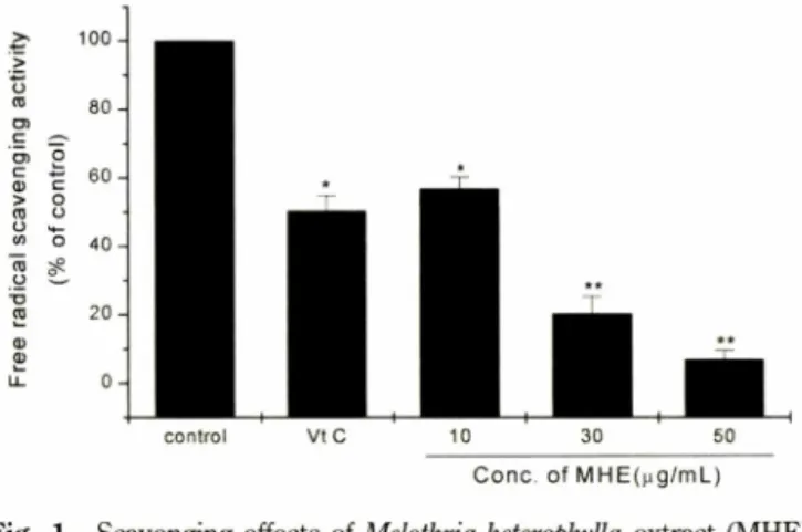
Conc. of MHE(pg/mL) - Scavenging effects of Melothria heterophylla extract (MHE)
on DPPH radicals. A solution of 150 \xlof 100 jiM DPPH solution in methanol was gently mixed with 150 |i/ of MHE for 10 min and the absorbance was measured at 565 nm.
The results were expressed as the average of triplicate samples with S.D. */><0.05, **/><0.01 compared with control.
J. Pharm. Soc. Korea
백렴추출물의항노화 361
으로 밝혀졌다.
UVA에의한 MMP-1 발현저해 효과
피부의 광노화에 있어 중요한역할을담당하고 있는 MMP-1
의발현은 UVA에의해 세포에서 JNK/p38 활성도가중가하고 전사인자인 AP-1 의활성도가 중가되는신호 전달경로를통해
MMP-1 발현을증가시켜 피부에서 교원질의 결핍을초래한다고
알려져 있다.22) 이러한 UVA에의해 발현이중가되는 MMP-1 에 백렴추출물이미치는영향을: 알아보고자 HDP게 UVA(6.3J/cm2) 를조사하고백렴추출물을첨가하여 24시간배양한후 MMP-1 발현저해 효과를 ELISAS. 측정하였다. 그결과 백렴 추출물은 농도 의존적으로 MMP-1 발현저해효과를나타내었다(Fig. 3).
또한, 실험에시용된 백렴추출물각각의 처리농도에서 세포 생 존률에는별다른영향을미치지 않는것으로확인되었다(Fig. 3).
백렴추출물을 1,10,lOOng/m/의농도로 처리한경우 MMP-1 의발현 저해 효과는 40.6%, 60.4%, 80.2%로 나타났으며,UV 에의한 c-Jun 단백질의발현증가를 억제하여 MMP-1 발현저해 효과가보고된23) retinoic acid의경우는 1.1 ng/m/에서 44.5% 발 현저해효과를나타내었다. 또한, HDF에서 UVA에의해 발현이
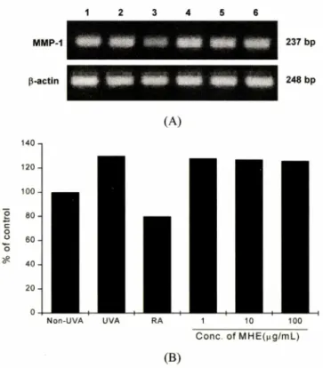
증가되는 MMP-1 mRNA에백렴 추출물이 미치는영향을 알아
보기 위해 RT-PCR을수행한 결과 양성 대조그룹으로사용된
retinoic acid에비해뚜렷한 저해효과를나타내지못히는 것으로 나타났다(Fig. 4). 결론적으로 백렴 추출물은 HDF에서 UVA에 의해발현이 증가되는 MMP-1 을단백질24"25) 수준에서만발현을 저해하는효과가있■음미확인되었으며,이는기존보고된 retinoic
conrol BHA 10 50 100
C one, o f M H E (n g /m L )
2 - Scavenging effects of Melothria heterophylla extract (MHE) on superoxide radicals. Superoxide radicals were generated by a xanthine/xanthine oxidase system and measured by NBT reduction method as described in the Materials and Methods. The results were expressed as the average of triplicate samples with S.D. */><0.05, **/><0.01 compared with control.
C one, o f M H E (나g/ml_)
Fig. 3 - Effect of Melothria heterophylla extract (MHE) on the production of MMP-1 in UV irradiated human dermal fibroblasts. The cells were treated with various concen
tration of MHE for 24 h. aMMP-l, matrix metalloproteinase- 1 (■ ). The MMP-1 contents in culture media were determined by ELISA as detailed under the Materials and Methods. bCytotoxicity was measured by MTT assay (♦ )•
The results were expressed as the average of triplicate samples with S.D. */><0.05, **/><0.01 compared with UVA-irradiated control.
출물에 의한항산화작용이보고되어 있다.19,20) 백렴추출물의 항산화효과를알아*보기위해 DPPH를이용하여 항 ^화 작용을 측정하였다. 양성 대조그룹으로는항산화작■§끼 있는것으로알 려진 vitamin C를이용하여 백렴 추출물의 항산화효과를비교 하였다. 그 결과 vitamin C는 11 ng/m/에서 50%의 DPPH radical을소거하였으며, 백렴 추출물은투여 농도의존적으로 DPPH radical 소거작용을 나타냈다(Fig. 1). 즉, 백렴 추출물을 10, 30, 50 ng/m/의농도로처리한경우각각의 DPPH radical 소 거능은 43.2%, 79.7%, 93.1%로나타났으며,S C # 13ng/mZ로 우수한 free radical 소거효과를나타내었다. 상기의결과로볼때 백렴추출물의 free radical 소거효과는단일성분인비타민 C에 뒤지지않는매우우수한효과를가지는것으로밝혀졌다•
Superoxide radical ;소^ 효고I
Xanthine/xanthine oxidase에의해생성되는 superoxide anion radical의저해작용은 superoxide radical 소거작용과 xanthine
oxidase의효소 활성 저해에 의해 나타난다.21) 백렴 추출물의
xanthine oxidase에의해생성되는 superoxide radical의소거결 과는 Fig. 2 에 나타내었다. 양성 대조그룹으로 3-t-butyl-4- hydroxyanisole(BHA)를 이용하여 백렴 주줄물의 superoxide
radical 소거효과를 비교하였다. 그결과백렴추출물은투여 농
도의존적으로 superoxide radical 소거작용을나타내 10,50, 100(ig/m/의농도로 처리한경우,각각의 superoxide radical 소 거능은 28.1%, 76.4%, 96.6%로나타났으며, SC^ 은 20|ig/mZ로 우수한 superoxide radical 소거효과를나타내었다. 양성 대조그 룹인 BHA는 32 ng/m/에서 50%의 superoxide radical을소거하 였다. 상기의결과로볼때백렴추출물은양성대:스그룹인 BHA
보다약1.6배정도높은 superoxide radical 소거능을가지는것 3
J
C8
o
%}
v s e
!
>
=90
o o
o o
o o
o o
6 4
2 0
8 6
4 2
0O4UOO
o
%}v-dlAIS
o
sluauioo
^l
>
ol
or
C
J)
.E6
u
a>)
co0
coI
CDO
'
-S
g j
a>
TJj
xᄋ
.l
a>d
rl
w
362 조영호 • 심관섭 . 김진희 . 박성민 . 이범천 . 표형배 . 윤여표 . 박흠•대
Conc. o f M HE(|ig/m L)
Fig. 5 - Inhibitory effect of Melothria heterophylla extract (MHE) on MMP-1 activity. A solution of 20 [il of DQ collagen (0.25 mg/m/) and 40 [il of MHE were gently mixed with 40 \\1 of 0.5 U collagenase for 20 min and the luminescence was measured at 495 nm (exitation wavelength) and 515 nm (emission wavelength). The results were expressed as the average of triplicate samples with S.D. */)<0.05,
**/><0.01 compared with control.
phenanthroline10)을이용하여 백렴추출물의 MMP-1 활성 저해 효과를비교하였다. 그 결과 1,10-phenanthroline는 24 ng/m/에 서 50%의 MMP-1 의활성을저해하였으며, 백렴추출물의 경우 투여농도의존적으로 MMP-1 의활성을저해하는것으로 나타 났다(Fig. 5). 즉,백렴추출물을 10,50,100 |파W의농도로처 리한경우 MMP-1 의활성을각각 50%, 83.3%, 96.7% 저해하는 것으로나타났으며,IC50은 10 로우수한 MMP-1 활성저 해효과를나타내었다. 상기의 결과로볼때백렴추출물은양성 대조그룹인 1,10-phenanthroline보다약 2.4배정도높은 MMP-
1활성 저해능을가지는것으로밝혀졌다.
결 론
본연구에서는백렴추출물에의한항산화효과, MMP-1 활성 저해효과및 HDF에서 UVA에의해발현이 중가되는 MMP-1 의발현에 미치는영향을관찰하였다. 백렴추출물의 DPPH와 superoxide radical 소거효과는처리농도가증가함에 따라효과 가 높아지는 농도 의존적인 경향을 나타냈으며, DPPH와 superoxide radical을 각각 50|ig/m/에서 93%, lOOjig/m/에서 97% 정도소거하여 우수한항산화효과를나타내었다. HDF에 서 UVA에의해발현이 증가되는 MMP-1 의발현저해 효과에 있어서 백렴추출물은 100ng/m/에서 80%로단백질 수준에서 우수한발현저해효과를나타냈지만, mRNA 수준에서는뚜렷한 발현저해효과를나타내지못하는것으로확인되었다. 또한,백렴 추출물의 MMP-1 활성저해효과는 100 에서약 97%로매 우우수한활성 저해효과를나타내었다. 이상의 결과를종합하 여볼때백렴추 '은 항산화효과및 MMP-1 활성 저해 효
MMP-1
p-actin
1 2 3 4 5 6
237 bp
248 bp
(A)
Non-UVA UVA RA 1 10 100
Conc. of MHE(ng/mL)
(B)
Fig. 4 Effect of Melothria heterophylla extract (MHE) on MMP-1 mRNA expression in UV irradiated human dermal fibro
blasts. (A) After irradiated UVA, HDF were treated with various concentration of MHE for 12 h. Total RNA extracted from human dermal fibroblasts was analyzed by RT-PCR. Lane 1; UVA non-irradiated, lane 2; UVA (6.3 J/
cm2) irradiated, lane 3; UVA+10 ^ig/m/ of retinoic acid, lane 4; UVA+1 [ig/ml of MHE, lane 5; UVA+10 |ig/m/ of MHE, lane 6; UVA+100 \xg/ml of MHE. (B) Each band was quantitated by a densitometer using P-actin bands as references.
acid보다우수한효과를가지는것으로확인되었다. 또한,HDF 에서 UVA로부터 ROS7} 생성되며,ROSS 인한 MMP발현26^
이촉진되는것을백렴추출물이 ROS 소거작용을통하여 MMP 발현을효과적으로조절할수있을것으로사료된다.
MMP-1 활성 저해효과
피부세포의 결합조직을구성하는성분들가운데콜라겐은피 부 건조중량의 약 90%에달하는주요구성 단백질이다. 따라서 콜라겐의분해는곧결합조직의 탄력 저하와피부의주름및처 짐에 직접적인 영향을 미친다. 체내에서 생성되는수십 종의
MMPs 가운데 MMP-1 은 콜라겐에 특이적으로 작용하는
proteinase로서 MMP-1 의활성을 억제하여 콜라겐을 보호하면 피부조직의 탄력을유지하고주름의 생성을 예방할수있는것 으로알려져 있다.9) 따라서 백렴추출물의 MMP-1 활성저해효 과를알아보기 위해형광물질이표지된 DQ gelatin, DQ-collagen 을이용하여 MMP-1 활성 저해작용을측정하였다. 양성 대조그 룹으로 MMP-1 활성 저해 작용이 있는 것으로 알려진 1,10-
o
o
o
o
o 0 8 6 4
0 2C O 3
00/)o A
«
>
o
(oT
a.s
s
'ouoijllul
J. Pharm. Soc. Korea
백렴추M :의항노화 363
과와 UVA에의한 MMP-1 의발현을효과적으로저해하는것으
로보아우수한항노화소재로써이•용될수있을것으로사^된다.
문 헌
1) Gilchrest, B. A. : Skin aging and photoaging: an overview. J.
Am. Acad. Dermatol. 21,610 (1989).
2) Cadenas, E. : Biochemistry of oxygen toxicity. Annu. Rev.
Biochem. 58,79 (1989).
3) Lavker, R. M. and Kligman, A. M. : Chronic heliodermatitis : a morphologic evaluation of chronic actinic dermal damage with emphasis on the role of mast cells. / Invest. Dermatol. 90, 325 (1988).
4) Davies, K. J. : Protein damage and degradation by oxygen radicals. J. Biol Chem. 262,9895 (1987).
5) Bailly, C., Dreze, S., Asselineau, D., Nusgens, B., Lapiere, C.
M. and Darmon, M. : Retinoic acid inhibits the production of collagenase by human epidermal keratinocytes./. Invest Derm.
94, 47 (1990).
6) Yaar,M. and Gilchrest, B. A. : Aging versus photoaging : postulated mechanisms and effectors. J. Investig. Dermatol Symp. Proc. 3,47 (1998).
7) Li, J. J., Dong, Z., Dawson, M. I. and Colburn, N. H .: Inhibition of tumor promoter-induced transformation by retinoids that transrepress AP-1 without transactivating retinoic acid response element. Cancer Res. 56,483 (1996).
8) Huang, C., Ma, W. Y., Dawson, M. I., Rincon, M .,Flavell, R. A.
and Dong, Z. : Blocking activator protein-1 activity, but not activation retinoic acid response element, is required for the antitumor promotion effect of retinoic acid. Proc. Natl Acad.
Sci USA 94, 5826 (1997).
9) Kondo, S. : The roles of cytokines in photoaging. J. Dermatol.
Sci. 23, S30 (2000).
10) Wang, Y, Johnson, A. R.’ Ye, Q. Z. and Dyer, R. D. : Catalytic activities and substrate specificity of the human membrane type 4 matrix metalloproteinase catalytic damain. J. Biol Chem. 274, 33043 (1999).
11) Baker, A. H.,Edwards, D. R. and Murphy, G. : Metallo
proteinase inhibitors : biological actions and therapeutic opportunities. J. Cell Sci. 115, 3719 (2002).
12) Brenneisen, R, Sies, H. and Scharffetter-Kochanek, K. : Ultraviolet-B irradiation and matrix metalloproteinases: from induction via signaling to initial events. Ann. N. Y. Acad. Sci.
973,31 (2002).
13) Park, J. H. : The encyclopedia of Chinese crude drugs.
Shinilbooks Publications, Seoul, 310 (2002).
14) Blois, M. S .: Antioxidant determinations by the use of a stable free radical. Nature 181, 1199 (1958).
15) Furuno, K.,Akasako, T. and Sugihara, N. : The contribution of
the pyrogallol moiety to the superoxide radical scavenging activity of flavonoids. Biol. Pharm. Bull. 25, 19 (2002).
16) Mosmann, T .: Rapid colorimetric assay for the cellular growth and survival: application to proliferation and cytotoxic assay. J.
Immunol Methods 65,55 (1983).
17) Dunsmore, S. E.,Rubin, J. S., Kovacs, S. O., Chedid, M., Parks, W. C. and Welgus, H. G. : Mechanisms of hepatocyte growth factor stimulation of keratinocyte metalloproteinase produc
tion. /• Biol Chem. 271,24576 (1996).
. 18) Kitahara, A., Matsumoto, U., Ueda, H. and Ueoka, R. : A remarkable antioxidation effect of natural phenol derivatives on the autoxidation of y-irradiated methyl linolate. Chem.
Pharm. Bull. 40,2208 (1992).
19) Hatano, T .: Constituents of natural medicines with scavenging effects on active oxygen species-tannins and related poly
phenols. Natural Medicines 49, 357 (1995).
20) Masaki, H., Sakaki, S., Atsumi, T. and Sakurai, H. : Active oxygen scavenging activity of plant extracts. Biol. Pharm. Bull.
18,162 (1995).
21) Kuppusamy, R and Zweier, J. L. : Characterization of free radical generation by xanthine oxidase. J. B iol Chem. 264, 9880 (1989).
22) Chun, J. H., Kang, S. W., Varani, J., Lin, J., Fisher, G. J. and Voorhees, J. J. : Decreased extracellular-signal regulated kinase and increased stress-activated MAP kinase activities in aged human skin in vivo. J. Invest Dermatol. 115,177 (2000).
23) Fisher, G. J., Talwar, H. S., Lin, J., Lin, R, McPhillips, R, Wang, Z. Q.,Li, X.,Wan, Y., Kang, S. W. and Voorhees, J. J. : Retinoic acid inhibits induction of c-Jun protein by ultraviolet radiation that occurs subsequent to activation of mitogen-activated protein kinase pathways in human skin in vivo. J. Clin. Invest.
101, 1432 (1998).
24) Seo, J. Y., Rhie, G. E. and Chung, J. H. : The effect of ultraviolet irradiation on the expression of type I procollagen and MMP-1 in human dermal fibroblast and human skin in vivo. Kor. J. Invest. Dermtaol. 8, 116 (2001).
25) Seo, J. Y, Choi, H. R., Rhie, G. E., Youn, C. S., Choi, W. W., Kim, J. A., Chung, J. H.,Kim, K. H., Cho, K. H. and Eun, H.
C .: The effect of retionic acid and vitamin C on the expression of the procollagen a l(I), tropoelastin, and MMP-1 in human dermal fibroblast. Kor. J. Invest Dermtaol. 8,23 (2001).
26) Kochanek, S. K.,Wlaschek, M., Briviba, K. and Sies, H. : Singlet oxygen induces collagenase expression in human skin fibroblast. FEBS Lett. 331,304 (1993).
27) Wlaschek, M., Brviba, K.,Stricklin, G. R, Sies, H. and Scharffetter-Kochanek, K. : Singlet oxygen may mediate the ultraviolet A induced synthesis of interstitial collagenase. J.
Invest Dermatol. 104,194 (1995).