777
Background and Purpose—The goal of reperfusion therapy in acute ischemic stroke is to limit brain infarction. The objective of this study was to investigate whether the beneficial effect of endovascular treatment on functional outcome could be explained by a reduction in post-treatment infarct volume.
Methods—The Endovascular Treatment for Small Core and Anterior Circulation Proximal Occlusion With Emphasis on Minimizing CT to Recanalization Times (ESCAPE) trial was a multicenter randomized open-label trial with blinded outcome evaluation. Among 315 enrolled subjects (endovascular treatment n=165; control n=150), 314 subject’s infarct volumes at 24 to 48 hours on magnetic resonance imaging (n=254) or computed tomography (n=60) were measured. Post- treatment infarct volumes were compared by treatment assignment and recanalization/reperfusion status. Appropriate statistical models were used to assess relationship between baseline clinical and imaging variables, post-treatment infarct volume, and functional status at 90 days (modified Rankin Scale).
Results—Median post-treatment infarct volume in all subjects was 21 mL (interquartile range =65 mL), in the intervention arm, 15.5 mL (interquartile range =41.5 mL), and in the control arm, 33.5 mL (interquartile range =84 mL; P<0.01). Baseline National Institute of Health Stroke Scale (P<0.01), site of occlusion (P<0.01), baseline noncontrast computed tomographic scan Alberta Stroke Program Early CT score (ASPECTS) (P<0.01), and recanalization (P<0.01) were independently associated with post-treatment infarct volume, whereas age, sex, treatment type, intravenous alteplase, and time from onset to randomization were not (P>0.05). Post-treatment infarct volume (P<0.01) and delta National Institute of Health Stroke Scale (P<0.01) were independently associated with 90-day modified Rankin Scale, whereas laterality (left versus right) was not.
Conclusions—These results support the primary results of the ESCAPE trial and show that the biological underpinning of the success of endovascular therapy is a reduction in infarct volume.
Clinical Trial Registration—URL: http://www.clinicaltrials.gov. Unique identifier: NCT01778335.
(Stroke. 2016;47:777-781. DOI: 10.1161/STROKEAHA.115.012424.)
Key Words: cerebrovascular disease/stroke ◼ infarct size ◼ ischemic stroke ◼ management
◼ modified Rankin Scale ◼ stroke
Insights From the ESCAPE Randomized Controlled Trial
Fahad S. Al-Ajlan, MD; Mayank Goyal, MD; Andrew M. Demchuk, MD;
Priyanka Minhas, MD; Farahna Sabiq, MD; Zarina Assis, MD; Robert Willinsky, MD;
Walter J. Montanera, MD; Jeremy L. Rempel, MD; Ashfaq Shuaib, MD; John Thornton, MD;
David Williams, MB, PhD; Daniel Roy, MD; Alexandre Y. Poppe, MD; Tudor G. Jovin, MD;
Biggya L. Sapkota, MD; Blaise W. Baxter, MD; Timo Krings, MD; Frank L. Silver, MD;
Donald F. Frei, MD; Christopher Fanale, MD; Donatella Tampieri, MD;
Jeanne Teitelbaum, MD; Cheemun Lum, MD; Dar Dowlatshahi, MD; Jai J. Shankar, MD;
Philip A. Barber, MD; Michael D. Hill, MD, MSc; Bijoy K. Menon, MD, MSc;
for the ESCAPE Trial Investigators
Received December 13, 2015; final revision received January 8, 2016; accepted January 11, 2016.
From the Department of Clinical Neurosciences and Department of Radiology, Cumming School of Medicine, University of Calgary, Calgary (F.S.A.-A., M.G., A.M.D., P.M., F.S., Z.A., P.A.B., M.D.H., B.K.M.); Department of Community Health Sciences and Department of Medicine, Cumming School of Medicine, University of Calgary, Calgary (M.D.H., B.K.M.); Department of Medical Imaging, UHN, Toronto Western Hospital, Toronto (R.W., W.J.M.);
Department of Surgery (Neurosurgery), University of Alberta, Edmonton (J.L.R.); Department of Medicine (Neurology), University of Alberta, Edmonton (A.S.); Department of Neuroradiology, Beaumont Hospital and the Royal College of Surgeons in Ireland (J. Thornton); Department of Geriatric and Stroke Medicine, Beaumont Hospital and the Royal College of Surgeons in Ireland (D.W.); Department of Radiology, CHUM, University of Montreal, Montreal (D.R.); Department of Neurosciences, CHUM, University of Montreal, Montreal (A.Y.P.); Department of Neurology, University of Pittsburgh Medical Center, Pittsburgh (T.G.J.); Division of Neurology, University of Tennessee, Chattanooga (B.L.S.); Department of Radiology, Erlanger Hospital, University of Tennessee, Chattanooga (B.W.B.); Division of Radiology, UHN, Toronto Western Hospital, Toronto (T.K.); Division of Neurology, Department of Medicine, UHN, Toronto Western Hospital, Toronto (F.L.S., C.F.); Colorado Neurological Institute, Engelwood (D.F.F., D.T.); Montreal Neurological Institute, McGill University, Montreal (J. Teitelbaum); Department of Radiology, The Ottawa Hospital, University of Ottawa, Ottawa (C.L.); Department of Neurology, The Ottawa Hospital Research Institute, University of Ottawa, Ottawa (D.D.); Department of Neuroradiology, Dalhousie University, Halifax (J.J.S.); and Hotchkiss Brain Institute, University of Calgary (M.G., A.M.D., P.A.B., M.D.H., B.K.M.).
Guest Editor for this article was Emmanuel Touzé, PhD.
Presented in part at the International Stroke Conference of the American Heart Association, Los Angeles, CA, February 17–19, 2016.
Correspondence to Bijoy K. Menon, MD, MSc, Foothills Medical Centre, 1403-29th St NW, Calgary, AB T2N 2T9. E-mail [email protected]
© 2016 American Heart Association, Inc.
Stroke is available at http://stroke.ahajournals.org DOI: 10.1161/STROKEAHA.115.012424
by guest on April 23, 2017http://stroke.ahajournals.org/Downloaded from
brain from death. Clinical trials in ischemic stroke test if these therapies are capable of reducing damage to the brain.
An ability to speak, listen, and understand; to move, walk, or run; to see and interact with the world around us are all examples of brain functions. Clinical trials use ordinal scales like the modified Rankin Scale (mRS), the National Institute of Health Stroke Scale (NIHSS), or the Barthel index to quan- tify these functions.1–3 Understandably, none of these scales ever capture the entirety of functions and capabilities of the human brain. Therefore, it is important for stroke therapies being tested within clinical trials to also be able to show that they save brain tissue.
Using a detailed post hoc analysis of the Endovascular Treatment for Small Core and Anterior Circulation Proximal Occlusion With Emphasis on Minimizing CT to Recanalization Times (ESCAPE) trial data, we analyze whether endovascular therapy administered to subjects with acute ischemic stroke and proximal anterior circulation occlusion is capable of sav- ing brain tissue when compared with standard care (intra- venous alteplase or best medical therapy).4 We also analyze clinical and imaging variables at baseline associated with post-treatment infarct volume (measured at 24–48 hours from stroke symptom onset). Finally, we analyze the relationship between post-treatment infarct volume and the subject’s func- tional ability at 90 days as captured by the mRS (the primary outcome in the ESCAPE trial).
Methods
The ESCAPE trial was an investigator-initiated multicenter random- ized controlled trial assessing the additional benefit of modern endo- vascular treatment when compared with guideline-based standard of care. The trial screened subjects fulfilling clinical eligibility criteria if they presented within 12 hours of stroke symptom onset and then included them only if they met prespecified neurovascular imaging criteria. The trial enrolled 316 subjects from 22 sites across 3 con- tinents between February 2013 and October 2014, with one subject excluded for improper consent procedures.4,5
All subjects enrolled in the trial had follow-up imaging (magnetic resonance imaging [MRI] or computed tomography [CT]) at 24 to 48 hours from stroke symptom onset. Magnetic resonance diffusion- weighted imaging was the modality of choice for measurement of post- treatment infarct volume. If magnetic resonance diffusion-weighted imaging was not available, a noncontrast computed tomographic scan (NCCT) was chosen for measurement. An expert (Dr Al-Ajlan) used Quantomo (Cybertrial Inc, Calgary)6 to delineate infarct and measure post-treatment infarct volumes (in milliliters) while being blinded to all other clinical and imaging information. Manual adjustments to delineate infarct boundaries were performed where necessary. If the infarct showed hemorrhagic conversion, the hemorrhage regions were incorporated within the boundaries of infarct. Early recanalization was defined using Thrombolysis in Cerebral Infarction (TICI) Score (2b or 3) on conventional angiography in the intervention arm and using the modified arterial occlusive lesion grade (2 or 3) on 2- to 8-hour CT angiography among subjects in the control arm of the trial.7
Statistical Analyses
Because post-treatment infarct volume had a non-normal distribution, we used the Wilcoxon Rank-Sum test to look for differences in the
ternal carotid artery versus M1 middle cerebral artery), intravenous alteplase (yes versus no), early recanalization status, and time from stroke onset to randomization. A cube root transformation of post- treatment infarct volume best satisfied the assumptions of this model (normality of residuals and homoscedasticity) and was used for this analysis. Because collateral status on baseline CT angiography was collinear with baseline ASPECTS (ρ>0.25), collateral status was not included in the analyses. The final models and related graphs only report main effects from variables that were statistically significant.
We used ordinal logistic regression to model the association be- tween predictor variables (post treatment infarct volume [mL], 24- hour NIHSS, delta NIHSS [baseline NIHSS−24 hour NIHSS], and side of stroke [left versus right]) and clinical outcome measured on the mRS at 90 days from stroke onset. Models satisfying the assump- tions of parallel regression (tested using Brant’s test) were compared using likelihood function tests (Akaike and Bayesian Information Criterion) to determine the model that provided the best fit to the data.
This model was used to provide predicted outcomes, and these are displayed using graphs and contour plots to show the relationship be- tween the adjusted probability of clinical outcome (mRS) at 90 days, NIHSS score change, and infarct volume. All statistical analyses were performed in Stata/MP version 14.0 (StataCorp LP). Statistical sig- nificance was assessed at α<0.05 in all analyses.
Results
Post-treatment infarct volume measured using MRI in 254 subjects and using NCCT in 60 subjects was not statistically different (P=0.19). Median post-treatment infarct volume in all subjects was 21 mL (interquartile range [IQR] 7–72 mL). Median post-treatment infarct volume in the interven- tion arm (15.5 mL, IQR 5–46.5 mL) was significantly lower than that in control arm (33.5 mL, IQR 11–95 mL; P<0.01).
Early recanalization (Treatment in Cerebral Ischemia 2b/3 in the intervention arm, modified arterial occlusive lesion 2–3 in the control arm) occurred in 72% of subjects in the interven- tion arm and 31% of subjects in the control arm. Median post- treatment infarct volume in those who achieved recanalization in both arms (14.5 mL, IQR 4–46 mL) was significantly lower than those who did not (35 mL, IQR 12–104 mL; P<0.01).
Median post-treatment infarct volume in the intervention arm in recanalizers was 13 mL (IQR 4–45 mL) versus 20 mL (IQR 9.5–67.5 mL) in the nonrecanalizers (P=0.05). Median post- treatment infarct volume in the control arm in recanalizers was 17.5 mL (IQR 4.5–48.5 mL) versus 47 mL (IQR 16–122 mL) in the nonrecanalizers (P=0.002). Distribution of post- treatment infarct volume by quartiles stratified by treatment type (intervention versus control) and recanalization achieved (yes versus no) is shown in Figure 1.
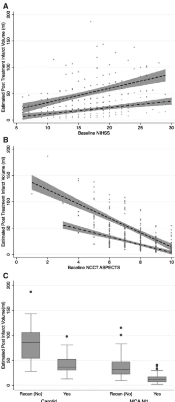
Baseline NIHSS (P<0.01), site of occlusion (P<0.01), baseline NCCT ASPECTS (P<0.01), and recanalization sta- tus (P<0.01) were independently associated with post-treat- ment infarct volume (cube root transformed) while age, sex, treatment type, intravenous alteplase, and time from onset to randomization were not (P>0.05). Treatment type was col- linear with recanalization status. Relationship between base- line NIHSS, baseline NCCT ASPECTS, and post-treatment infarct volume (from final adjusted model) stratified by
by guest on April 23, 2017http://stroke.ahajournals.org/Downloaded from
early recanalization status are shown in Figure 2A and 2B.
Distribution of post-treatment infarct volume by site of occlu- sion (from final adjusted model) stratified by early recanaliza- tion status is shown in Figure 2C.
Finally, when assessing the relationship between post- treatment infarct volume, 24-hour NIHSS, delta NIHSS (baseline NIHSS−24 hour NIHSS), side of stroke (left ver- sus right), and mRS at 90 days, the model that best fit the data (Akaike Information Criterion 1031 and Bayesian Information Criterion 1061) included post-treatment infarct volume (P<0.01) and delta NIHSS (P<0.01) as independently associated with 90-day mRS. A model with delta NIHSS (Akaike Information Criterion 1054 and Bayesian Information Criterion 1080) was a better fit than a model with final infarct volume (Akaike Information Criterion 1123 and Bayesian Information Criterion 1149) in association with mRS at 90 days. No interaction was noted between post-treatment infarct volume and delta NIHSS (P>0.05) in predicting 90-day mRS.
Side of occlusion (left versus right) was not a significant pre- dictor of 90-day mRS. Models with the addition of age did not increase model fit substantially. Figure 3A shows the rela- tionship between post-treatment infarct volume and estimated probability of excellent (mRS 0–1) outcome, independent (mRS 0–2) outcome, and death (mRS 6) at 90 days adjusted for laterality (quadratic best fit). Figure 3B and 3C are contour plots showing the relationship between post-treatment infarct volume, delta NIHSS, and estimated probability of achiev- ing independent outcome (mRS 0–2) and death (mRS 6) at 90 days.
Discussion
Analysis from the ESCAPE trial shows that endovascular treatment in subjects with acute ischemic stroke and proxi- mal anterior circulation occlusions was associated with sig- nificantly smaller infarct volumes. This effect of endovascular treatment on post-treatment infarct volumes was only seen when recanalization was achieved. Small infarct volumes were similarly noted in the control arm of the trial when early recanalization was achieved (Figure 1). These results provide supportive evidence for the physiological hypothesis that early recanalization saves brain.
Easily measured clinical and imaging variables at baseline (stroke severity, NCCT ASPECTS, and site of occlusion) are significant independent predictors of post-treatment infarct
volume together with early recanalization status. Importantly, age itself is not a predictor of post-treatment infarct volume in this analysis, and it is probable that the influence of age on brain physiology is captured by baseline stroke severity and NCCT ASPECTS. Time to randomization was not a signifi- cant predictor of post-treatment infarct volume in our analy- sis, but we hypothesize that this is because of study design;
imaging selection used in the ESCAPE trial (subjects selected using favorable imaging criteria independent of time from onset to randomization) may have contributed to these results.
Stroke onset time is also imprecisely measured with many subjects either waking up after stroke onset or only last seen normal at a certain time.
Previous studies have examined the association between post-treatment infarct volume (measured at 24–48 hours) and functional ability at 90 days as measured by the mRS.8,9 Our results show that post-treatment infarct volume is a strong independent predictor of 90-day mRS. The mRS at 90 days is best predicted when information on acute treatment response (change in NIHSS from baseline to 24 hours) is available along with post-treatment infarct volumes. These results are not sur- prising; subjects with large brain infarcts after treatment could have better outcomes on the mRS if treatment succeeded in sav- ing small but eloquent brain (significant reduction in NIHSS from baseline to 24 hours). Patients with small brain infarcts after treatment could have worse outcomes on the mRS if treat- ment did not succeed in saving small but eloquent brain (mini- mal change in NIHSS from baseline to 24 hours). Interestingly, no relationship was found between side of stroke and mRS at 90 days in our study. This lack of effect could be because the effect of side of stroke (and consequent eloquence of affected brain) on 90-day mRS may have been captured by delta NIHSS.
Our study has limitations. It is a post hoc explor- atory analysis. Infarct volume was measured on NCCT in
≈20% of subjects. This modality may be less precise than diffusion-weighted imaging. Although we used diffusion- weighted imaging to measure infarct volume versus fluid attenuation inversion recovery,10–12 there is debate in the stroke literature on the optimal MRI sequence and timing of imaging for the assessment of post-treatment infarct. We measured post-treatment infarct volume at 24 to 48 hours, but there remains a possibility of infarct growth after 24 to 48 hours.8–12 Neither CT nor MRI has been compared with brain histology as methods measuring brain infarct in
Figure 1. Post-treatment infarct volumes by quartiles (data stratified by treatment type and early recanalization status).
by guest on April 23, 2017http://stroke.ahajournals.org/Downloaded from
humans, and it is therefore not well known how meaningful are the inherent differences between brain CT and MRI for measuring discrete infarct volumes.
In summary, our results support the primary results of the ESCAPE trial and show that the biological underpinning of
the success of arterial recanalization and brain tissue reperfu- sion therapies is reduction in infarct volume.
Acknowledgments
Drs Menon and Hill had full access to all the data in the study and take responsibility for the integrity of the data and the accuracy of the data analysis. All authors fulfill ICMJE criteria for authorship.
Figure 2. Model estimated post-treatment infarct volume in mL (24–48 hours) by baseline National Institutes of Health Stroke Scale (NIHSS; A), baseline Alberta Stroke Program Early CT score (ASPECTS) (B), and site of occlusion at baseline (C); strati- fied by recanalization status (dash line=no recanalizers, dash and dot=recanalizers, grey boundaries indicate 95% confidence intervals, scatter plot in the background shows data distribution).
MCA indicates middle cerebral artery; and NCCT, noncontrast computed tomographic scan.
Figure 3. A, Laterality (left vs right) adjusted probability of achieving modified Rankin Scale score (mRS) 0 to 1, mRS score 0 to 2, and death at 90 days estimated using post-treatment infarct volume. Estimated probability of achieving mRS score 0 to 2 at 90 days estimated using post-treatment infarct volume (measured at 24–48 hours) and delta National Institutes of Health Stroke Scale (NIHSS; from baseline to 24 hours) (B) and achieving death at 90 days (C).
by guest on April 23, 2017http://stroke.ahajournals.org/Downloaded from
Sources of Funding
The study sponsor was the University of Calgary. Covidien Inc pro- vided major funding through an unrestricted grant to the University of Calgary. Additional active and in-kind support for the trial is from a consortium of funding public and charitable sources (Heart
& Stroke Foundation Canada, Alberta Innovates Health Solutions, Alberta Health Services, CSPIN network through CIHR) and the University of Calgary (Hotchkiss Brain Institute, Department of Clinical Neurosciences, Department of Radiology, and Calgary Stroke Program). The sponsor of the trial was the Governors of the University of Calgary. The sponsor had no role in the design, data gathering, analysis, or reporting of the trial. The University of Calgary received unrestricted grants from Medtronic (Covidien), Alberta Innovates Health Solutions, Heart & Stroke Foundation Canada, Canadian Institute for Health Research through the CSPIN Network, and Alberta Health Services. The University of Calgary provided in- ternal funding from the Hotchkiss Brain Institute, the Department of Clinical Neurosciences, and Department of Radiology.
Disclosures
Dr Roy reports grants and personal fees from University of Calgary during the conduct of the study. Dr Williams reports personal fees from Boehringer Ingelheim, Bayer, Bristol Myers Squibb, and Daliichi Sankyo outside the submitted work. Dr Demchuk reports research support from Covidien/Medtronic, unrestricted grant for ESCAPE trial, and no compensation. Speaker’s Bureau: Medtronic:
Significant >10K compensation. Dr Poppe reports personal fees from Covidien and Pfizer-BMS outside the submitted work. Dr Frei reports personal fees from Stryker (modest), personal fees from Penumbra (modest), honoraria from the Neuro-critical Care Society (modest), stocks in Penumbra (modest), and consultant relationship with Penumbra. Dr Thornton reports personal fees from Neuravi, Galway, and Ireland outside the submitted work. Dr Baxter reports personal fees from Penumbra, Stryker Neurovascular, Covidien (MedTronics), Rapid Medical, and Silk Road Medical outside the submitted work. Dr Jovin reports being a consultant for Neuravi, Codman Neurovascular, Blockade Medical, Silk Road, Stryker, and Covidien/Medtronic. Michael Hill reports grants from Covidien (Medtronic), Alberta Innovates Health Solutions, Heart & Stroke Foundation, Hotchkiss Brain Institute, CSPIN Network (ICRH- CIHR), Calgary Stroke Program, DCNS, University of Calgary, and nonfinancial support from Alberta Health Services, during the conduct of the study; personal fees from Merck, nonfinancial sup- port from Hoffmann-La Roche Canada Ltd, outside the submitted work, In addition, Dr Hill has a patent Systems and Methods for Assisting in Decision-Making and Triaging for Acute Stroke Patients pending to US Patent office Number: 62/086,077 and owns stock in Calgary Scientific Incorporated, a company that focuses on medical imaging software, and reports board membership of QuikFlo Health Inc. Dr Menon reports membership of the Steering and Executive Committee, ESCAPE trial that received support from Covidien Inc, Site Principal Investigator, SOCRATES Trial, sponsored by Astra Zeneca, honoraria from Penumbra Inc, a provisional patent 62/086077 for triaging systems in ischemic stroke, research fund- ing from CIHR, HSFC, AIHS, HBI, and the Faculty of Medicine, University of Calgary and reports board membership of QuikFlo Health Inc. Dr Goyal reports partial support for ESCAPE trial pro- vided to University of Calgary. Dr Goyal also helped in design and conduct of SWIFT PRIME trial; Compensation: Significant (>$10K
or 5%). In addition, Dr Goyal has received compensation for speak- ing engagements from Covidien Inc. (significant) and Stryker Inc.
(modest). He also has a patent for Systems of stroke diagnosis li- censed to GE Healthcare (compensation significant). The other au- thors report no conflicts.
References
1. Duncan PW, Jorgensen HS, Wade DT. Outcome measures in acute stroke trials: a systematic review and some recommendations to improve prac- tice. Stroke. 2000;31:1429–1438.
2. Quinn TJ, Dawson J, Walters MR, Lees KR. Functional outcome mea- sures in contemporary stroke trials. Int J Stroke. 2009;4:200–205. doi:
10.1111/j.1747-4949.2009.00271.x.
3. Banks JL, Marotta CA. Outcomes validity and reliability of the modi- fied Rankin scale: implications for stroke clinical trials: a literature review and synthesis. Stroke. 2007;38:1091–1096. doi: 10.1161/01.
STR.0000258355.23810.c6.
4. Goyal M, Demchuk AM, Menon BK, Eesa M, Rempel JL, Thornton J, et al; ESCAPE Trial Investigators. Randomized assessment of rapid endo- vascular treatment of ischemic stroke. N Engl J Med. 2015;372:1019–
1030. doi: 10.1056/NEJMoa1414905.
5. Demchuk AM, Goyal M, Menon BK, Eesa M, Ryckborst KJ, Kamal N, et al; ESCAPE Trial Investigators. Endovascular treatment for Small Core and Anterior circulation Proximal occlusion with Emphasis on minimizing CT to recanalization times (ESCAPE) trial: methodology.
Int J Stroke. 2015;10:429–438. doi: 10.1111/ijs.12424.
6. Kosior JC, Idris S, Dowlatshahi D, Alzawahmah M, Eesa M, Sharma P, et al; PREDICT/Sunnybrook CTA ICH study investigators. Quantomo:
validation of a computer-assisted methodology for the volumetric analy- sis of intracerebral haemorrhage. Int J Stroke. 2011;6:302–305. doi:
10.1111/j.1747-4949.2010.00579.x.
7. Higashida RT, Furlan AJ, Roberts H, Tomsick T, Connors B, Barr J, et al; Technology Assessment Committee of the American Society of Interventional and Therapeutic Neuroradiology; Technology Assessment Committee of the Society of Interventional Radiology. Trial design and reporting standards for intra-arterial cerebral thrombolysis for acute ischemic stroke. Stroke. 2003;34:e109–e137. doi: 10.1161/01.
STR.0000082721.62796.09.
8. Zaidi SF, Aghaebrahim A, Urra X, Jumaa MA, Jankowitz B, Hammer M, et al. Final infarct volume is a stronger predictor of outcome than recanalization in patients with proximal middle cerebral artery occlu- sion treated with endovascular therapy. Stroke. 2012;43:3238–3244. doi:
10.1161/STROKEAHA.112.671594.
9. Yoo AJ, Chaudhry ZA, Nogueira RG, Lev MH, Schaefer PW, Schwamm LH, et al. Infarct volume is a pivotal biomarker after intra- arterial stroke therapy. Stroke. 2012;43:1323–1330. doi: 10.1161/
STROKEAHA.111.639401.
10. Albers GW, Thijs VN, Wechsler L, Kemp S, Schlaug G, Skalabrin E, et al; DEFUSE Investigators. Magnetic resonance imaging profiles predict clinical response to early reperfusion: the diffusion and perfusion imag- ing evaluation for understanding stroke evolution (DEFUSE) study. Ann Neurol. 2006;60:508–517. doi: 10.1002/ana.20976.
11. Campbell BC, Purushotham A, Christensen S, Desmond PM, Nagakane Y, Parsons MW, et al; EPITHET–DEFUSE Investigators. The infarct core is well represented by the acute diffusion lesion: sustained reversal is infrequent. J Cereb Blood Flow Metab. 2012;32:50–56. doi: 10.1038/
jcbfm.2011.102.
12. Lansberg MG, Lee J, Christensen S, Straka M, De Silva DA, Mlynash M, et al. RAPID automated patient selection for reperfusion therapy:
a pooled analysis of the Echoplanar Imaging Thrombolytic Evaluation Trial (EPITHET) and the Diffusion and Perfusion Imaging Evaluation for Understanding Stroke Evolution (DEFUSE) Study. Stroke.
2011;42:1608–1614. doi: 10.1161/STROKEAHA.110.609008.
by guest on April 23, 2017http://stroke.ahajournals.org/Downloaded from
A. Barber, Michael D. Hill, Bijoy K. Menon and for the ESCAPE Trial Investigators
Print ISSN: 0039-2499. Online ISSN: 1524-4628
Copyright © 2016 American Heart Association, Inc. All rights reserved.
is published by the American Heart Association, 7272 Greenville Avenue, Dallas, TX 75231 Stroke
doi: 10.1161/STROKEAHA.115.012424
2016;47:777-781; originally published online February 18, 2016;
Stroke.
http://stroke.ahajournals.org/content/47/3/777
World Wide Web at:
The online version of this article, along with updated information and services, is located on the
http://stroke.ahajournals.org//subscriptions/
is online at:
Stroke Information about subscribing to Subscriptions:
http://www.lww.com/reprints
Information about reprints can be found online at:
Reprints:
document.
Permissions and Rights Question and Answer process is available in the
Request Permissions in the middle column of the Web page under Services. Further information about this Once the online version of the published article for which permission is being requested is located, click
can be obtained via RightsLink, a service of the Copyright Clearance Center, not the Editorial Office.
Stroke in
Requests for permissions to reproduce figures, tables, or portions of articles originally published Permissions:
by guest on April 23, 2017http://stroke.ahajournals.org/