I N T RO D U CT I O N
Idiopathic facial paralysis (Bell’s palsy) is the most common cause of facial weakness. It is equally common in men and women and can be seen in patients of any age. The clinical presen- tation of Bell’s palsy is a rapid onset of unilateral paresis that occurs over a period of a few hours to a day. Most cases of Bell’s palsy are neu- rapraxic and recovery is rapid and complete.
Electrodiagnostic tests have been developed to
estimate the degree of facial nerve injury during the acute phase and to provide a prognostic indi- cator for spontaneous recovery of voluntary movement. Side-to-side amplitude comparison with the affected side expressed as a percentage of the nonaffected side has been one of the most valuable electrophysiologic methods of assessing facial nerve functioning .1
In facial nerve palsy, most neurophysiologic laboratories cover the frontalis, orbicularis oculi, orbicularis oris, and nasalis muscles routinely.
Sometimes electromyographers choose some among the four muscles. However, it is unknown how the changes of amplitudes and terminal latencies recorded in each muscle on facial nerve stimulation are in the facial nerve palsy. The aim of this study was to know whether there is any difference in the side-to-side comparison of
얼굴마비에서 얼굴근육의 전기생리학적 양상
충북대학교 의과대학 신경과학교실, 관동대학교 의과대학 신경과학교실
이상수・신동익
Electrophysiologic Pattern of Facial Muscles in Bell ’ s Palsy
Sang-Soo Lee, M.D., Dong-Ick Shin, M.D.
Department of Neurology, College of Medicine, Chungbuk National University, Department of Neurology, College of Medicine, Kwandong University
Backgrounds: Electrodiagnostic tests have been developed to estimate the degree of facial nerve injury during the acute phase. Side-to-side amplitude comparison with the affected side expressed as a percentage of the nonaffected side has been one of the most valuable electrophysiologic methods of assessing facial nerve functioning. This study was designed to know whether there is any difference in the side-to-side comparison of amplitudes and terminal latencies of the compound muscle action potentials (CMAP) of the facial muscles in the patients with Bell’s palsy.
M e t h o d s: Electroneurographic recordings with surface electrodes on the frontalis, orbicularis oculi, nasalis, and orbicularis oris muscles were made within 2 weeks post-onset (mean, day 7) in 39 patients.
Results: Of the 39 Bell’s palsy patients, 38 patients (97.4%) recovered satisfactorily within 6 months. The amplitude of CMAP in all patients was not reduced to 10% or less of that of the contralateral healthy muscle. The correlation of amplitude change between four facial muscles was relatively strong, but the correlation of latency change was weak.
When the electroneurographic values were compared in the four muscle groups, the general linear models procedure did not show any significant difference for CMAP amplitude and latency changes (p=0.62-0.63).
Conclusions: This study did not show any significant clinical advantage of electroneurographic recordings in more than one facial muscle at the early stage of Bell’s palsy.
Key Words: Bell’s palsy, Facial muscle, Facial nerve, Electroneurography
Address for correspondence Sang-Soo Lee, M.D.
Department of Neurology, College of Medicine, Chungbuk National University,
12 Gaeshin-dong, Heungduk-gu, Cheongju-si, Chungbuk, 361-711, Korea Tel: +82-43-269-6336 Fax: +82-43-275-7591
E-mail : [email protected]
amplitudes of the CMAP and terminal latencies of the four tested facial muscles in the patients with facial nerve palsy.
M AT E R I A LS AND METHODS
Thirty-nine consecutive adults patients (18 men and 21 women) with acute idiopathic facial nerve palsy were studied within 2 weeks post-onset (mean, day 7). Their ages ranged from 21 to 74 years (mean 44.8±14.9). Each of 15 patients had a right-sided palsy and each of 24 a left-sided palsy; none had bilateral palsy or a previous his- tory of facial palsy. The Ethics Committee, Chungbuk National University Hospital approved the investigation. Informed consent was obtained from each patient before the enrollment.
The first clinical examination was performed on mean day 7. All patients showed unilateral facial muscle weakness including the frontalis, orbicu- laris oris, and orbicularis oculi muscles. Regular visit was made at outpatient clinic to follow the prognosis and last follow-up examinations were performed at 6 months after the onset.
The electrophysiological study was carried out on the same days as the clinical examination. A handheld bipolar prong stimulator was used to provide single square wave pulses 0.1 msec in duration at a frequency of less than 1 Hz. The cathode was placed just below and anterior to the lower tip of the mastoid, beneath the earlobe. The
anode was inferior to the cathode. Active elec- trode of the recording electrodes was placed over the nasalis muscle just lateral to and 1 cm above the external nares, directly beneath the pupil.
The frontalis electrode was placed 2 cm above the eyebrow and 2 cm from the midline. The orbicu- laris oculi electrode was placed on the lateral third on the lower eyelid. The perioral electrode for the orbicularis oris was placed 2 cm lateral to the midline and 2 cm below the lower lip. The reference electrode was in the same position as the recording electrode, but on the opposite side of the face. The ground electrode was placed on the chin. The measurements were made for the terminal latency and CMAP using the Excel EMG instrument (Cadwell). To estimate axonal loss, side-to-side evoked amplitude comparison with the affected side expressed as a percentage of the nonaffected side was made. The latency differ- ence between affected and nonaffected side was calculated to know the conduction time along the fastest-conducting fibers of the distal segment of the facial nerve.
The general linear model was used to compare the electroneurographic values in the four indi- vidual facial muscles. Pearson correlation coeffi- cients were calculated to know the relationship between the values in the four muscles.
Table 1. Correlation coefficients of electroneurographic values (n=39) of amplitude and terminal latency in the frontalis, orbicularis oculi, nasalis, and orbicularis oris recordings
Frontalis Orbicularis
Nasalis Orbicularis
oculi oris
Amplitude
Frontalis 0.5216* 0.5730* 0.5174*
Orbicularis 0.5216* - 0.2645 0.6040*
oculi
Nasalis 0.5730* 0.2645 - 0.5918*
Orbicularis
oris 0.5174* 0.6040* 0.5918* -
Latency
Frontalis - 0.2140 0.1724 0.0951
Orbicularis 0.2140 - 0.3576* 0.0597
oculi
Nasalis 0.1724 0.3576* - 0.3112
Orbicularis
oris 0.0951 0.0597 0.3112 -
*p<0.05
R E S U LTS
Thirty-nine patients were subject to facial nerve electroneurography. The amplitude of CMAP in all patients was not reduced to 10% or less of that of the contralateral healthy muscle. The correlation coefficients for the electroneurographic values obtained in the four different facial muscles are shown in the Table 1. The correlation values of amplitude change between the muscles were
strong. However, the value between the nasalis and orbicularis oculi muscles was relatively weak (r=0.2645). The correlation values of latency change between muscles were weak. Statistically signifi- cant value was found only between the orbicularis oculi and nasalis muscles (r=0.3576, p=0.0254).
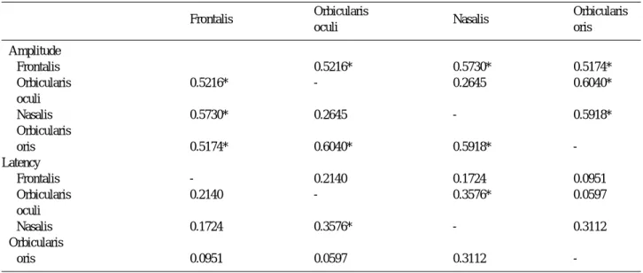
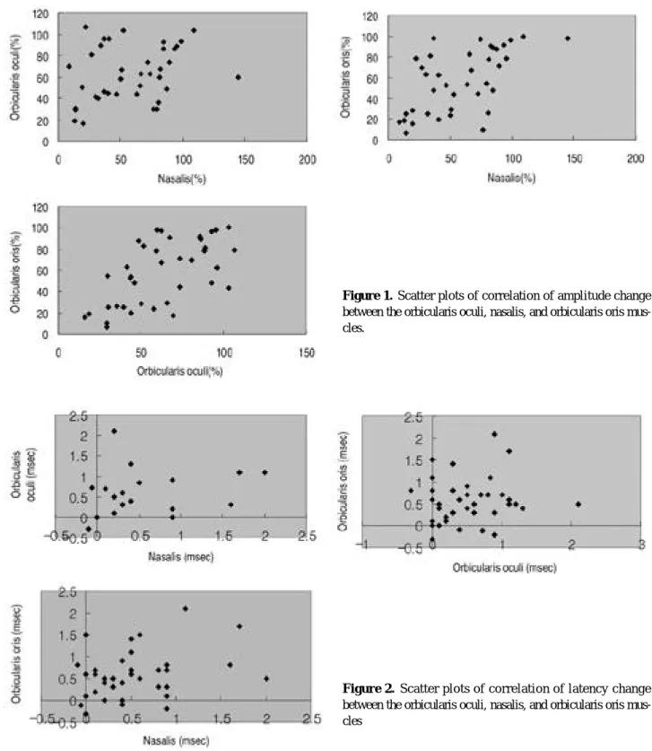
The scatter plots show the correlation between the nasalis, orbicularis oculi, and orbicularis oris recordings for amplitude and latency changes (Fig. 1,2). It appeared that the correlation of
Figure 1. Scatter plots of correlation of amplitude change between the orbicularis oculi, nasalis, and orbicularis oris mus- cles.
Figure 2. Scatter plots of correlation of latency change between the orbicularis oculi, nasalis, and orbicularis oris mus- cles
amplitude change between the individual muscles was stronger than that of latency change in this plotting as statistical analysis showed.
When the electroneurographic values were compared in the four muscle groups, the general linear models procedure did not show any signifi- cant difference for CMAP amplitude and latency changes (p=0.62-0.63).
Of the 39 Bell’s palsy patients, 38 patients (97.4%) recovered satisfactorily within 6 months.
Only a patient of the 39 Bell’s palsy patients (2.6%) had an unfavorable outcome.
D I S C U S S I O N
Although the correlation value between the nasalis and orbicularis oculi muscles was rela- tively weak (r=0.2645), the electrophysiologic pattern was similar in all four muscles studied in the 39 patients with Bell’s palsy at least for amplitude changes. There are some reports on electroneurographic recordings from different facial muscles in Bell’s palsy. One of them showed that electroneurography over the nasal alae and orbicularis oris muscle had reliable val- ues among other muscles and it was the best sin- gle recording in predicting patients with favorable o u t c o m e .2 , 3 , 4 In facial nerve palsy, most elec- tromyographers obtain electroneurographic recordings in the frontalis, orbicularis oculi, orbicularis oris, and nasalis muscles routinely to initially assess facial nerve functioning, prognos- ticate a functional return and follow acute, suba- cute and chronic stages of all facial nerve lesion.
However, the general linear models procedure in this study did not show any significant difference for CMAP amplitude and latency changes, when the electroneurographic values were compared in the four muscle groups. These results were con- sistent with the suggestions by Laskawi and Damenz that the lesion in Bell’s palsy is located in a portion of the facial nerve with no somato- topic organization.5 Our findings are also similar to the conclusion by Engstrom et al.6 They were reluctant to record in more than one facial muscle region for the clinical advantage in Bell’s palsy.
Aside from this result, the correlation values between the muscles were weak in their initial examination, which is different from ours.
However, the correlation was improved at follow-
up examinations in their study. Although we did not perform the follow-up study, the correlation was relatively strong in the early electroneuro- graphic recordings. There would be some reasons why there were somewhat different findings in those studies. Thirty-eight patients among our 39 patients had a favorable outcome at 6 months.
Some studies indicated that if 90% of the nerve fibers degenerate within the first 2 weeks of the acute paralysis, a severe injury had occurred and the chances of complete recovery were less than 5 0 % .7They suggested a response of 10% or less as a prognostic factor,1 whereas there were just 2 patients among 39 patients satisfied with these criteria in our study. Most patients in our study had less degeneration of facial nerve fibers than others at the initial examination. Thus, the sta- tistical power was insufficient to assess the advantage and predictive effect of specific muscle recording among four muscles. Likewise clinical status might affect the electroneurographic fea- tures when it was recorded in the early stage of B e l l’s palsy in particular. There was no statement of the initial status of the patients in the previous studies, which could make the direct comparison with other studies impossible. Furthermore, there is a study showing that the amplitude ratio is not constant in every healthy individual at repeated m e a s u r e m e n t s .8 In addition, Bell’s palsy is one of the acute disorders that can be expected to recover without any measure in many patients and show variable features according to the stage of the assessment because of potentially dynamic lesion. Even 50% of patients with 95% to 98%
degeneration will still have a satisfactory recov- ery. It is also noteworthy to check the facial CMAP 4~7 days after facial weakness develops so that enough time will have passed for Wallerian degeneration to occur. It seems that those aspects could make correlation value of this study differ- ent from others. It is also likely that too many patients with favorable outcome in our study affected the correlation values. In addition, benign clinical course yielded poor correlations with functional return of a few patients in this study and are of limited usefulness in the prog- nostication of Bell’s palsy. Likewise, some study showed that of the 18 patients who recovered completely, 13 had a total palsy at presentation.9 These unavoidable natures of Bell’s palsy could
explain the gap between clinical outcome and ini- tial electrophysiologic values in many studies.
The only patient with unfavorable outcome in our study had CMAP amplitude more than 10% when it was recorded 3 days after the onset.
Given the changes of terminal latencies of facial nerve, they were of very limited value in reflect- ing the features of facial nerve lesion at least in B e l l’s palsy. In many cases, normal and even less value of latencies were measured as often as pro- longed terminal latencies for four muscles in the affected side. It seems that a normal latency would be expected for as long as even a small number of axons remained. The correlation values of terminal latencies in our study were also weak except the value between the nasalis and orbicu- laris oculi muscle (r=0.3576). We did not classify the patients according to the severity of the weakness, because the symptoms of Bell’s palsy in the acute stage could change day by day. It was not easy to assess the symptom at the same day after onset.
In conclusion, the correlation of amplitude change between the different facial muscle elec- troneurographic recordings was relatively strong in Bell’s palsy patients, but it was weak for ter- minal latency change. This study did not show any significant clinical advantage of electroneu- rographic recordings in more than one facial muscle at the early stage of Bell’s palsy.
REFERENCES
01. Dumitru D, Walsh NE, Porter LD. Electrophysiologic evaluation of the facial nerve in Bell’s palsy. Am J Phys Med Rehabil 1998;67:137-144.
02. Yasukawa M, Yasukawa K, Ohnuma H. Prognostic diag- nosis of facial palsy with electroneuronography. M a s u i 1995;44:378-387.
03. Kartush JM, Lilly DJ, Keminik JL. Facial electroneurogra- phy: Clinical and experimental investigations. Otolaryngol Head Neck Surg 1985;93:516-523.
04. Coker NJ. Facial electroneurography: Analysis of tech- niques and correlation with degenerating motoneurons.
Laryngoscope 1992;102:747-759.
05. Laskawi R, Damenz W. Elektrophysologische Untersuchun- gen zum Schadigungs-muster der mimischen Muskulatur bei der Bellschen Lähmung. L a r y n g o - R h i n o - O t o l 1 9 9 3 ; 7 2 : 1 9 7 - 2 0 3 .
06. Engstrom M, Jonsson L, Grindlund M, Stalberg E.
Electroneurographic facial muscle pattern in Bell’s palsy.
Otolaryngol Head Neck Surg 2000;122:290-297.
07. Fisch U. Surgery for Bell’s palsy. Arch Otolaryngol Head Neck Surg 1981;107:1-11.
08. Sittel C, Guntinas-Lichius O, Streppel M, Stennert E.
Variability of repeated facial nerve electroneurography in healthy subjects. Laryngoscope 1998;108:1177-1180.
09. Smith IM, Maynard C, Mountain RE, Barr-Hamilton R, Armstrong M, Murray JA. The prognostic value of facial electroneurography in Bell’s palsy. Clin Otolaryngol 1994;19:201-203.